Fischer W.B. Viral Membrane Proteins: Structure, Function, and Drug Design
Подождите немного. Документ загружается.

alone was seen to drive the formation of VLP at the plasma membrane and the presence of
Vpu enhanced the release of VLP into culture medium by approximately 13-fold. HMA
(10 M) inhibited the release of VLP from Gag/Vpu expressing cells by more than 90%. It
can be seen that the budding in Figure 15.4A is depressed in the presence of 10 M HMA
(Figure 15.4B).
2.3.3. HMA Inhibits HIV-1 Replication in Monocytes and Macrophages
As a consequence of the demonstrated ability to both block the Vpu ion channel and
inhibit VLP budding, HMA and DMA were tested for anti-HIV-1 activity and were found to
repress replication of the laboratory-adapted macrophage-tropic strain HIV
BaL
in cultured
human monocyte-derived macrophages (Ewart et al., 2004). At concentrations in the range of
1–10 M, HMA and DMA strongly inhibited production of extracellular virus as measured
by p24 antigen ELISA, as illustrated in Figure 15.5.
In addition to measuring p24 in culture supernatants, the level of HIV-1 viral DNA
inside the cells was assessed using semiquantitative (Q) PCR to amplify a 320 bp LTR/Gag
fragment. In these experiments, some inhibition of HIV DNA accumulation in the cells was
observed but it was clearly not as marked as the corresponding inhibition of p24 antigen.
These findings indicated that the primary inhibitory target of HMA is at a stage of the repli-
cation cycle after reverse transcription of viral RNA and are consistent with an effect of HMA
on the Vpu ion channel and virus budding.
In summary, Vpu from HIV-1, like M2 from influenza A, belongs to a growing family
of virus ion channels. Recombinant and synthetic Vpu polypeptides in planar lipid bilayers
form Na
- & K
-selective channels that show gating and have a minimal conductance of
about 14 pS. The Vpu channel activity is inhibited by HMA and DMA, but not by the parent
compound, amiloride. The ion channel activity of Vpu has been linked to the protein’s func-
tion in augmenting virus particle release: Virion budding is strongly suppressed by either
Virus Ion Channels Formed by Vpu of HIV-1 215
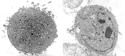
AB
Figure 15.4. Inhibition of budding of virus-like particles (VLP) by HMA. HeLa cells at 60–70% confluence were
infected with the T7 RNA polymerase expressing vaccinia virus vector vTF7.3 and then transfected with pcDNA3.1
based plasmids for expression of HIV-1 Vpu and Gag proteins under control of T7 promoters. For observation by
electron microscopy, the cells were grown on glass coverslips and subsequently fixed before staining with uranyl
acetate and encasing in Spurr’s resin from which thin sections were cut and stained with lead citrate. The cell in
micrograph A is representative of control cultures incubated in the absence of HMA and shows a distinctly
“unhealthy” cell surrounded by many VLP. Micrograph B is a representative cell from a parallel culture incubated in
the presence of 10 M HMA showing a large reduction in the number of VLP associated with the cell membrane.
genetic mutations that ablate Vpu ion channel activity or chemical inhibitors of the channel,
HMA and DMA. Though it is not currently understood how changes in cellular ion gradients
contribute to the budding process, nevertheless, the rate and efficiency of budding of new
virions from the plasma membrane is clearly of fundamental importance to the replication of
HIV-1. Vpu plays a number of roles in the molecular biology of HIV-1, and while expression
of Vpu is not essential in some in vitro systems, this protein does enhance the efficiency of
virus replication, particularly in macrophages. At present the presumably critical roles played
by Vpu in human HIV-1 infections remain elusive. However, the ability of HIV-1 to infect
and efficiently replicate in macrophages is thought to be essential in AIDS pathogenesis
(Schuitemaker, 1994). In fact, it has been suggested that macrophage-tropic HIV isolates may
be necessary and sufficient for the development of AIDS (Mosier and Sieburg, 1994).
Therefore, drugs such as HMA and DMA that block the Vpu channel and inhibit HIV-1 repli-
cation in macrophages may ultimately have potential therapeutic use as anti-AIDS agents.
Clearly, there is a need for further fundamental research to define the structure of the Vpu ion
channel and to understand its mechanism so that a rational approach to designing better
channel-blocking drugs can be undertaken. The ultimate aim would be to design a drug of
such high specificity for the Vpu channel that it would act like a magic bullet to slow or even
prevent the progression from HIV-1 infection to AIDS.
3. Alphavirus 6K Proteins
Alphaviruses are members of the Togaviridae family, which includes flaviviruses, the
smallest enveloped animal viruses. This family contains a number of important animal and
human pathogens that are extremely adaptable and are found in a range of habitats on every
216 Peter W. Gage et al.
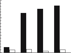
36914
days after infection
0
6000
12000
18000
virus (p24 pg/ml)
Macrophages
Figure 15.5. Inhibition of HIV-1 replication by HMA. Monocyte-derived macrophages (MDM) cultured
from human HIV seronegative blood, were infected at a multiplicity of infection of 0.02 with the laboratory adapted
R5 M-tropic HIV
BaL
strain: 10
6
MDM per well in a 48-well tissue culture plate were incubated in the presence of
virus for 1 hr, then the medium was replaced and cultures were maintained for variable periods up to 14 days after
infection. Compounds were added immediately after infection of cells with the virus and media—containing the
appropriate concentration of HMA—were replenished every 3 or 4 days. Culture media were sampled at the days
indicated in the graph and the concentration of HIV p24 antigen in the supernatants was quantified by ELISA using
the Coulter HIV-1 p24 antigen assay. The filled and open bars show the levels of HIV in the control (no drug) and
HMA (10 M)-treated cultures.
continent except Antarctica. Alphaviruses are unusual in their ability to cross phyla in the
normal course of the infection cycle (i.e., vertebrate to nonvertebrate).
3.1. Replication of Alphaviruses
Much is known about the cell biology of alphavirus replication in vertebrate cells
through the study of the model viruses Sindbis virus (SINV) and Semliki Forest virus (SFV).
The cellular infection process can be summarized as follows: binding and endocytosis, fusion
with endosomes and release of genome to cytoplasm, transcription and translation of the viral
genome, viral assembly, and budding.
3.1.2. Viral Budding
Alphavirus budding has been extensively studied, and some of the molecular mecha-
nisms have been described. The “early” budding model was based on observations with SFV
(Garoff and Simons, 1974); it was proposed that protein–protein interactions between the
cytoplasmic domain of the spike proteins and the nucleocapsid drive budding. Initially, a pre-
assembled nucleocapsid at the plasma membrane interacts with just a few spike proteins. The
reduced mobility of the complex attracts more spike proteins to interact with the nucleocap-
sid. This causes the membrane to curve, eventually encircling the entire nucleocapsid with
glycoprotein spikes. Budded virions are predominantly seen exterior to cells, appearing as
electron-dense regions of diameter 40–60 nm. SFV genomes containing various deletions
were used to show that budding is dependent on nucleocapsid–spike interactions. Genomes
lacking either the spike protein or capsid genes were unable to generate particles individually.
However, co-expression of spike proteins with capsid protein allowed nucleocapsids to
assemble into particles at the plasma membrane. The molecular mechanism of the spike–C
interaction have been identified using molecular modeling, and reconstructions using anti-
idiotype antibodies and fitting the X-ray crystal structures of capsid and E2 into cryoelectron
microscopy-derived electron density maps (Garoff et al., 1998).
3.2. The 6K Protein of Alphaviruses
The importance and role(s) of the 6K protein in cellular infection are not well charac-
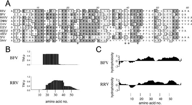
terized. The 6K proteins are not highly conserved between alphaviruses (Figure 15.6A),
having only 58% sequence identity but presumably have a common structure and roles.
The small size of the 6K protein (60 amino acids for Ross River virus (RRV) and 58 for
Barmah Forest virus (BFV)), fatty acylation (SINV 6K is palmitoylated on cysteine residues
23, 35, 36, and 39), and hydrophobicity (Figures 15.6B and C) are typical characteristics of
membrane-associated proteins.
These reasons, coupled with its toxicity when expressed in E. coli are probably why a
purification strategy for 6K had not been published earlier (Melton et al., 2002). Examination
of hydrophobicity plots of 6K proteins (see Figure 15.6C) suggests two TM domains. This is
supported by evidence for the topology of the polypeptide from which 6K protein is derived.
However, TM prediction of BFV 6K (using a hidden Markov model) to identify residues typ-
ically found at membrane-aqueous junctions shows a single TM domain covering amino acid
residues 14–32 (Sonnhammer et al., 1998). The prediction for RRV 6K is not as definitive,
but nevertheless indicates a single TM domain. If the protein were to cross the membrane
Virus Ion Channels Formed by Vpu of HIV-1 217
twice, the second domain would be squeezed in very tightly, or would have to make an
intra-membranous loop.
3.3. The 6K Protein and Virus Budding
To investigate the role of the 6K protein in virus replication, a range of site-directed
mutations have been made in SFV and SINV. Removal of the SFV 6K gene affects the effi-
ciency of glycoprotein processing, and the cleavage of p62 and its subsequent transport to the
plasma membrane (Schlesinger et al., 1993; Sanz and Carrasco, 2001). The efficiency of SFV
budding from BHK cells is dramatically decreased when the 6K gene is deleted (Loewy et al.,
1995). It is notable that the relative importance of the 6K protein for budding is highly depend-
ent on the cell type examined. For example, SFV 6K virus yield ranged from 3–50% of
wild-type SFV (Loewy et al., 1995). This is suggestive of some interaction of 6K protein with
plasma membrane components. SINV with residues 35 and 36 of the 6K protein mutated from
cysteine to alanine and serine make virions of a multi-cored appearance (Gaedigk-Nitschko
et al., 1990). The altered appearance of virions suggests some involvement of 6K protein with
budding or membrane fusion. How the 6K protein exerts its effects on budding is not known.
It has been reported to be present at low levels in virions and yet it is not contained in any
virion reconstructions. Nor does it seem to have any role in docking/internalization processes,
as 6K gene deletion has no effect on the infectivity of virions (Liljestrom et al., 1991).
3.4. Ion Channels Formed by BFV and RRV 6K Protein
In order to test the hypothesis that 6K proteins can form ion channels, we expressed the
6K protein gene sequences of RRV and BFV in E. coli, purified the 6K protein and examined
218 Peter W. Gage et al.
Figure 15.6. A: Homology sequence alignment of alphavirus 6K proteins. Identical residues are shown in gray
boxes, homologous residues are shown in plain boxes. Cysteine residues that become palmitoylated in SINV 6K
protein are indicated with asterisks (*). B: Transmembrane prediction for BFV and RRV 6K proteins. The probability
of a residue being located in a transmembrane helix (TM p) is plotted against amino acid sequence no. using the
TMHMM algorithm (Sonnhammer et al., 1998). C: Hydrophobicity plot for BFV and RRV 6K proteins.
Hydrophobic regions are above the baseline, hydrophilic regions are below.
Virus Ion Channels Formed by Vpu of HIV-1 219
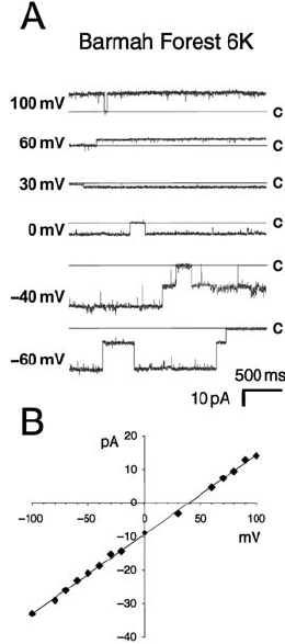
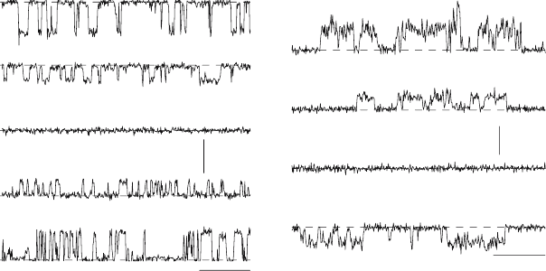
Figure 15.7. BFV 6K ion channels. A: Channel activity is shown for a range of potentials. The closed state is
shown as a solid line. Openings are deviations from this line. Scale bars are 500 ms and 10 pA. Solutions contained
cis: 510 mM NaCl, 10 mM TES (N-tris[Hydroxymethyl] methyl-2-aminoethanesulfonic acid), pH 7.0; trans:60mM
NaCl, 10 mM TES (N-tris[Hydroxymethyl] methyl-2-aminoethanesulfonic acid), pH 7.0. B: Current–voltage
relationship for bacterially expressed BFV 6K. Largest single opening events of a single channel are plotted for each
holding potential. Cis and trans solutions were identical to those in part A.
whether it formed ion channels in planar lipid bilayers (Melton et al., 2002). Examples of
typical current traces caused by BFV 6K protein are shown in Figure 15.7A and the currents
can be seen to reverse at 40 mV in Figure 15.7B.
Solutions used in these experiments were 510 mM NaCl in the cis chamber and 60 mM
NaCl in the trans chamber. The currents can be seen to reverse between 30 and 60 mV,
indicating a preference for Na
over Cl
. The average reversal potential in the presence of a
510 mM cis/60 mM trans NaCl gradient was 49.1 / 0.7 mV. When no protein was added
to the cis solution, no activity was seen (n 20, average waiting time 7 min).
The open probability of RRV 6K channels was found to be voltage-dependent. Channels
were much more active at 100 mV than at 100 mV. A channel that was closed at positive
holding potentials could often be reactivated by switching to negative holding potentials.
The permeability of RRV 6K channels to cations other than Na
was tested by
changing the solution in the cis bath to either KCl or CaCl
2
while maintaining the holding
potential at 100 mV. When the cis chamber contained potassium or calcium ions, ionic
currents were observed. Thus, the channels were also permeable to the cations K
and Ca
2
.
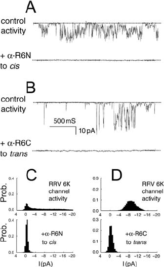
3.5. Antibody Inhibition of RRV 6K Channels
It is not possible to obtain proteins at 100% purity from expression systems or by
synthesis with a peptide synthesizer. Because only a few protein molecules are needed to form
an ion channel, it is important to be able to demonstrate that it is indeed the predominant
protein of interest, not some contaminant that is forming the ion channel. One way of doing
this is to demonstrate effects of specific antibodies to the protein of interest on the ion
channel activity. Polyclonal antibodies raised in rabbits immunized with synthetic peptides
corresponding to the N- or C-termini of RRV 6K were used to confirm that the 6K protein
was indeed the channel-forming molecule in preparations of the purified recombinant protein.
When antibody recognizing the N-terminal 20 amino acids of RRV 6K (-R6N) was added
to the cis chamber, a reduction in current to baseline levels occurred. An example of this is
shown in Figure 15.8A and was seen in eight bilayers. The -R6N antibody had no effect
when added to the trans chamber (n 5, data not shown).
Conversely, antibody against the C-terminal of RRV 6K (-R6C) inhibited channel
openings when added to the trans bath (n 6, Figure 15.8B), but not the cis bath (n 7, not
shown). All points histograms of currents recorded before and after addition of antibody are
shown in Figures 15.8C and D.
The chamber-dependent effect of antibodies demonstrates that channel inhibition was
specific to the particular epitope recognized by the antibody.
The antibody inhibition experiments also indicate that the RRV 6K protein is stably ori-
ented in bilayers. This is a corollary of the specific topological requirements of vesicle fusion
with planar lipid bilayers, that is, proteoliposomes placed in the cis solution, will fuse
with a bilayer so that the intra-vesicular domains of TM proteins will be exposed to the trans
solution.
The use of affinity-purified antibodies to specifically inhibit channel currents from both
sides of the bilayer (Figure 15.8) supports the conclusion that the 6K protein forms an ion
channel. The chamber-specific effect of antibody inhibition suggests further that 6K proteins
are oriented in bilayers with the N-terminal facing the cis bath, and the C-terminal facing the
trans bath. Given the length of the 6K polypeptide chain, location of N- and C-termini on
opposite sides of the membrane suggests that the hydrophobic domain consists of a single TM
-helix. Earlier reports have suggested that 6K crosses the ER membrane twice, with both
termini in the lumen. However, these data do not exclude the possibility that the C-terminus
of 6K is only transiently located in the ER lumen. The C-terminus of the E2 protein of SINV
has been shown to retract through the ER membrane following cleavage by the signalase
enzyme. A similar retraction of the C-terminal of the RRV and BFV 6K proteins may occur
following cleavage by signalase. The structures of most virus-encoded ion channels discov-
ered to date consist of a single TM domain. Thus, it seems likely that the biologically active
form of 6K protein has a single TM domain.
Following the onset of viral RNA translation in alphavirus-infected cells, the plasma
membrane becomes more permeable to monovalent cations (Ulug et al., 1984). This is fol-
lowed by an increase in permeability to larger molecules, such as translation inhibitors
(Munoz et al., 1985). It is possible that the ion channels formed by 6K proteins are responsi-
ble for both of these changes. This hypothesis is supported by previous experiments on the
220 Peter W. Gage et al.
fate of 6K protein in infected cells (Lusa et al., 1991). During alphavirus infection, the cell’s
secretory pathway is used to export the structural proteins of the virus (excepting capsid
protein) to the plasma membrane. The topology of the membrane-bound proteins is such
that the N-terminal of the 6K protein becomes exterior to the cell: fusion of ER-derived vesi-
cles with the plasma membrane results in a cell-exterior location for residues in the ER lumen.
The bilayer experiments with 6K proteins at a holding potential of 100 mV approximate the
voltage and orientation conditions for 6K proteins at the plasma membrane of infected cells.
Thus, the dramatic activation of 6K ion channels by negative voltages (see Figure 15.6) would
also occur at the plasma membrane of infected cells.
If the 6K protein is indeed located at the cell surface then it is also possible that
the increased permeability of infected cells to monovalent cations (Ulug et al., 1984) is due
to the preference of 6K channels for monovalent cations over divalent cations. The wide range
Virus Ion Channels Formed by Vpu of HIV-1 221
Figure 15.8. Antibody inhibition of RRV 6K channels. A: Channel activity is shown before (control), and after
(-R6N) addition of anti-RRV 6K N-terminus antibody to the cis chamber. B: Channel activity is shown before
(control), and after (-R6C) addition of anti-RRV 6K C-terminus antibody to the trans chamber. A dashed line
represents the closed (baseline) state. Channel openings are downward deflections from the baseline. C,D: All points
histograms showing the effect of antibodies on RRV 6K channels. Paired histograms are shown before (above, “RRV
6K channel activity”), and after addition of antibody to the stated chamber (below). C: Anti-RRV 6K N-terminus
(-R6N) was added to the cis chamber. D: Anti-RRV 6K C-terminus (-R6C) was added to the trans chamber.
Current amplitude probability histograms were generated from 30 s of record. NB: baseline current 0 pA.
of conductance values obtained for 6K channels may reflect the protein’s ability (as infection
proceeds) to form channels with larger pores and conductances.
4. NB of Influenza B Virus
In 1984 NB was first described (Shaw and Choppin, 1984) and is unique to the
influenza B virus. It was thought to be the homolog of the influenza A virus M2 protein (Shaw
and Choppin, 1984; Williams and Lamb, 1988). NB is abundantly expressed in virus-infected
cells (Shaw and Choppin, 1984) and found in low copy numbers in the influenza B virus
(Betakova et al., 1996; Brassard et al., 1996).
Using [
35
S]cysteine and [
3
H]isoleucine, Shaw (Shaw and Choppin, 1984) detected the
presence of NB in cells infected with three different strains of influenza B virus. The pres-
ence of NB in all three strains of influenza B indicated that this protein was not unique to any
one influenza B strain. Shaw and Choppin (1984) used pulse-chase experiments to study the
kinetics, synthesis, and stability of NB in virus infected cells. Their observations showed that
both NA and NB accumulated in approximately equal quantities in the cells 4 hr after infec-
tion. It was observed that after 8–10 hr of infection, NB was the major protein and that there
was less NA than NB during this stage of infection. They reported that NB disappeared from
the cells 4 hr after it was synthesized. This result is contrary to other findings (Betakova et al.,
1996) showing that the protein is stable for up to 12 hr after infection.
Shaw and Choppin (1984) used hyper-immune mouse serum to demonstrate the pro-
duction of NB during a respiratory infection of influenza B in mice. They showed that mice
infected with the influenza B virus develop antibodies to the NB protein. The B/Lee/40 strain
seems to have a stronger anti-NB response than was detected with the NA or M1 proteins.
Hyper-immune mouse serum was used to detect the presence of NB in cells infected with dif-
ferent strains of influenza B. The serum reacted with all the strains that were tested suggest-
ing that the NB had retained its antigenic determinants sites over a period of 43 years
separating the oldest and newest isolates studied.
4.1. Structure of NB
It has been found that NB is an integral membrane protein associated with the same
membrane fractions as hemagglutinin (HA) and neuraminidase (NA). Like other viral and
cellular membrane proteins 2% TritonX-100 and 0.5M KCl are required to solubilize NB
(Williams and Lamb, 1986). NB is a 100 amino acid protein and has a molecular weight of
12,000 Da whereas the glycosylated protein has a molecular weight of 18,000 Da. The
nucleotide sequence of NB contains 7 cysteine residues, 18 isoleucine residues, and 4 poten-
tial glycosylation sites (Asn-X-Ser/Thr), of which two are found on either side of the TM
region. Asn at residue 3 and Asn at position 7 have been identified as the precise glycosyla-
tion sites. NB contains carbohydrate side chains of the high-mannose type that are processed
to complex sugars in either MDCK or CV1 cells.
NB is a Class III integral membrane protein. The N-terminal domain of 17 amino acids
is extracellular and the large C-terminus of 64 amino acids is intracellular (Williams and
Lamb, 1988). The amino acid sequence of NB shows that it has a region of uncharged
hydrophobic residues (19–40) that could span a membrane. This region has a hydropathic
index greater than two indicating that the protein interacts with membranes.
222 Peter W. Gage et al.
The NB protein has two stretches of hydrophobic amino acids the first one at the
N-terminus, residues 19–40, and at the C-terminus, residues 84–95. NB is anchored in
the membrane via its N-terminal hydrophobic domain. William and Lamb (1986) used
site-directed mutagenesis involving the deletion of the N-terminal region to show that the
N-terminus of 18 amino acids is exposed at the cell surface.
The sequence of the NB protein is shown below.
A
MNN A TF NCTN INPI T15
HIRGSI IITICVSLI30
VILIVFGCIAKI FI N45
KNN C TNNVIRVHKRI60
KCPDCE PFCNKRDDI75
STPRAGVDIPSFILP90
GLNLSEGTPN
4.2. Similarities Between M2 and NB
It was suggested that NB is the influenza B homolog of the influenza A M2 protein
(Shaw et al., 1983; Williams and Lamb, 1986). Recently there have been suggestions that the
BM2 protein of the influenza B is the counterpart of the M2 protein of influenza A and that
BM2 forms ion channels that conduct protons (Mould et al., 2000). Nevertheless, the NB pro-
tein of influenza B virus is analogous to the M2 protein of influenza A virus in many respects.
There is no amino acid sequence homology between the two proteins but they share a num-
ber of common characteristics. Both are small proteins, NB 100 amino acids and M2 96
amino acids. Both are class III integral membrane proteins (von Heijne, 1988) with a single
highly hydrophobic TM region. This region in both proteins has a hydropathic index greater
than 2, indicating that the proteins interact with the membrane. The proteins are anchored
in the membrane via their hydrophobic regions and are oriented with a short extracellular
N-terminal region (M2 24, NB 18) and a long cytoplasmic tail (M2 53 amino acids and
NB 60). The oligomeric structure of both proteins is similar; like M2, NB runs as a dimer or
tetramer on nonreducing gels. The level of expression of both proteins in virus-infected cells
is the same. Both proteins are abundantly expressed on virus-infected cells and are found
associated with the same membrane fractions as HA and NA. On the other hand both proteins
are found in low copy numbers in the viral envelope. The number of molecules per virion is
14 to 68 molecules of M2, 15 to 100 molecules of NB.
The function of NB in the virus still remains uncertain. As NB is located at the host cell
surface it has been suggested that it may be involved in transcription or replication of RNA, or
that it may be involved in organizing proteins on the cell surface during budding. The function of
NB is probably important to the viral life cycle, otherwise the overlapping reading frame would
have been lost by natural mutation. A single mutation in the initiation codon of NB can lead to
elimination of the NB reading frame. This mutation would not affect the coding of NA but this
has not occurred and NB is found both in virus-infected cells and the virus particle.
There is direct evidence that M2 forms a proton channel and since NB is similar in
so many respects to M2, it is possible that NB may also function as an ion channel. To test
the hypothesis that NB forms an ion channel like the M2, we cloned the NB protein from the
strain influenza/B/Lee/40 into bacterial expression vectors, and then purified and incorpo-
rated NB into artificial lipid bilayers to determine whether it functions as an ion channel.
Virus Ion Channels Formed by Vpu of HIV-1 223
4.3. Channel Activity of the NB Protein
We cloned the cDNA for NB into two expression systems (Sunstrom et al., 1996).
In the first the cDNA was cloned into the plasmid vector pQE-70 (Qiagen) so that the
C-terminus of the protein had a tag of six histidines at the end of the open reading frame. In
the second expression system the NB cDNA was cloned into the BamH1 site of the plasmid
vector pGEX-2T. In this plasmid the NB open reading frame is fused to the C-terminus of
GST (Glutathione-S-transferase). The NB produced by the GST-fusion protein was truncated
at the C-terminus. The protein failed to react with the C-terminal polyclonal antibody. The
protein was, however, detected on silver-stained gels. Both the polyHis and GST purified NB
showed channel activity.
4.4. Currents at pH 6.0
The planar lipid bilayer technique was used to see whether the NB protein formed ion
channels. The purified protein was incorporated into artificial bilayers by adding 10 to 50 l
of the fractions containing the protein (as detected from Western blots) to the cis chamber.
The contents of the chamber were stirred until channel activity was seen. Channel activity was
observed between 1 and 30 min after addition of the protein at a potential of 0 mV. The cis
and trans chambers contained 150–500 mM NaCl and 50 mM NaCl solutions, respectively,
at pH 6.0. Typical currents recorded are shown in Figure 15.9A.
The average reversal potential obtained with 150/50NaCl mM in three experiments
was found to be 20 2.1 mV. With 500/50 NaCl mM in four experiments the reversal
potential was found to be 36.8 1.85 mV. The sodium equilibrium potentials for solutions
containing 500 mM NaCl in the cis chamber and 50 mM in the trans chamber would be
53 mV and that for 150 mM NaCl in the cis chamber and 50 mM in the trans chamber
224 Peter W. Gage et al.
0.4 s
0
80
60
40
20
2 pA
40
–40
–20
20
2 pA
0.2 s
pH 6.0 pH 2.5
AB
Figure 15.9. Channels formed by NB. Current traces recorded after the addition of NB to the cis chamber
containing 500 mM NaCl and the trans chamber contained 50 mM NaCl. The broken lines indicate the zero current
level. A. Currents recorded at pH 6. B. Currents recorded at pH 2.5.