Cui Dongmei. Atlas of Histology: with functional and clinical correlations. 1st ed
Подождите немного. Документ загружается.

CHAPTER 14
■
Oral Cavity
275
Name of
Tissue
Productive
Cells
Inorganic
(Mineral) %
Organic % Water % Sequence
of Hard
Tissue
Formation
Sequence
of
Productive
Cell
Formation
Activity Embryological
Origin
Dentin Odontoblasts 70 20 10 First Odonto-
blasts second
Lifetime Dental papilla
(mesoderm)
Enamel Ameloblasts 96 3 1 Second Amelo-
blasts fi rst
Before
tooth
eruption
Enamel organ
(ectoderm)
Cementum Cementoblasts 65 23 12 Third Cemento-
blasts third
Lifetime Dental sac/
follicle
(mesoderm)
Alveolar
bone
Osteoblasts 60 25 15 — — Lifetime;
respond
to stresses
and
tensions
Mesoderm
TABLE 14-3 Dental Hard Tissue
CLINICAL CORRELATIONS
Figure 14-16A.
V
itamin D–Resistant Rickets. H&E, 476
Vitamin D–resistant rickets, commonly known as X-linked
hypophosphatemic rickets, is characterized by resistance to con-
ventional vitamin D treatment, decreased reabsorption of phos-
phate by the renal tubules, abnormalities in bones and teeth,
osteomalacia, and hypocalcemia. It is an X-linked autosomal
dominant disorder. Patients with these rickets disorders do not
respond to high-dose vitamin D treatment. Signs include rickets,
short stature, bowing of lower limbs, seizures, congestive cardiac
failure, and tooth defects including calcifi cation defects in dentin,
enlarged pulp chambers, pulpitis, and pulp necrosis. Treatment
with 1, 25-vitamin D (which is not dependent on normal hor-
monal mechanisms or organ systems to activate) plus controlled
phosphate therapy may improve the condition.
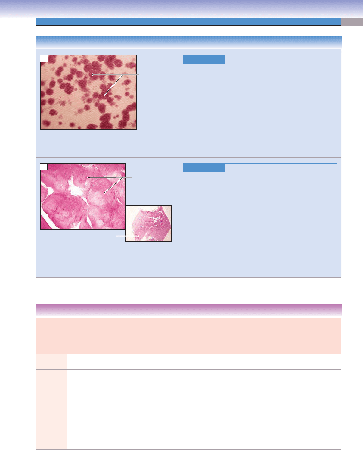
Figure 14-16B.
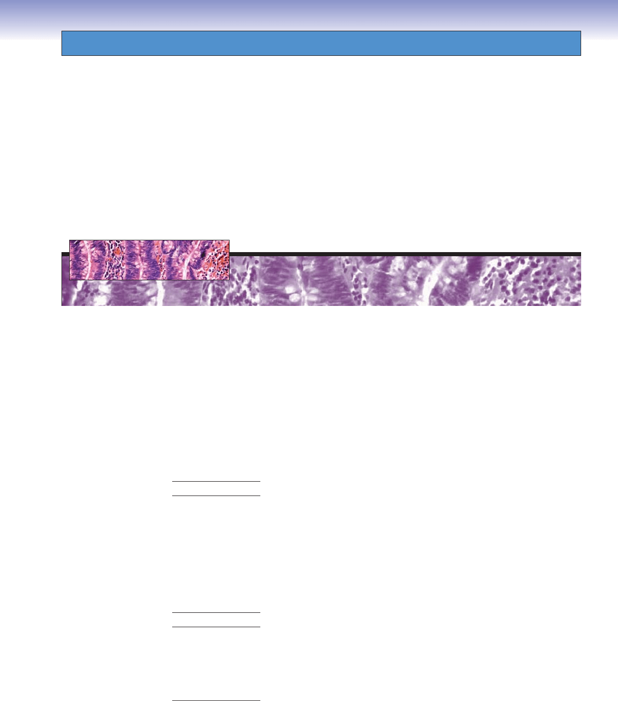
Dentin Dysplasia. H&E, 35; inset 7
Dentin dysplasia is an autosomal-dominant tooth disorder
. Teeth
in this disorder are sometimes called rootless teeth, because they
often have very short and conical roots. Dentin dysplasia can be
classifi ed into types I and II. Type I, also called the radicular type,
is characterized by short roots and pulp obliteration. Type II, also
called the coronal type, is characterized by a “thistle tube” pulp.
Radiographically, extension of the pulp chamber into the root is
usually observed in dentin dysplasia. Pulp stones and sudden con-
striction of the pulp chamber are common. Dentin dysplasia type
II shares some similarities with dentinogenesis imperfecta type II,
but in dentin dysplasia the permanent dentition has normal color
or only slight discoloration. Histologically, globules of dentin and
disorganized dentinal tubules are characteristic of this condition.
The left image shows globules of dentin; the right image shows a
low-power photograph of a dentin dysplasia.
Calcification
defect in dentin
A
Dentin
Dentin
Broad
root
Globules
of dentin
Dentin
B
CUI_Chap14.indd 275 6/2/2010 8:23:55 AM
276
UNIT 3
■
Organ Systems
T. Yangi
Odontoblasts
Predentin
Dentin
Cell-free
Cell-free
zone
zone
Cell-free
zone
Cell-rich
zone
Pulp core
Blood
vessel
Apical foramen
Apical foramen
Apical foramen
Radicular
Radicular
pulp
pulp
Radicular
pulp
Coronal
Coronal
pulp
pulp
Coronal
pulp
Blood vessels
Blood vessels
and nerves
and nerves
Blood vessels
and nerves
A
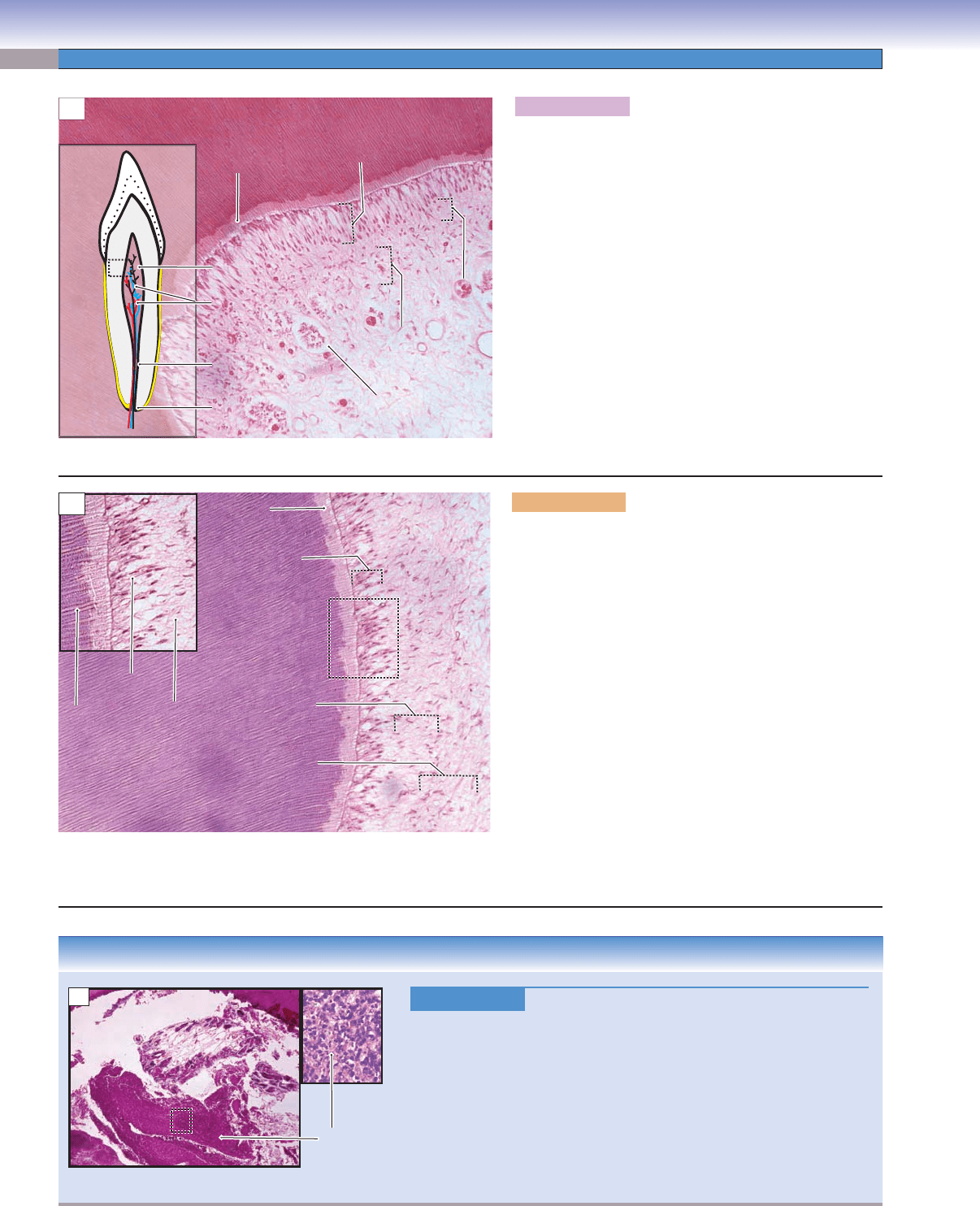
Figure 14-17A. Dental pulp. H&E, 136
Dental pulp is a specialized loose, cellular mucous connective
tissue that fi lls the pulp chamber in the central core and
root canals of the tooth. Fibroblasts are the most numer-
ous cells in the pulp; they produce connective tissue fi bers
(mainly type I and III collagen, fi bronectin, and elastin)
and ground substance. They maintain the pulp matrix. The
second most numerous cells are odontoblasts ( producing
dentin). Other defense cells, such as macrophages and
lymphocytes, may be found in the pulp. No mast cells or
adipocytes (fat cells) are found in the pulp. The pulp con-
tains many blood vessels and nerve fi bers, which enter and
leave at the apical foramen. Most of the nerve fi bers are
afferent fi bers; they are either C fi bers or Ad fi bers, and
both carry pain information through the trigeminal system
to the brain. The efferent fi bers are autonomic nerve fi bers
and innervate the smooth muscle of the blood vessels.
Dental pulp plays an important role in producing dentin
and providing nutrients and sensory input for the dentin.
Dental pulp
Predentin
3. Cell-rich zone
1. Odontoblast
layer
2. Cell-free zone
(zone of Weil)
Odontoblasts
Cell-free
zone
Dentin
Dentinal
tubules
B
Figure 14-17B. Dental pulp. H&E, 136; inset 190
Dental pulp is derived from mesenchymal tissue that forms
the dental papilla during tooth development. The papilla
becomes pulp in the mature tooth. The pulp can be divided
into four zones from the periphery to the center. (1) The
odontoblast layer forms a single cell layer along the periph-
eral edge of the pulp. These cells have processes extending
into the dentin. (2) The cell-free zone, also called the zone
of Weil, is directly under the odontoblast layer. This layer
has fi bers, cellular processes, axons, and capillaries running
through it but contains no cell nuclei. (3) The cell-rich zone
is beneath the cell-free zone, and has many cells and nuclei
of cells densely packed in rows. This layer has fi broblasts,
undifferentiated mesenchymal cells, neural plexuses, and
capillaries. If some odontoblasts die, the undifferentiated
mesenchymal cells in this layer will differentiate into new
odontoblasts. (4) The pulp proper (pulp core) is the cen-
tral part of the pulp and contains blood vessels and nerves
within the loose, mucous connective tissue. The layers of
both cell-free and cell-rich zones are more visible in the
coronal than the radicular pulp region (Fig. 14-17A).
CLINICAL CORRELATION
Figure 14-17C.
Pulp Abscess. H&E 23; inset 168
Pulp abscess refers to an abscess involving the pulp tissue of a tooth.
Pulp abscesses are usually sequelae of dental caries, but they can also
develop in teeth showing no detectable lesions. Pulp abscesses can also
occur after restoration work has been performed. They are characterized
by severe, intermittent pain that may intensify when a patient reclines.
Sharp pain may also be triggered by cold liquids, tapering off to a dull,
pulsating pain. Periapical tissues may not be involved, and the affected
tooth may not show any difference from other healthy teeth in percussion
or pressure tests. T
reatment includes using antibiotics and undergoing
root canals and even tooth extraction.
Pulp cavity
Abscess with
neutrophils
C
CUI_Chap14.indd 276 6/2/2010 8:23:57 AM
CHAPTER 14
■
Oral Cavity
277
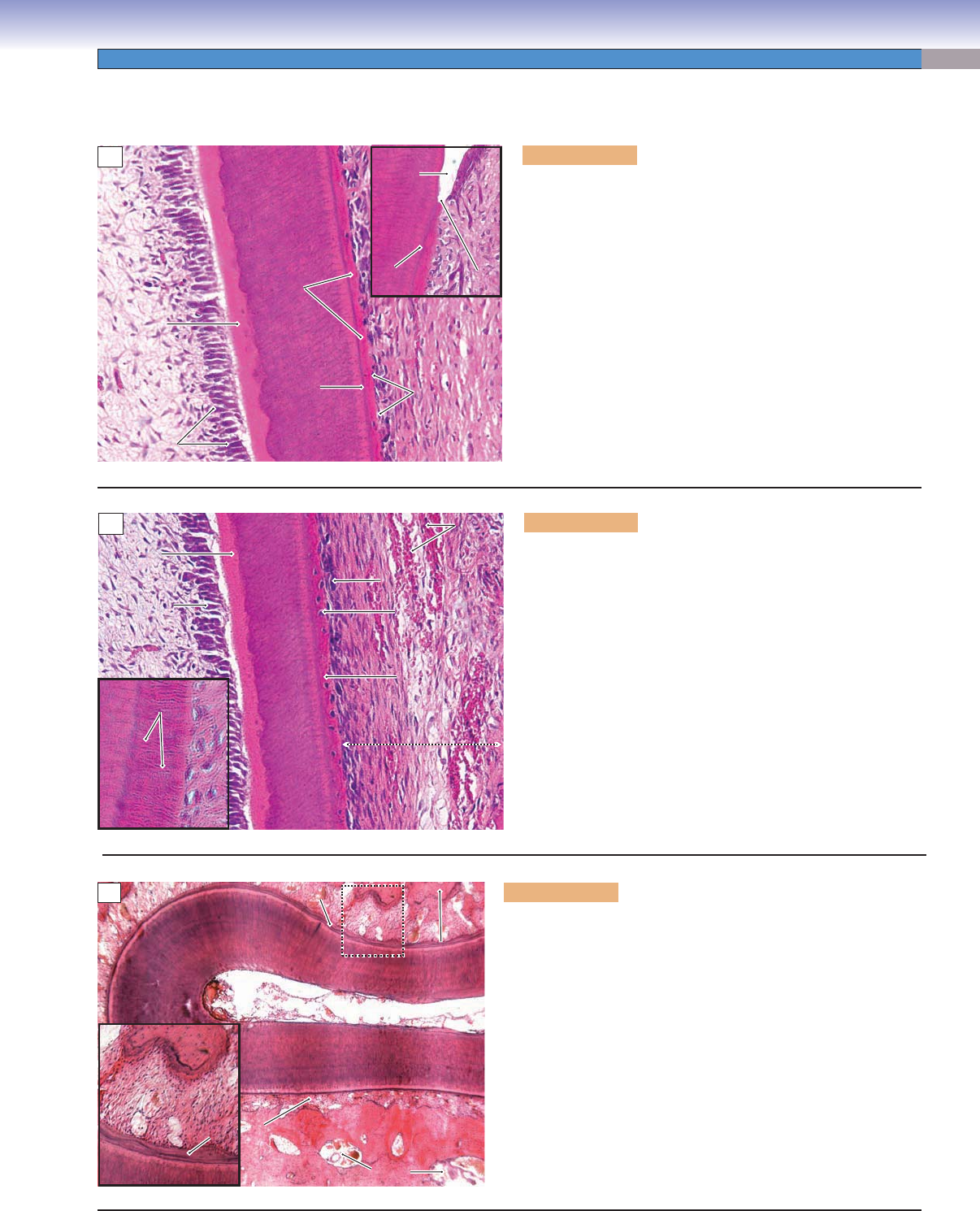
Figure 14-18C. PDL and alveolar bone. H&E, 34; inset 72
The PDL is a dense fi brous connective tissue with a direct nerve
and blood supply. It is located between the cementum and
the alveolar bone, which surrounds the tooth root. Fibroblasts are
the main cells responsible for the formation of the PDL. The PDL
supports the tooth root by forming a strong attachment between
the cementum and alveolar bone by Sharpey fi bers (Fig. 14-19A).
The functions of the PDL are to provide sensory information
regarding pain and pressure, to provide signals to the alveolar
bone regarding bone remodeling, and to maintain tooth position
in the dental arch. The alveolar bone (also called the alveolar pro-
cess) surrounds and supports the tooth root and is part of the
maxilla and mandible. It is constantly remodeled and is built up
by osteoblasts and absorbed by osteoclasts. Alveolar bone has the
general characteristics of bones. It consists of both compact and
cancellous bones (see Chapter 5, “Cartilage and Bone”).
Enamel
Enamel
space
space
Enamel
space
DCJ
DCJ
DCJ
CEJ
CEJ
CEJ
Acellular
Acellular
cementum
cementum
Acellular
cementum
Dentin
Dentin
Dentin
DCJ
DCJ
DCJ
Cementoblasts
Cementoblasts
Cementoblasts
Predentin
Predentin
Predentin
Dental
Dental
pulp
pulp
Dental
pulp
Odontoblasts
Odontoblasts
Odontoblasts
A
Figure 14-18A. Acellular cementum, cervical region of tooth
root. H&E, 181; inset 158
The periodontium includes the cementum, the PDL, and the
alveolar bone. These structures surround and support the
tooth in the tooth socket. Cementum is a thin layer of hard tis-
sue ( calcifi ed matrix) that does not have a direct blood supply.
It covers the dentin and seals the dentinal tubules at the root
region. Cementum is thicker at the root apex than at the cervix
region and can be divided into acellular and cellular cementum.
Acellular cementum has no cementocytes embedded within the
matrix and is often found at the cervical two thirds of the root,
attached to the dentin. The junction between the dentin and
cementum is called the dentinocemental junction (DCJ), and the
junction between the cementum and enamel is the CEJ. There are
three forms of the CEJ: (1) the cementum overlaps the enamel
(60%); (2) the cementum meets the enamel end-end (30%); and
(3) there is a gap between the cementum and the enamel (10%).
Periodontium
Blood
Blood
vessels
vessels
Blood
vessels
Cellular
Cellular
cementum
cementum
Cellular
cementum
Cementocyte
Cementocyte
Cementocyte
Periodontal ligament (PDL)
Periodontal ligament (PDL)
Periodontal ligament (PDL)
Dentin
Dentin
Dentin
Predentin
Predentin
Predentin
Odotoblast
Odotoblast
Odontoblast
Dental
Dental
pulp
pulp
Dental
pulp
Cementoblast
Cementoblast
Cementoblast
Sharpey fibers
Sharpey fibers
in cementum
in cementum
Sharpey fibers
in cementum
B
Figure 14-18B. Cellular cementum, apical region of tooth
root. H&E, 181; inset 408
Cellular cementum is often found at the apical third of the tooth
root and is similar to bone with a calcifi ed intercellular matrix.
During the process of formation of cementum some cemento-
blasts are trapped in the matrix, and each one is surrounded by
a lacuna. These cementoblasts then become cementocytes. The
matrix of cellular cementum is deposited more rapidly than that
of acellular cementum. Acellular cementum has a slower forma-
tion rate and formation continues throughout life. The slower
growing cementum allows fi bers (Sharpey fi bers, left inset) from
the PDL to become trapped in the matrix of the cementum to
form the tooth attachment. Cementum is much more resistant
to reabsorption than bones. This characteristic helps maintain
root integrity and prevents exposure of the root dentin, which
is important as teeth are moved around during orthodontic
procedures.
Alveolar
Alveolar
bone
bone
Alveolar
bone
Dentin
Dentin
Dentin
PDL
PDL
PDL
PDL
PDL
PDL
Cementum
Cementum
Cementum
Dental pulp
Dental pulp
Dental pulp
Alveolar bone
Alveolar bone
Alveolar bone
Bone
Bone
marrow
marrow
Bone
marrow
PDL
PDL
PDL
Cementum
Cementum
Cementum
PDL
PDL
PDL
Alveolar bone
Alveolar bone
Alveolar bone
Dentin
Dentin
Dentin
C
CUI_Chap14.indd 277 6/2/2010 8:24:04 AM
278
UNIT 3
■
Organ Systems
T. Yang
Horizontal fibers
Alveolar crest fibers
Gingival fibers
Oblique fibers
Apical fibers
Alveolar
bone proper
Supporting
bone
Fig. 14-18A
Fig. 14-18B
Fig. 14-18B
Fig. 14-18B
Fig. 14-18B
Fig. 14-19A,B
PDL
Sharpey fibers
Sharpey fibers
in alveolar bone
in alveolar bone
Sharpey fibers
in alveolar bone
Dentin
Dentin
Dentin
Alveolar bone
Alveolar bone
Alveolar bone
Cementum
Cementum
Cementum
PDL
PDL
PDL
A
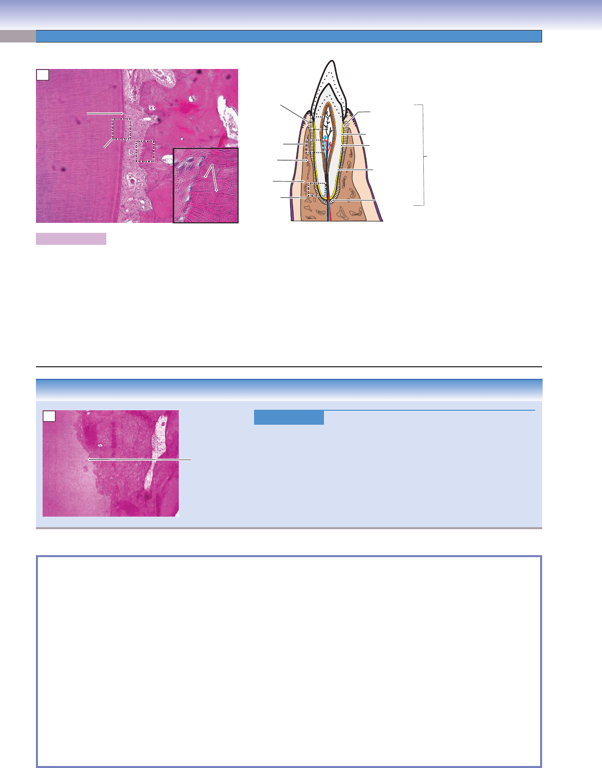
Figure 14-19A. PDL and alveolar bone, tooth root. H&E, 68; inset 408
The cementum and PDL are derived from the dental sac. During root formation, some fi bers from the PDL are trapped within the
cementum and the alveolar bone. These Sharpey fi bers bridge the space between the cementum and the alveolar bone and serve to
suspend the tooth within the alveolar socket. The principal fi bers of the PDL are organized in several groups based on their orientation
and location: apical, oblique, horizontal, alveolar crest, interradicular, and gingival fi bers. The interradicular fi bers are only present
between multirooted teeth. The gingival fi bers attach the gingiva to the hard tissue of the tooth. The alveolar bone provides support
and protection for the tooth root. The alveolar bone includes the alveolar bone proper and supporting bone. The alveolar bone proper
is a thin layer of compact bone which lines the tooth socket and has Sharpey fi bers embedded in it; it is remodeled constantly to adapt
to stresses and tensions. The supporting bone is composed of compact bone and cancellous bone. The compact bone forms the cortical
plate, which provides surface strength. The cancellous bone makes up the central core of the alveolar bone and contains bone marrow.
CLINICAL CORRELATION
Figure 14-19B.
T
ooth Ankylosis. H&E, 68
Tooth ankylosis is the fusion of a mineralized root surface to its surround-
ing alveolar bone. It is characterized by a typical metallic percussion sound
and loss of the PDL space in radiographs. Periapical infl ammation and
subsequent tissue repair are believed to be the cause of ankylosis. Ankylo-
sis is more common in deciduous teeth and usually results in impaction of
a subjacent permanent tooth. Impaction of incisors is much less common
than it is in the mandibular third molars. This slide shows impaction of a
central incisor with focal loss of the PDL the incisor.
SYNOPSIS 14-1 Pathological and Clinical Terms for the Oral Cavity
Microdontia ■ : disproportionately small teeth (disturbances occur at bud stage).
Macrodontia
■ : disproportionately large teeth (disturbances occur at bud stage).
Dens invaginatus (dens in dente)
■ : developmental anomaly in tooth formation at cap stage in which the epithelium invaginates
into the pulp space to form enamel and dentin, creating a “tooth within a tooth” as shown in the radiographic imaging.
Misalignment
■ : a condition in which teeth are too crowded and incorrectly positioned.
Tooth decay (caries)
■ : bacterial destruction of teeth, including erosion of the enamel and damage to the pulp tissue.
Hypersensitivity
■ : a painful condition, especially to touch, sweetness, and cold drinks caused by exposed dentin.
Periodontal disease
■ : a chronic infl ammatory condition of gingival and surrounding periodontal tissues. Calculus and
plaque are the major causes of the disease.
Gingivitis
■ : infection and infl ammation of the gingival tissue.
Periodontitis
■ : infection and infl ammation affecting the periodontium, characterized by gingivitis, destruction of the alveolar
bone and periodontal ligament, and the formation of periodontal pockets.
Pulpitis
■ : an infl ammation of the pulpal tissue of a tooth.
White lesions
■ : lesions on the mucosa of the oral cavity that have a white coating such as leukoplakia.
Red lesions
■ : chronic red oral mucosal lesions in which underlying vascular structures become more visible (as in
erythroplakia).
Alveolar
Alveolar
bone
bone
Alveolar
bone
Dentin
Dentin
Dentin
Junction between
alveolar bone
and dentin
(absence of PDL)
B
CUI_Chap14.indd 278 6/2/2010 8:24:11 AM
279
15
Digestive Tract
Introduction and Key Concepts for the Digestive Tract
Figure 15-1 Overview of the Digestive Tract
Figure 15-2 Orientation of Detailed Digestive Tract Illustrations
Figure 15-3 General Structure of the Wall of the Digestive Tract
Esophagus
Figure 15-4A Overview of the Esophagus
Figure 15-4B Upper Esophagus
Figure 15-4C Middle Esophagus
Figure 15-5A Lower Esophagus, Esophagogastric Junction
Figure 15-5B Clinical Correlation: Barrett Esophagus
Figure 15-5C Clinical Correlation: Esophageal Carcinoma
Stomach
Figure 15-6 Overview of the Stomach
Figure 15-7A Cardiac Region, Esophagogastric Junction
Figure 15-7B Fundic Region of the Stomach
Figure 15-7C Body Region of the Stomach
Figure 15-8A Pyloric Region of the Stomach
Figure 15-8B Clinical Correlation: Gastric Ulcer (Peptic Ulcer)
Figure 15-8C Clinical Correlation: Gastrinoma (Zollinger-Ellison Syndrome)
Small Intestine
Figure 15-9 Overview of the Small Intestine
Figure 15-10A,B Duodenum, Small Intestine
Figure 15-10C Clinical Correlation: Peptic Duodenitis
Figure 15-11A Plicae Circulares, Villi, and Microvilli
CUI_Chap15.indd 279 6/2/2010 3:23:19 PM

280
UNIT 3
■
Organ Systems
Figure 15-11B Villi of the Small Intestine
Figure 15-12A Columnar Absorptive and Goblet Cells of the Small Intestine
Figure 15-12B Goblet, Columnar Absorptive Cells, and Microvilli
Figure 15-13A Paneth Cells, Small Intestine
Figure 15-13B Enteroendocrine Cells, Small Intestine
Figure 15-14A Submucosal/Meissner Plexus, Small Intestine
Figure 15-14B Muscularis Externa, Small Intestine
Figure 15-14C Myenteric/Auerbach Plexus, Muscularis Externa of the Small Intestine
Figure 15-15A Jejunum, Small Intestine
Figure 15-15B Ileum with Peyer Patches, Small Intestine
Figure 15-15C Mucosa of the Ileum, Small Intestine
Large Intestine
Figure 15-16 Overview of the Large Intestine
Figure 15-17A,B Colon, Large Intestine
Figure 15-17C Mucosa of the Colon, Large Intestine
Figure 15-18A Clinical Correlation: Colon Polyps
Figure 15-18B Clinical Correlation: Colorectal Cancer
Figure 15-18C Clinical Correlation: Meckel Diverticulum
Figure 15-19A Appendix and Cecum
Figure 15-19B Anorectal Junction
Figure 15-19C Clinical Correlation: Hemorrhoids
Figure 15-20A Clinical Correlation: Ulcerative Colitis
Figure 15-20B Clinical Correlation: Crohn Disease
Synopsis 15-1 Pathological and Clinical Terms for the Digestive Tract
Table 15-1 Digestive Tract
Introduction and Key Concepts
for the Digestive Tract
The digestive system is composed of the oral cavity, digestive
tract, and digestive glands with associated organs. The “Oral
Cavity” is discussed in Chapter 14; the “Digestive Glands and
Associated Organs” are discussed in Chapter 16. The diges-
tive tract is discussed in this chapter. It includes the esopha-
gus, stomach, small intestine, and large intestine. The digestive
tract is a continuation of the oral cavity, and its main functions
are to ingest food and to digest the food as it passes along the
tract. In this process, nutrients and water are absorbed, and
waste materials are prepared for elimination from the body.
Each section of the digestive tract has its unique histological
features, which are closely associated with the function of that
part of the tract, although there are some common character-
istics: (1) Organs of the digestive tract are all hollow; (2) they
are composed of four general tunic layers: mucosa, submu-
cosa, muscularis externa, and adventitia or serosa; (3) they
are innervated by the enteric portion of the autonomic ner-
vous system, also known as the enteric nervous system (or the
“second brain”); (4) they include epithelium, connective tissue,
muscle, blood and lymphatic vessels, lymphatic nodules, and
nerve fi bers; and (5) they contain glands in the lamina propria
or submucosa.
General Structure of the
Digestive Tract
Based on its histological organization, the wall of the digestive
tract can be divided into four tunics (Fig. 15-3).
1. Mucosa is the innermost layer of the digestive wall. It
includes epithelium, lamina propria, and muscularis muco-
sae. The epithelium consists of simple columnar epithelium
lining most of the tract and stratifi ed squamous epithelium
lining the two ends, the esophagus and anal canal. The
lamina propria is a loose connective tissue that contains
abundant ground substance, many fi bers, and numerous
connective tissue cells such as fi broblasts, macrophages,
mast cells, plasma cells, and leukocytes (see Chapter 4,
“Connective Tissue”). Various types of glands are found in
the lamina propria depending on the region of the digestive
tract. The muscularis mucosae is a very thin layer of smooth
muscle, which is the boundary between the mucosa and the
submucosa. It is usually arranged in an inner circular and
outer longitudinal layer. However, the muscularis mucosae
varies in different regions, and it is often diffi cult to distin-
guish between the muscle layers.
2. Submucosa is a thick layer of dense irregular connective
tissue. This layer contains blood vessels, lymphatic vessels,
CUI_Chap15.indd 280 6/2/2010 3:23:32 PM

CHAPTER 15
■
Digestive Tract
281
and submucosal (Meissner) plexuses, which contain nerve
fi bers and neurons of the enteric nervous system. In some
regions of the digestive tract, this layer is characterized by
mucous glands or lymphatic nodules.
3. Muscularis externa is composed of two or three oblique,
circular, and longitudinal muscle layers, which vary from
region to region. Most of the muscularis externa consists
of smooth muscle fi bers, but the upper and middle esophagi
contain some skeletal muscle. The myenteric (Auerbach)
plexuses (nerve fi bers and neurons of the enteric nervous sys-
tem) are located between the muscle layers. They innervate
and control contraction of the muscularis externa.
4. Serosa and adventitia are coverings of the outermost wall
of the digestive tract. Most parts of the digestive tract are
covered by serosa, a thin layer of loose connective tissue
lined by mesothelium. The mesothelium produces a lubri-
cating fl uid that reduces friction during movement of the
organs against each other (see Chapter 3, “Epithelium and
Glands,” Fig. 3-2A,B). The serosa is the visceral layer of the
peritoneum and covers the wall of the digestive tract where
it connects to the mesentery in the peritoneal cavity (intrap-
eritoneal organs). The adventitia is a layer of loose connec-
tive tissue without mesothelium that covers the upper region
of the esophagus, part of the duodenum, and the lower part
of the digestive tract, such as the rectum and anal canal.
Adventitia covers regions of the digestive tract where it is
connected to other organs or to the body wall (e.g., retro-
peritoneal organs).
Esophagus
The esophagus is the upper part of the digestive tract, con-
necting the oral cavity to the stomach. The major function of
the esophagus is to provide passage for food from the mouth
to the stomach. The luminal surface of the esophagus is lined
by nonkeratinized stratifi ed squamous epithelium. Mucous
glands called esophageal glands are located in the submucosa
of the esophagus. The muscularis externa consists of two lay-
ers of muscle: inner circular and outer longitudinal layers. Both
skeletal and smooth muscle fi bers are found in the muscularis
externa of the esophagus. The proportions of skeletal and
smooth muscle fi bers are different in different regions of the
esophagus. The esophagus can be divided into three regions:
the upper esophagus, middle esophagus, and lower esophagus
(Figs. 15-4A to 15-5A).
1. The upper esophagus connects the oropharynx to the mid-
dle esophagus. This segment contains numerous esophageal
glands in the submucosa. These glands secrete mucus to
lubricate the esophageal wall so that food will pass through
easily. The upper esophagus contains only skeletal muscle
fi bers in the muscularis externa. These are voluntary muscle
fi bers and are innervated by the glossopharyngeal nerve (cra-
nial nerve [CN] IX) (see Fig. 15-4B).
2. The middle esophagus has mucosa similar to that of the
upper esophagus. The esophageal glands in the submucosa
are less numerous than in the upper esophagus. The mus-
cularis externa contains both skeletal and smooth muscles
(Fig. 15-4C).
3. The lower esophagus connects the esophagus to the car-
dia of the stomach. This region contains large numbers of
mucous glands in the lamina propria and submucosa. These
are called esophageal cardiac glands and produce mucous
secretions to protect the lower esophagus from being dam-
aged by refl ux of acidic gastric juices from the stomach.
The lower esophagus contains only smooth muscle fi bers in
the muscularis externa. These are controlled by the enteric
branches of the vagus nerve (CN X) (see Fig. 15-5A).
Stomach
The stomach is a “J”-shaped sac (hollow) organ. It temporarily
stores food, mixes food with gastric juice, and initiates the pro-
cessing of food by breaking it down into simpler substances that
are easier to digest. The stomach can be divided into the cardia,
fundus, body, and pylorus. The inner surface of the stomach
is lined by simple columnar epithelium composed mainly of
surface mucous cells. The surface epithelium of the stomach is
invaginated into the lamina propria to form gastric pits. These
pits serve as ducts for the glands in the lamina propria, which
vary from region to region in the stomach.
1. The cardiac region connects to the lower esophagus at the
esophagogastric junction, which is characterized by a change
from the nonkeratinized stratifi ed squamous epithelium of
the esophagus to the simple columnar epithelium of the
stomach. A thickened smooth muscle ring called the gastroe-
sophageal sphincter (lower esophageal sphincter) or cardiac
sphincter surrounds the opening at the junction of the lower
esophagus and cardiac region of the stomach. This smooth
muscle contracts to prevent the acidic stomach contents from
entering the esophagus. The glands in the lamina propria of
the cardia are called cardiac glands and are branched tubu-
lar glands with coiled secretory portions. The cardiac gland
contains mainly mucus-secreting cells and some stem cells,
enteroendocrine cells, and, occasionally, parietal cells. The
mucus-secreting cells mainly produce mucus and lysozymes.
The mucus protects the stomach wall from acidic gastric
juices; lysozymes destroy bacterial membranes, preventing
bacterial infections (Fig. 15-7A).
2. The fundic and body regions form the largest portions of the
stomach. Their mucosa has similar histological characteris-
tics, including short gastric pits and long branched tubular
glands in the lamina propria. The glands are called fundic or
gastric glands in both the fundus and the body regions. The
gastric glands contain mainly parietal cells and chief cells,
along with some stem cells, mucous neck cells, and enteroen-
docrine cells. Parietal cells are more numerous in the superior
regions of the glands; these cells produce large quantities of
hydrochloric acid (HCl), creating an acidic environment to
help digestion. Parietal cells also secrete intrinsic factor (IF),
which is required for the absorption of vitamin B12. Chief
cells are located in the more inferior regions of the glands;
they secrete precursor enzymes such as pepsinogen, which is
activated by HCl and becomes pepsin. Pepsin helps to break
down proteins (particularly protein collagen) into simpler,
more absorbable compounds. Chief cells also secrete precur-
sors of lipases, which help in lipid digestion (Fig. 15-7B,C).
3. The pyloric region is the lower end of the stomach, which
connects with the duodenum. Its mucosa is similar to that of
the cardia, with long gastric pits and short, coiled secretory
portions. A
circular smooth muscle ring called the pylorus
sphincter (pyloric valve) surrounds the end of the pylorus
region. This valve controls the entry of stomach contents
into the duodenum. The glands in the lamina propria of
the pylorus are called pyloric glands and contain primarily
CUI_Chap15.indd 281 6/2/2010 3:23:32 PM

282
UNIT 3
■
Organ Systems
mucus-secreting cells and two special types of enteroendo-
crine cells: gastrin-secreting cells (G cells) and somatostatin-
secreting cells (D cells), as shown in Figure 15-8A. These
enteroendocrine cells regulate gastric (HCl) secretion.
Small Intestine
The small intestine (Figs. 15-9 to 15-15C) is a hollow organ of
small diameter that is typically 6 to 7 m long. It is the major
site for the absorption of nutrients. Important features of the
small intestine are villi and microvilli, which increase surface
area for absorption. Intestinal glands called glands (crypts)
of Lieberkühn are located in the lamina propria of the small
intestine. Villi project into the lumen of the intestine; the glands
of Lieberkühn open into the mucosa at the base of the villi
(Fig. 15-11A,B). The small intestine can be divided into three
parts: the duodenum, jejunum, and ileum.
1. The duodenum is the shortest segment of the small intestine,
about 20 to 25 cm long. It has small openings called duodenal
papillae (minor and major [Figs. 16-9A and 16-15A]), which
allow pancreatic juice and bile to enter the digestive tract.
It has a similar general structure to other parts of the small
intestine (Fig. 15-9). However, the Brunner glands (mucus-
secreting gland) in the submucosa are a unique feature of the
duodenum (Fig. 15-10A,B).
2. The jejunum is much longer than the duodenum, about
2.5 m long (two fi fths of the rest of the small intestine). It has
long villi and a somewhat increased number of goblet cells. It
has neither Brunner glands nor Peyer patches (Fig. 15-15A).
3. The ileum is the longest segment, about 4 m long (three fi fths
of the rest of the small intestine). It has short villi with sig-
nifi cantly increased numbers of goblet cells on the surface
of the mucosa. There are clusters of lymphatic nodules in
the lamina propria of the ileum; sometimes these lymphatic
nodules extend into the submucosal layer. These clusters of
lymphatic nodules are called Peyer patches and are unique to
the ileum (Fig. 15-15B,C).
Large Intestine
The large intestine (Figs. 15-16 to 15-19B) is a hollow organ
with a relatively large diameter compared to the small intestine
and is about 1.5 m long. It is the last region of the digestive tract
and is the major site for absorption of water and salts. It also
forms, stores, and eliminates feces. Most of the regions of the
large intestine have tunics that are similar to those of the small
intestine, but there are no villi in the mucosa. There are large
numbers of goblet cells in the large intestine. These cells produce
mucus, which helps in the formation of the feces and protects and
lubricates the surface of the intestinal wall. The large intestine
includes the cecum, appendix, colon, rectum, and anal canal.
1. The cecum is the most proximal region of the large intestine.
It is a small, blind pouch of the large intestine where the
ileum connects to the ascending colon. A sphincter muscle, a
thickening of the muscularis mucosae, is called the ileocecal
valve and is located at the junction of the ileum and cecum.
It prevents the contents of the large intestine from backing
up into the small intestine (Fig. 15-16).
2. The appendix is a small, blind tube that attaches to the
posterior-medial wall of the cecum. It has the general tunic
structure of the intestine and a small irregular lumen. There
are many lymphatic nodules in the lamina propria (Fig.
15-19A).
3. The colon is the longest segment of the large intestine. It
includes the ascending colon, transverse colon, descend-
ing colon, and sigmoid colon. The proximal half of the
colon is responsible for the majority of the absorption
of water and salt; the distal half of the colon has only a
small absorptive function and is predominantly for pro-
cessing and storing feces. The colon does not have villi; it
has a smoother surface than the small intestine. Columnar
absorptive cells and goblet cells line the mucosa. The large
intestinal glands, the glands (crypts) of Lieberkühn, con-
tain primarily goblet cells, columnar cells, enteroendocrine
cells, and stem cells. Lymphatic nodules may also be found
in the lamina propria. The muscularis externa consists
of inner circular layers of muscle; the outer longitudinal
smooth muscle layer becomes three teniae coli (Figs. 15-16
and 15-17A–C).
4. The rectum and anal canal are the last segments of the large
intestine. The junction between the rectum and the anal
canal is called the “anorectal junction.” The mucosa of the
rectum is similar to that of the colon but has fewer glands
of Lieberkühn. The main function of the rectum is the tem-
porary storage of feces. The sensory receptors in the rectum
send signals to the brain when feces need to be evacuated.
The anal canal is the distal end of the large intestine. Most
of the anal canal is lined by stratifi ed squamous epithelium,
although simple cuboidal epithelium may be present at the
anorectal junction. Sebaceous glands and hair follicles may
be found at or near the anal opening. There are many veins
in the lamina propria and submucosa of the anal canal. The
term hemorrhoids refers to the condition in which these
veins become chronically swollen and infl amed in the rectal
and anal regions (Fig. 15-19B).
CUI_Chap15.indd 282 6/2/2010 3:23:32 PM
CHAPTER 15
■
Digestive Tract
283
Esophagus
Small intestine
Small
Small
intestine
intestine
Small
intestine
Stomach
Large intestine
Rectum
Rectum
Rectum
Sigmoid
Ascending
colon
colon
colon
Descending
colon
Transverse
colon
Cecum
Appendix
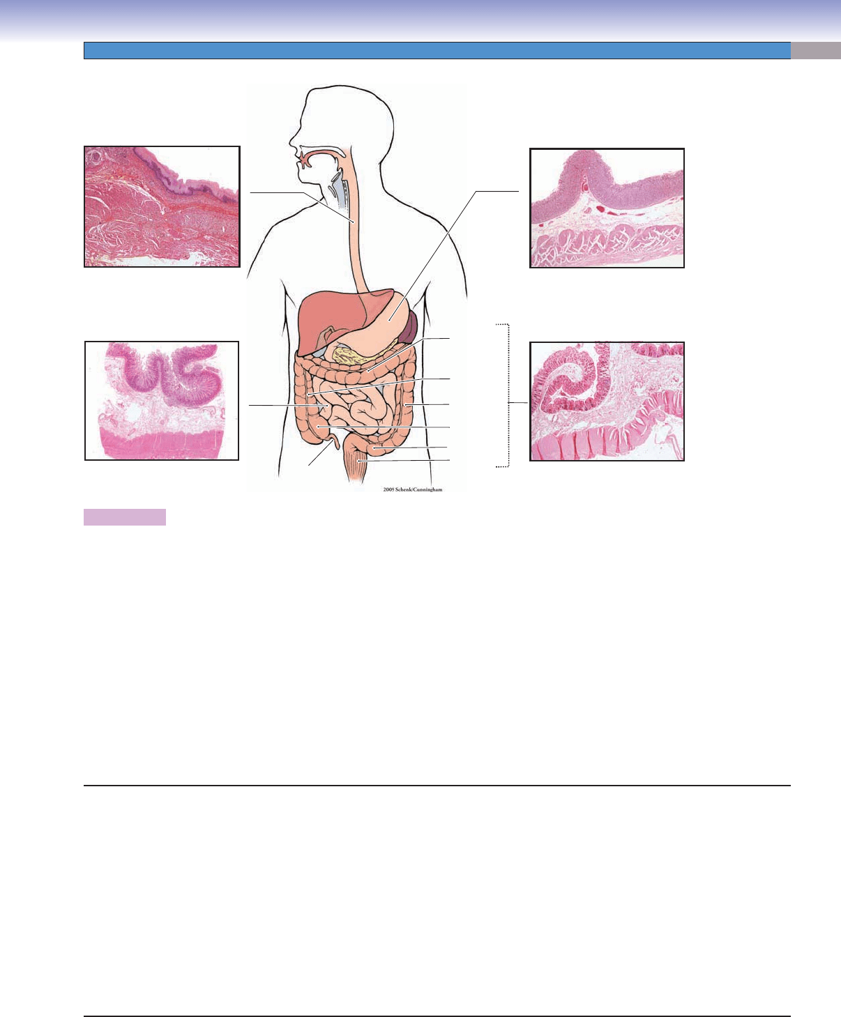
Figure 15-1. Overview of the digestive tract. H&E, 5 to 6
The digestive tract, also called the alimentary canal, includes the esophagus, stomach, small intestine, and large intestine. (1) The
esophagus transports food to the stomach. Its luminal surface is lined by stratifi ed squamous nonkeratinized epithelium and contains
mucous glands in the lamina propria. It has both skeletal and smooth muscles in the muscularis externa. (2) The stomach temporarily
stores and digests food. Its luminal surface is lined by simple columnar epithelium and contains gastric glands in the lamina propria.
It has three layers of smooth muscle in the muscularis externa. (3) The small intestine digests and absorbs carbohydrates, proteins,
and lipids. The small intestine includes the duodenum, jejunum, and ileum. The luminal surface is lined by simple columnar epithe-
lium and contains the glands (crypts) of Lieberkühn in the lamina propria. There are two layers of smooth muscles (inner circular
and outer longitudinal muscles) in the muscularis externa. (4) The large intestine includes the cecum, appendix, colon, rectum, and
anal canal. The main functions of the large intestine are the absorption both of a large volume of the water that enters it (90%)
and of electrolytes (e.g., Na
+
and Cl
−
) and the formation of the feces. Most parts of the large intestine are lined by simple columnar
epithelium, but stratifi ed squamous epithelium lines the anal canal. The glands of Lieberkühn are located in the lamina propria. The
muscularis externa contains an inner layer of circular smooth muscle and an outer layer of longitudinal smooth muscle, which forms
three teniae coli (see Fig. 15-16). In general, the submucosa of the digestive tract is a thick layer of connective tissue containing blood
vessels, lymphatic vessels, and submucosal (Meissner) plexuses consisting of nerve fi bers and neuron cell bodies in the enteric nervous
system. The myenteric (Auerbach) plexuses located in the muscularis externa are also components of the enteric nervous system.
Digestive Tract
I. Esophagus
A. Upper esophagus (skeletal muscle)
B. Middle esophagus (mixed skeletal and smooth muscles)
C. Lower esophagus (smooth muscle)
II. Stomach
A. Cardia
B. Fundus
C. Body
D. Pylorus
III. Small Intestine
A. Duodenum
B. Jejunum
C. Ileum
IV. Large Intestine
A. Cecum
B. Appendix
C. Colon (ascending, transverse, descending, and sigmoid)
D. Rectum
E. Anal canal
CUI_Chap15.indd 283 6/2/2010 3:23:33 PM
284
UNIT 3
■
Organ Systems
Digestive Tract with Figure Numbers
Esophagus
Figure 15-4A
Figure 15-4B
Figure 15-4C
Figure 15-5A
Figure 15-5B
Figure 15-5C
Stomach
Figure 15-6
Figure 15-7A
Figure 15-7B
Figure 15-7C
Figure 15-8A
Figure 15-8B
Figure 15-8C
Small intestine
Figure 15-9
Figure 15-10A
Figure 15-10B
Figure 15-10C
Figure 15-11A
Figure 15-11B
Figure 15-12A
Figure 15-12B
Figure 15-13A
Figure 15-13B
Figure 15-14A
Figure 15-14B
Figure 15-14C
Figure 15-15A
Figure 15-15B
Figure 15-15C
Large intestine
Figure 15-16
Figure 15-17A
Figure 15-17B
Figure 17-17C
Figure 15-18A
Figure 15-18B
Figure 15-18C
Figure 15-19A
Figure 15-19B
Figure 15-19C
Figure 15-20A
Figure 15-20B
Fig. 15-15A
Fig. 15-15B,C
Fig. 15-4A
Fig. 15-4A
Fig. 15-4B
Fig. 15-5A
Fig. 15-7A
Fig. 15-7B
Fig. 15-7C
Fig. 15-8A
Fig. 15-17A,B,C
Fig. 15-9B
Fig. 15-9B
Fig. 15-19B
Fig. 15-4C
Fig. 15-10A,B
Fig. 15-19A
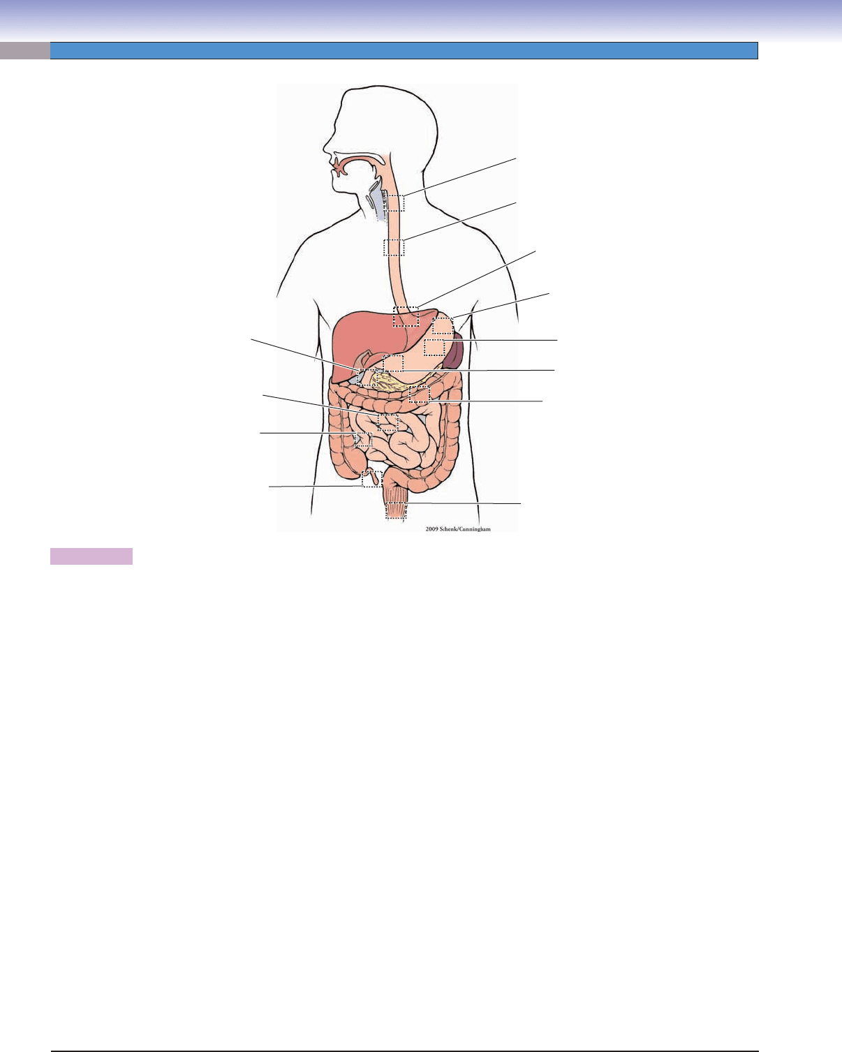
Figure 15-2. Orientation of detailed digestive tract illustrations.
CUI_Chap15.indd 284 6/2/2010 3:23:37 PM