Cui Dongmei. Atlas of Histology: with functional and clinical correlations. 1st ed
Подождите немного. Документ загружается.

CHAPTER 13
■
Integumentary System
245
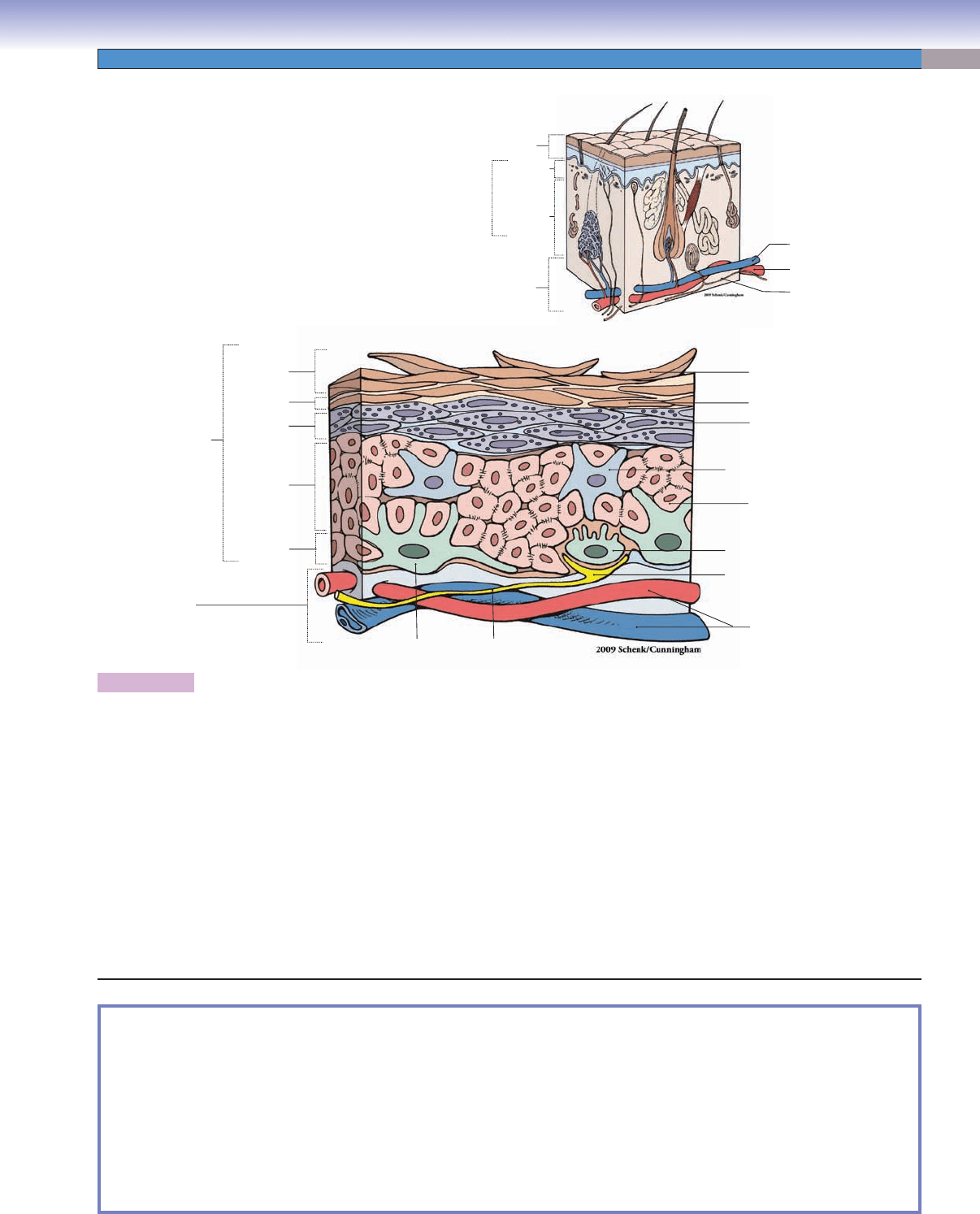
Figure 13-2. Overview of the layers of the epidermis.
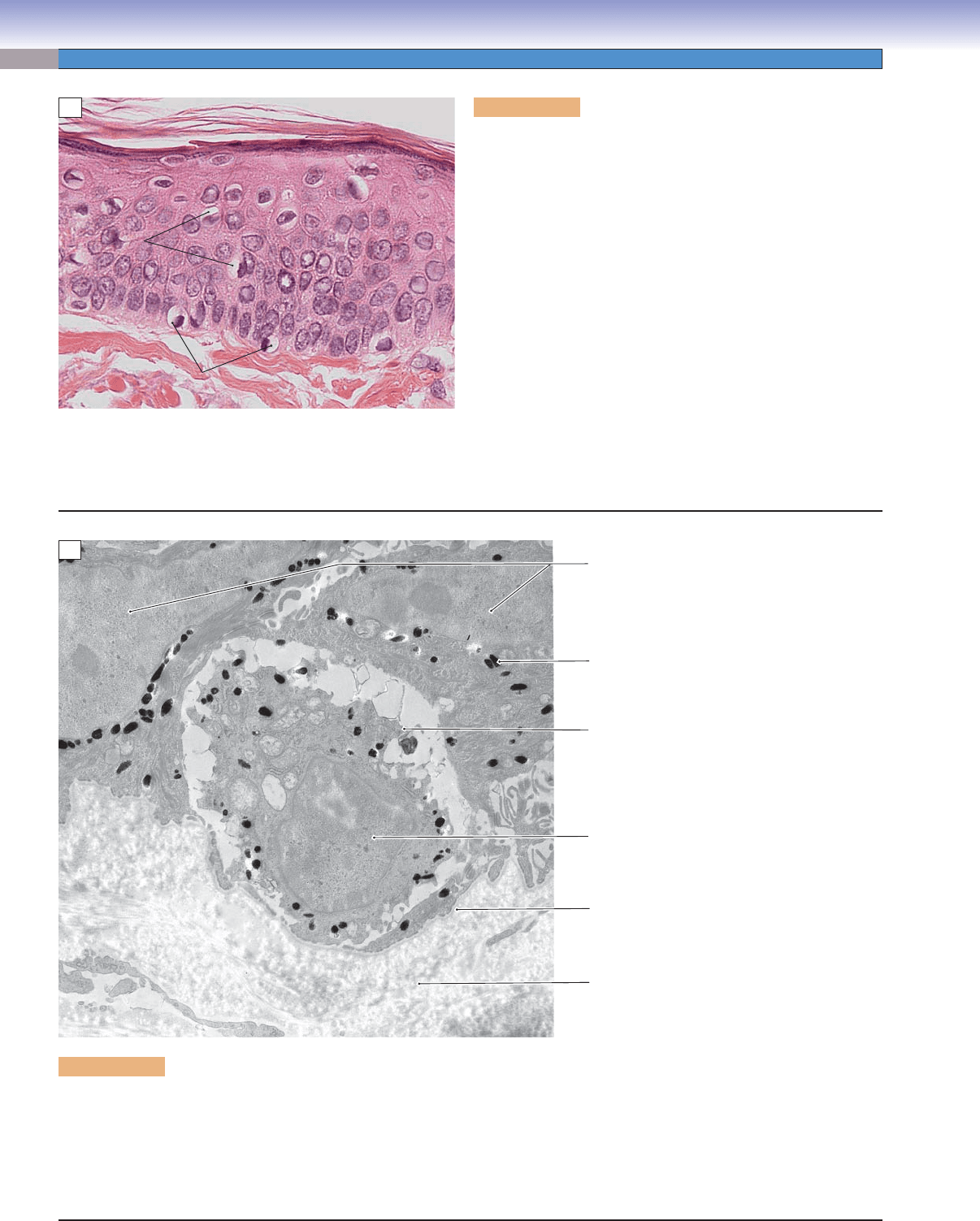
The epidermis is composed of fi ve cell layers. (1) The stratum basale is composed of a single layer of cuboidal or tall cuboidal cells,
melanocytes and Merkel cells which are also called Merkel cell neurite complexes or Merkel disks. Many of these cells are actually
stem cells; they divide continuously and migrate from the basal layer toward the surface and give rise to keratinocytes in the
other layers. (2) The stratum spinosum contains polygon-shaped keratinocytes with many tonofi lament bundles in their cytoplasm.
These cells are interconnected with each other by desmosomes. Langerhans cells are often found in this layer. The stratum basale
and the stratum spinosum are the only layers with mitotically active cells, and, together they are also called the Malpighian layer.
(3) The stratum granulosum contains three to fi ve layers of keratinocytes with fl attened nuclei. The cytoplasm of the cells is fi lled
with basophilic keratohyalin granules from which the name derives. The cytoplasm also contains lamellar granules, which can
release their contents into the intercellular spaces to help seal the skin, preventing water loss. This layer is more obvious in thick
skin; only a single cell layer is visible in thin skin. (4) The stratum lucidum is a very thin, clear layer that contains keratinocytes with
pycnotic nuclei. This layer is found only in thick skin. (5) The stratum corneum is the top layer of the epidermis. It contains many
layers of fl attened cells fi lled with mature keratin. These are dead cells with no nuclei or organelles. The cells in this layer, which are
constantly replaced by cells from deeper layers, form a barrier to prevent loss of water and entry of pathogens. This layer is much
thicker in thick skin than in thin skin.
SYNOPSIS 13-1 Functions of the Skin
Protection ■ of body from invasion of pathogens; prevention of tissue damage by toxic chemicals and ultraviolet light
Prevention
■ of dehydration and loss of body fl uids (impermeable to water)
Regulation
■ of body temperature (production and excretion of sweat, vascular shunts)
Sensation
■ of touch, pain, temperature, pressure, and vibration; important for communication, dexterity, and injury
prevention
Immunological function
■ of Langerhans cells (antigen-presenting cells) present antigens to lymphocytes in the immune
responses (See Chapter 10, “Lymphoid System.”)
Production
■ of vitamin D from precursors under the effects of steroids and sunlight
5. Stratum
corneum
3. Stratum
granulosum
Vein
Artery
Nerve
2. Stratum
spinosum
Merkel cell (Merkel disk)
Langerhans cell
Horny cell (dead cell)
Flattened keratinocyte
Keratinocyte with
keratohyalin granules
Keratinocyte
Sensory nervous fiber
1. Stratum
basale
Melanocyte
Nerve fiber
4. Stratum
lucidum
Small blood vessels
Epidermis
Dermis
Epidermis
Dermis
Hypodermis
(subcutaneous layer)
Reticular
layer
Papillary
layer
CUI_Chap13.indd 245 6/2/2010 8:20:50 AM
246
UNIT 3
■
Organ Systems
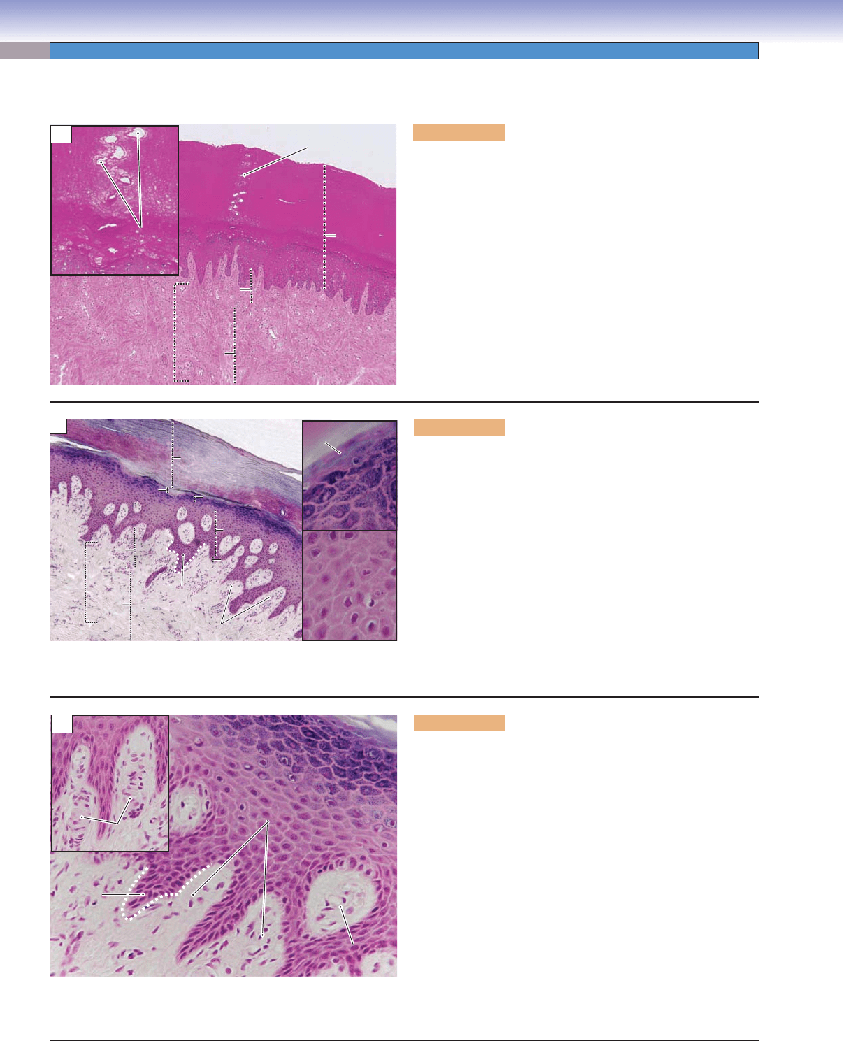
Figure 13-3A. Thick skin, palm. H&E, 34; inset 116
The skin can be classifi ed into thick skin and thin skin based on
the thickness of the epidermis. Thick skin has a thick epidermis
(400–600 μm) with fi ve distinct cell layers. The stratum corneum
is extremely thick in this skin. Thick skin covers the palms of
the hands and soles of the feet. Thick skin has abundant eccrine
sweat glands and lacks hair follicles. The epidermis is a stratifi ed
squamous epithelium. Because it is an avascular tissue (no direct
blood supply), nutrients are delivered to the tissue by fl uid diffu-
sion from the dermis (connective tissue). The dermis is composed
of a superfi cial papillary layer, a layer of loose connective tissue,
and a deeper reticular layer, which is a thick layer of dense irregu-
lar connective tissue. This section shows thick epidermis containing
the duct of an eccrine sweat gland.
Duct of eccrine
Duct of eccrine
sweat gland
sweat gland
Duct of eccrine
sweat gland
Duct of eccrine
Duct of eccrine
sweat gland
sweat gland
Duct of eccrine
sweat gland
Stratum
Stratum
corneum
corneum
Stratum
corneum
Epidermis
Epidermis
Epidermis
Reticular
Reticular
layer
layer
Reticular
layer
Papillary
Papillary
layer
layer
Papillary
layer
Dermis
Dermis
Dermis
A
Figure 13-3B. Layers of the epidermis, palm. H&E, 68;
insets 422
The epidermis of thick skin has fi ve layers. (1) The stratum basale
contains a single layer of cuboidal/tall cuboidal cells (stem cells)
and sits upon the basement membrane. This layer forms a dividing
line between the epidermis and dermis (dotted line). (2) The stra-
tum spinosum contains 5 to 10 layers of polyhedral keratinocytes,
fl attened toward the surface. These cells are also called prickle
cells. (3) The stratum granulosum contains three to fi ve layers of
fl attened keratinocytes fi lled with keratohyaline granules, which
appear dark blue here. (4) The stratum lucidum is a very thin layer
containing extremely fl attened and tightly packed keratinocytes
fi lled with keratin fi laments. Their nuclei are beginning to be elimi-
nated. (5) The stratum corneum is a layer of dead, nonnucleated
cells, which form the most superfi cial layer of the skin. Cells in this
layer are constantly sloughed off and replaced by new cells.
Dermal
Dermal
papilla
papilla
Dermal
papilla
Epidermal
Epidermal
rete ridge
rete ridge
Epidermal
rete ridge
Stratum
Stratum
corneum
corneum
Stratum
corneum
Stratum
Stratum
lucidum
lucidum
Stratum
lucidum
Stratum
Stratum
granulosum
granulosum
Stratum
granulosum
Stratum
Stratum
spinosum
spinosum
Stratum
spinosum
Stratum
Stratum
basale
basale
Stratum
basale
Papillary
Papillary
layer
layer
Papillary
layer
Dermis
Dermis
Dermis
Reticular
Reticular
layer
layer
Reticular
layer
Stratum
Stratum
granulosum
granulosum
Stratum
granulosum
Stratum
Stratum
spinosum
spinosum
Stratum
spinosum
Stratum
Stratum
lucidum
lucidum
Stratum
lucidum
B
Figure 13-3C. Dermal papilla, palm. H&E, 272; inset
192
The border between the epidermis and dermis (dotted white line) is
expanded into folds. The dermis is a connective tissue layer, which
contains blood vessels, nerves, and sensory receptors (free nerve
endings and Meissner corpuscles). The portion of the epidermis
that projects into the dermis is termed the epidermal rete ridge, and
the portion of the dermis that projects into the epidermis is called
the dermal papilla. This unique feature increases the contact area
between these two layers, preventing the epidermis from detaching
from the dermis. The dermal papilla contains loose connective tis-
sue that includes many capillaries, free nerve endings, and encapsu-
lated sensory receptors. Meissner corpuscles are shown here. The
nerve fi bers cannot be seen in H&E stains; demonstration of nerve
fi bers requires special stains (Fig. 13-1; see also Chapter 7, “Ner-
vous Tissue,” Fig. 7-8A,B). Meissner corpuscles are responsible for
discriminative touch and are more numerous in thick skin such
as at the tips of the fi ngers. These receptors help us to distinguish
between, for example, different coins by touch alone.
C
Dermal
Dermal
papilla
papilla
Dermal
papilla
Meissner
Meissner
corpuscle
corpuscle
Meissner
corpuscle
Meissner
Meissner
corpuscles
corpuscles
Meissner
corpuscles
Epidermal
Epidermal
rete ridge
rete ridge
Epidermal
rete ridge
Dermis
Dermis
Dermis
Thick Skin
CUI_Chap13.indd 246 6/2/2010 8:20:52 AM
CHAPTER 13
■
Integumentary System
247
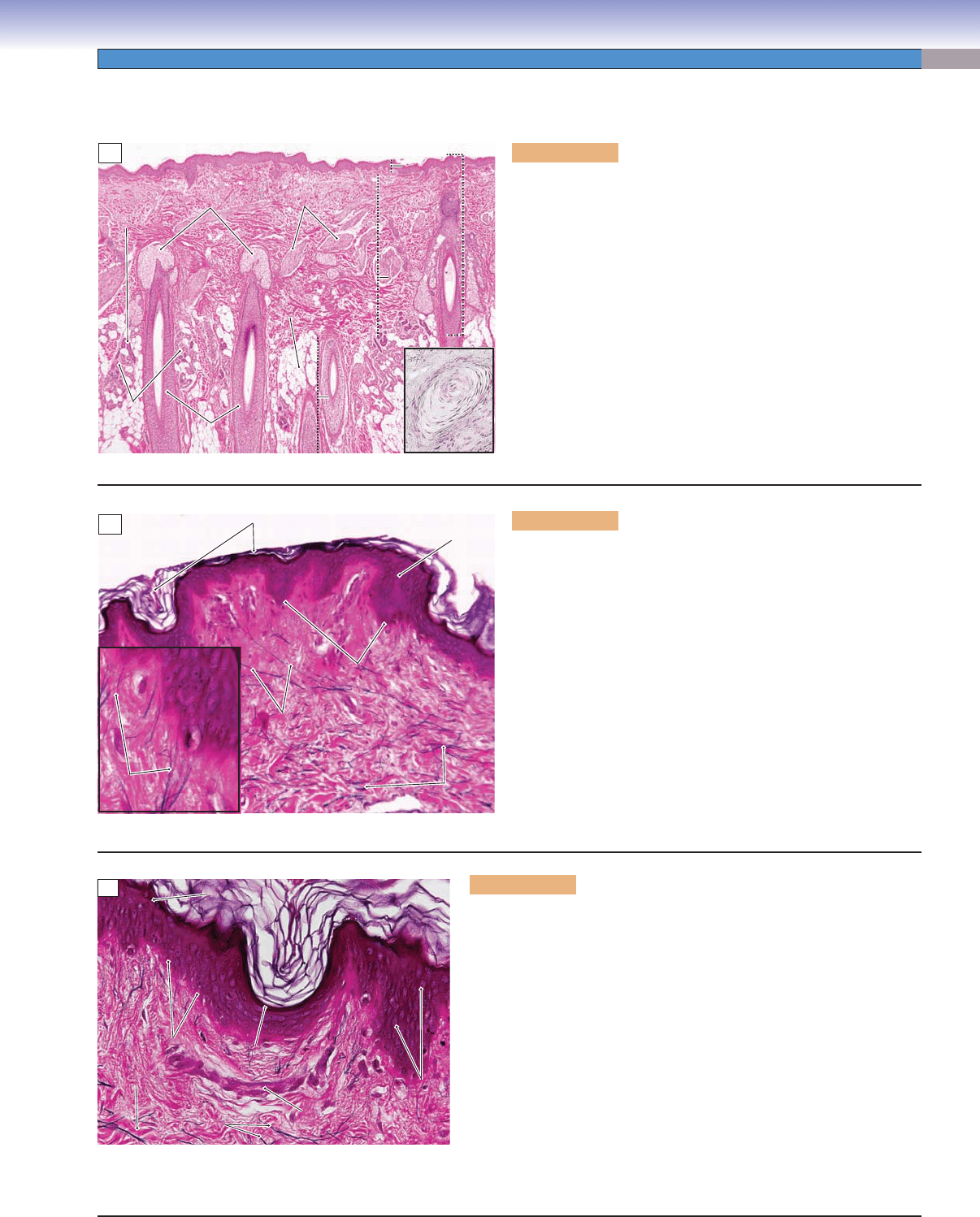
Thin Skin
Figure 13-4A. Thin skin, scalp. H&E, 25; inset 84
Thin skin covers the entire body surface except for the palms of
the hands and the soles of the feet. Thin skin has a thin epidermis,
largely because its stratum corneum is much reduced compared to
that of thick skin. In contrast to thick skin, thin skin contains hair
follicles and their associated sebaceous glands. This section shows
the epidermis and dermis of the skin and a deeper layer of sub-
cutaneous tissue called the hypodermis. The hypodermis is a layer
of loose connective tissue, which contains adipose tissue, nerves,
arteries, and veins. The nerves give off branches, which provide the
various types of sensory and autonomic nerve endings in the dermis.
Pacinian corpuscles, sensory receptors that respond to vibration
stimuli, are found in the hypodermis of both thin and thick skin.
They are found in many regions of the body but are more numerous
in the tips of the fi ngers and toes than in other areas (Fig. 13-1). The
hypodermis serves as a transition layer, providing the dermis with a
fl exible attachment to the underlying muscles and other structures.
Sebaceous
Sebaceous
glands
glands
Sebaceous
glands
Arrector
Arrector
pili muscles
pili muscles
Arrector
pili muscles
Duct of the
Duct of the
sweat glands
sweat glands
Duct of the
sweat glands
Skin
Skin
Skin
Epidermis
Epidermis
Epidermis
Dermis
Dermis
Dermis
Hypodermis
Hypodermis
Hypodermis
Pacinian
Pacinian
corpuscle
corpuscle
Pacinian
corpuscle
Hair
Hair
follicles
follicles
Hair
follicles
Adipose
Adipose
tissue
tissue
Adipose
tissue
Eccrine
Eccrine
sweat
sweat
glands
glands
Eccrine
sweat
glands
A
Figure 13-4B. Thin skin. Elastic fi ber stain, 142; inset 487
The epidermis of thin skin consists of four layers, including
the stratum basale, stratum spinosum, stratum granulosum, and
stratum corneum. (The stratum lucidum is absent in thin skin.)
The stratum granulosum is very thin, often only a single cell layer,
and it is not easily distinguished in thin skin. The stratum cor-
neum is thin but varies in thickness from region to region. This
section is stained with an elastic fi ber stain, which shows the elas-
tic fi bers in the dermis. These fi bers become very fi ne toward the
epidermis. The dermis contains type I collagen fi bers and elas-
tic fi bers, which give the skin fl exibility and strength. The inset
shows a few very fi ne fi bers called oxytalan fi bers. The elastic
fi bers can be classifi ed into three types based on their microfi -
bril and elastin content: (1) elastic fi bers, the largest fi bers, con-
taining predominantly elastin; (2) elaunin fi bers, intermediate
in size, containing small amounts of amorphous elastin; and (3)
oxytalan fi bers, the smallest fi bers, containing only microfi brils.
Oxytalan
Oxytalan
fibers
fibers
Oxytalan
fibers
Elastic fibers
Elastic fibers
Elastic fibers
Stratum basale
Stratum basale
Stratum basale
Elaunin fibers
Elaunin fibers
Elaunin fibers
Stratum
spinosum
Stratum corneum
B
Figure 13-4C. Stratum corneum, thin skin. Elastic fi ber stain, 284
Fine grooves (sulci cutis) and elevated areas (cristae cutis) are the basis of
the varying surface contours characteristic of specifi c areas of both thin skin
and thick skin. The orientation of the grooves varies from region to region.
Fingerprints (dermatoglyphics) are a good example of a skin pattern, which
is distinctive. The top layer of the epidermis, the stratum corneum, is com-
posed of several layers of fl attened and cornifi ed keratinocytes. These cells
have no nuclei and are fi lled with keratin, which helps to stabilize the cells
against physical stress. This layer of cells is constantly sloughed off and
replaced by differentiating cells from beneath. In this section, the extensive
spaces between the dead cells of the stratum corneum are artifacts of speci-
men preparation. Some of the cuboidal cells in the stratum basale are stem
cells capable of cell division. Some cells derived by division of the stem cells
remain in the stratum basale as stem cells and some begin differentiation
in the stratum spinosum. Keratinocytes undergo an orderly sequence of
differentiation (keratinization) and cell death (apoptosis) as they move up
toward the surface of the epidermis.
Collagen
Collagen
fibers
fibers
Collagen
fibers
Cuboidal cells
Cuboidal cells
in stratum basale
in stratum basale
Cuboidal cells
in stratum basale
Elastic
Elastic
fibers
fibers
Elastic
fibers
Capillary
Capillary
Capillary
Keratinocytes
Keratinocytes
in stratum
in stratum
spinosum
spinosum
Keratinocytes
in stratum
spinosum
Sulci cutis
Sulci cutis
Sulci cutis
Stratum corneum
Stratum corneum
Stratum corneum
Crista cutis
Crista cutis
Crista cutis
C
CUI_Chap13.indd 247 6/2/2010 8:21:00 AM

248
UNIT 3
■
Organ Systems
CLINICAL CORRELATION
Figure 13-5B.
Squamous Cell Carcinoma. H&E,
108
Squamous cell carcinoma (SCC) is the second most
common form of skin neoplasm. It originates from
keratinocytes of the epidermis. This carcinoma is
characterized by a slow-growing reddish or ulcerated
lesion with hard, raised borders. It is often found in
sun-exposed areas, with a high incidence in elderly
male Caucasians. Prolonged sun exposure, chronic
infl
ammatory lesions, and genetic factors, especially
p53 tumor suppressor gene mutations, contribute to
the development of the disease. SCCs called actinic
keratoses commonly arise in premalignant lesions on
sun-damaged skin. Carcinoma cells have enlarged and
hyperchromatic nuclei with variable differentiation,
some lesions producing abundant keratin. Treatment
includes surgical excision, cryosurgery, electrosurgery,
radiation therapy, and topical treatment.
Eosinophilic
squamous
cell carcinoma
cells
B
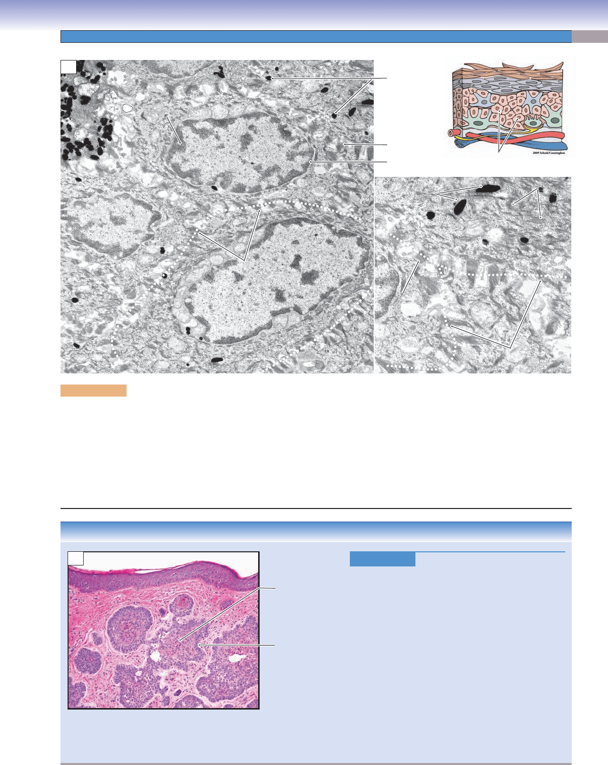
Figure 13-5A. Stratum corneum of the epidermis, thin skin. EM, 8,065; inset 21,889
The stratum corneum is the fi nal product of the proliferation and differentiation that take place in the deeper layers of the epidermis. The
dead cells (horny cells) of the stratum corneum have lost the usual organelles and have become fi lled with mature keratin, a tough net-
work of keratin intermediate fi laments that are cross-linked by the protein fi laggrin. Keratin and the persisting desmosomes between cells
account for the mechanical strength of the epidermis. Contributing to the relative impermeability of the stratum corneum is a coating of
involucrin linked to the inner surfaces of the plasma membrane and the presence of lipid that has been secreted into the spaces between
cells. In this specimen of thin skin, the stratum granulosum is only one cell thick, and some of its characteristic features (keratohyalin gran-
ules and lamellar granules) are not clearly discernible. Bundles of tonofi laments, melanin granules, and desmosomes can be distinguished.
Remaining
Remaining
desmosomes
desmosomes
Remaining
desmosomes
Nucleus of stratum
spinosum cell
Cornified keratinocytes
Cornified envelope
Stratum granulosum
A
CUI_Chap13.indd 248 6/2/2010 8:21:06 AM
CHAPTER 13
■
Integumentary System
249
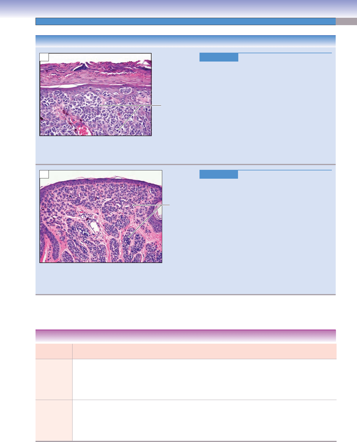
Figure 13-6A. Keratinocytes in stratum spinosum, thin skin. EM, 7,097 (left); 12,390 (right)
The stratum spinosum gets its name from the many small processes that seem to join neighboring cells with one another. The basis for
these spines is obvious in this transmission electron micrograph. Each cell is joined to its neighbors by numerous maculae adherens
(desmosomes), and the spines refl ect the persistence of these connections after some cell shrinkage has occurred during processing of
the tissue. The electron dense tuft of material evident on either side of each desmosome is a bundle of tonofi laments anchored into
the attachment plaques at the cytoplasmic faces of the desmosomes. The desmosomes and tonofi lament bundles are numerous and
more easily seen in the higher magnifi cation view in the right-hand panel. As indicated by the extensive euchromatin in the nuclei of
these cells, the keratinocytes are actively synthesizing proteins, most prominently subunits of keratin fi laments, which will function
in establishing a tough, impermeable barrier layer at the surface of the skin.
Desmosomes
Desmosomes
Desmosomes
Desmosomes
Keratin filaments
Keratin filaments
Keratin filaments
Keratin
Keratin
filaments
filaments
Keratin
filaments
Desmosomes
Desmosomes
Desmosomes
Melanin
granules
Melanin
Melanin
granules
granules
Melanin
granules
Euchromatin
Euchromatin
Euchromatin
Heterochromatin
Heterochromatin
Heterochromatin
Keratinocyte
Keratinocytes in
stratum spinosum
A
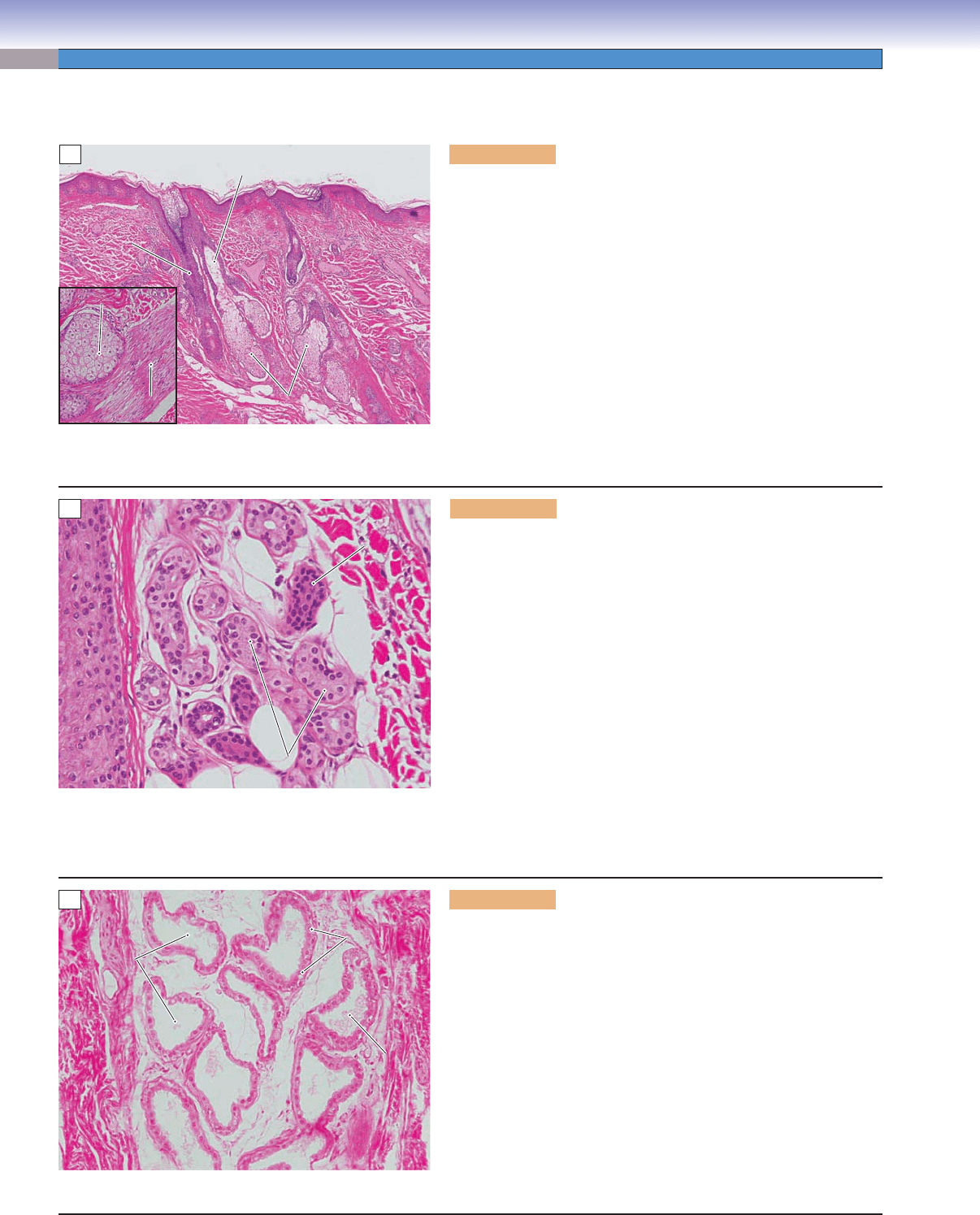
CLINICAL CORRELATION
Figure 13-6B.
Basal Cell Carcinoma. H&E, 50
Basal cell carcinoma is the most common form of malig-
nant skin neoplasm. It originates from the basal layer
of epidermis and often occurs on sun-exposed areas.
Basal cell carcinoma rarely metastasizes and is usually
non–life-threatening if addressed early. Local invasion
may damage surrounding tissues causing cosmetic con-
cerns. Genetics and long-term exposure to ultraviolet
light and arsenical compounds contribute to the dis-
ease. Clinically, basal cell carcinoma appears as pearly
white nodules or waxy bumps on the face or neck with
telangiectatic blood vessels. Subtypes of basal cell car-
cinoma include nodular, superfi cial, pigmented, and
fi brosing. Histologic features include a lobular growth
pattern of malignant basal cells with peripheral pali-
sading and retraction of lobules from the surrounding
stroma. Treatment includes surgical excision, cryosur-
gery, curettage, and electrodessication.
B
Peripheral
palisading
Lobe of basal cell
carcinoma with
squamous
diffentiation
CUI_Chap13.indd 249 6/2/2010 8:21:09 AM
250
UNIT 3
■
Organ Systems
Figure 13-7A. Special types of cells in the epidermis, thin skin.
H&E, 281
Keratinocytes make up the bulk of the epidermis, but there are
additional cell types: melanocytes, Merkel cells, and Langerhans
cells. All three types of cells have clear cytoplasm and are some-
times called clear cells. It is diffi cult to distinguish among them
in H&E-stained sections. Melanocytes and Merkel cells are both
located in the stratum basale where they are scattered among the
basal cuboidal cells. In contrast, Langerhans cells are typically
found in the stratum spinosum. The functions of the three cells
are quite different: (1) Melanocytes produce melanin granules and
insert them into keratinocytes. (2) Merkel cells are receptor cells,
that establish synaptic contacts with sensory nerve terminals;
they have cytoplasmic granules, which contain neurotransmitters
(Fig. 13-2). (3) Langerhans cells are monocyte derivatives, which
play an important role in capturing antigens and presenting them
to lymphocytes, thereby participating in the cutaneous immune
response. Langerhans cells make no desmosome junctions with
neighbors; indeed, their function requires that they be motile so
that they can transport any captured antigens from the epidermis
to lymph nodes deep to the skin.
Langerhans
Langerhans
cells
cells
Langerhans
cells
Melanocytes
Melanocytes
Melanocytes
A
Figure 13-7B. Melanocyte. EM, 8,129
Melanocytes, along with Merkel cells, are the two clear cell types found in the stratum basale of the epidermis. The two cells cannot
be distinguished in conventionally prepared sections for light microscopy, but each has distinctive ultrastructural features. The mel-
anocyte generates melanin granules and injects them into nearby keratinocytes. This is a continuous process because keratinocytes
are constantly replaced as they differentiate and move toward the surface. The melanin protects the cells, particularly the nucleus,
from the mutagenic effects of ultraviolet irradiation. As shown here, the melanocyte contacts the basal lamina of the epidermis, but
it does not establish desmosome junctions with the keratinocytes.
B
Melanocyte
processes
Melanin granules
Nuclei of keratinocytes
Basal lamina of epidermis
Collagen
fibrils in
the dermis
Nucleus of the
melanocyte
CUI_Chap13.indd 250 6/2/2010 8:21:13 AM
CHAPTER 13
■
Integumentary System
251
CLINICAL CORRELATIONS
Figure 13-8A.
Malignant Melanoma. H&E, 198.
Melanoma is an aggressive malignant skin neoplasm
originating from melanocytes of the skin. It is charac-
terized by signifi cant morphologic diversity, with skin
lesions of irregular shapes and various degrees of pig-
mentation. Melanoma metastasizes via the lymphatic
system. Malignant melanoma is less common than basal
cell or SCCs, but it causes the majority of deaths from
skin cancer. Genetic factors and sun exposure contribute
to the development of the disease. The most common
forms of melanoma include superfi cial spreading mela-
noma and nodular malignant melanoma. Melanoma
cells contain large nuclei with irregular contours, often
with prominent nucleoli. Treatment includes surgical
excision, chemotherapy, radiation therapy, and immu-
notherapy.
Figure 13-8B.
Melanocytic Nevus. H&E, 22
Melanocytic nevi, commonly referred to as “moles,”
may be congenital or acquired, and are composed of
melanocytes in nests at the dermo-epidermo junction, in
the dermis, or both. If the nevus cells are restricted to the
dermis, the lesion is referred to as a dermal melanocytic
nevus. If the nevus cells are present only at the dermo-
epidermo junction, the lesion is referred to as a junctional
nevus. If the nevus cells are present in both locations, the
term compound nevus is used. Moles typically appear as
raised tan to brown soft lesions on sun-exposed or sun-
restricted skin. Dysplastic nevi, which may transform
to malignant melanoma, are typically larger than most
nevi, may have irregular borders, and pigmentary varia-
tion. Microscopically
, dysplastic nevi may show larger
junctional nests that fuse to adjacent nests and cytologic
atypia. This image shows a dermal melanocytic nevus
with no evidence of dysplasia or atypia.
Melanoma
(tumor) cells
A
Nests of
nevus cells
B
Type of Skin Epidermis Hair/Hair
Follicles
Glands Sensory
Receptors
Location/
Distribution
Special Features
Thick skin Five layers;
thick stratum
corneum;
thick stratum
granulosum
No Lack of
sebaceous
glands;
more eccrine
sweat glands
More receptors Palms of the
hand and soles
of the feet
Thick epidermis:
thick stratum
corneum; stratum
lucidum present;
several cell layers of
stratum granulosum
Thin skin Four layers; no
stratum lucidum;
single layer of
or no stratum
granulosum
Present in most
areas (except a
few places, such
as lips, labia
minora, and
glans penis)
Many
sebaceous
glands;
fewer
eccrine
sweat glands
Fewer receptors Entire body
except thick
skin areas
Thin epidermis: thin
stratum corneum;
stratum lucidum
absent; one layer
or no stratum
granulosum
TABLE 13-1 Comparison of Thick and Thin Skin
CUI_Chap13.indd 251 6/2/2010 8:21:16 AM
252
UNIT 3
■
Organ Systems
Accessory Structures
Figure 13-9A. Sebaceous gland, thin skin (scalp). H&E, 35;
inset 66
Sebaceous glands are found in thin skin, usually associated with hair
follicles. They are most numerous in the skin of the scalp and face.
Sebaceous glands are classifi ed as simple branched acinar glands (see
Chapter 3, “Epithelium and Glands”). The secretory cells are lipid-
producing cells, arranged into several acini, which open into a short
duct. Usually, the ducts of sebaceous glands empty their oily secre-
tion, called sebum, into a hair follicle (Fig. 13-1); however, the ducts
sometimes open directly onto the surface of the skin. Sebaceous glands
release their products by holocrine secretion, that is, by the disintegra-
tion of entire cells. Sebum lubricates the skin and coats and protects
hair shafts from becoming brittle. The inset shows an acinus of a seba-
ceous gland and a nearby arrector pili muscle. Arrector pili muscles
are bundles of sympathetically innervated smooth muscle cells that
span between hair follicles and the papillary layer of the dermis. They
contract to stand the hair up in response to cold or fear (Fig. 13-1).
Sebaceous gland
Sebaceous gland
duct
duct
Sebaceous gland
duct
Hair
Hair
follicle
follicle
Hair
follicle
Sebaceous glands
Sebaceous glands
Sebaceous glands
Sebaceous glands
Sebaceous glands
Sebaceous glands
Arrector pili
Arrector pili
muscle
muscle
Arrector pili
muscle
A
Figure 13-9B. Eccrine sweat gland, thin skin (scalp). H&E, 248
Eccrine (merocrine) sweat glands can be found in both thin and thick
skin over most of the body. They are more numerous in the palms
and soles. Eccrine sweat glands produce a clear watery product called
sweat. These glands are simple glands in which the secretory cells are
arranged into coiled tubules. These glands have long unbranched, but
coiled, ducts, which are lined by two layers of cuboidal cells and open
directly onto the surface of the skin. Sweat consists mainly of water
(99%), some ions (K
+
, Na
+
, and Cl
−
), waste, and metabolic prod-
ucts. Releasing sweat onto the surface of the skin helps adjust body
temperature as well as aiding in the excretion of metabolic wastes.
The secretory units of eccrine sweat glands contain three cell types.
(1) Dark cells are pyramid-shaped cells containing dark secretory
granules. These cells lie toward the lumen of the tubule. (2) Clear
cells are also pyramid shaped and have no secretory granules, but
have ultrastructural features of ion pumping cells. They are located
toward the basement membrane. (3) Myoepithelial cells are not
secretory cells. They are spindle-shaped contractile cells, which help
to push secretory products into and along the lumen.
Eccrine sweat glands
Eccrine sweat glands
Duct of eccrine
Duct of eccrine
sweat gland
sweat gland
Duct of eccrine
sweat gland
Eccrine sweat glands
B
Figure 13-9C. Apocrine sweat gland, labia. H&E, 248
Apocrine sweat glands are simple coiled tubular glands like the
eccrine sweat glands, but their lumens are larger (about 10 times
larger than those of the eccrine sweat glands) and their ducts empty
into the superfi cial regions of the hair follicles. The secretory cells of
the apocrine glands release their products by shedding part of their
apical cytoplasm; this is called apocrine secretion. The tubules of the
glands are lined by cuboidal or columnar epithelial cells, depending
on the secretory stage. These glands are infl uenced by hormones and
start to function at puberty. They are also called sexual scent glands.
They are restricted in location to some specifi c regions of thin skin,
such as the axilla, the areola (nipple), and the perianal and genital
areas. Their product is a viscous, thick, milky fl uid that contains pro-
tein, ammonia, lipids, and carbohydrates. These secretory fl uids are
odorless when they are released but may have an axillary body odor
after degradation by bacteria.
Secretory cells
Secretory cells
(cuboidal cells)
(cuboidal cells)
Secretory cells
(cuboidal cells)
Lumen of the
Lumen of the
apocrine
apocrine
sweat gland
sweat gland
Lumen of the
apocrine
sweat gland
Secretion
Secretion
inside lumen
inside lumen
Secretion
inside lumen
C
CUI_Chap13.indd 252 6/2/2010 8:21:18 AM
CHAPTER 13
■
Integumentary System
253
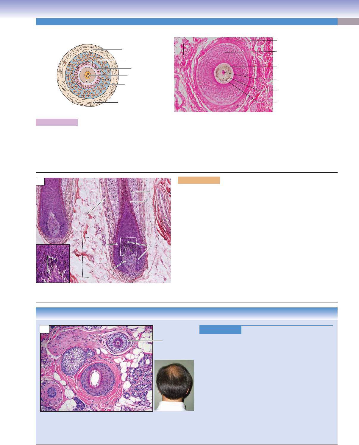
CLINICAL CORRELATION
Figure 13-10C.
Androgenetic Alopecia. H&E, 50.
Androgenetic alopecia is the most common form of hair
loss, affecting 30% to 40% of the adult population.
Males and females have a similar incidence in developing
this type of hair loss, characterized by varying degrees of
partial hair thinning from the vertex and frontal areas of
the scalp. In females, it rarely leads to total baldness. In
males, the cause is both genetic and androgen dependent.
Patients usually have higher levels of 5-alpha-reductase
and androgen receptors. 5-alpha-reductase increases
production of dihydrotestosterone, which binds to the
androgen receptors in susceptible follicles to trigger the
genes to miniaturize the follicles and weaken hair growth.
T
reatment options include the oral medication fi nasteride
and topical solutions of minoxidil. This cross section
of scalp tissue shows variation in hair follicle size with
miniaturized follicles and the absence of infl ammation.
Miniaturized
follicle
C
D. Cui /T. Yang
Hair medulla
Connective
tissue sheath
Hair cuticle
Inner root
sheath
Outer root
sheath
Hair cortex
Blood vessel
Blood vessel
Blood vessel
Hair medulla
Hair medulla
Hair medulla
Hair cuticle
Hair cuticle
Hair cuticle
inner root sheath
inner root sheath
Inner root sheath
outer root sheath
outer root sheath
Outer root sheath
Hair cortex
Hair cortex
Hair cortex
Connective
Connective
tissue sheath
tissue sheath
Connective
tissue sheath
A
Figure 13-10A. Hair follicle. H&E, 95
A cross section of a hair follicle is shown on the left and a photomicrograph of a cross section of a hair follicle from the scalp on
the right. The structures of the hair follicle containing a hair shaft include (from inside to outside) the hair medulla (thin core of the
hair shaft), the hair cortex (keratinized cells surrounding the medulla), the hair cuticle (outermost layer of the hair shaft), the inner
root sheath (cellular sheath that extends from the hair bulb and surrounds and grows along with the hair), the outer root sheath
(a cellular sheath which is a continuation of the epidermis), and the connective tissue sheath (dermal root sheath).
Figure 13-10B. Hair follicles, thin skin (scalp). H&E, 78; inset
172
Hair follicles are the structures that produce the hair and maintain
hair growth. They are cellular structures extending from the epider-
mis into the dermis or hypodermis. The basal region of the hair fol-
licle forms a balloon-shaped structure called the hair bulb, which is
composed of the hair root and the dermal papilla. The hair root con-
tains melanocytes and a group of epithelial cells called the matrix
or germinal matrix. These cells are capable of cell division and give
rise to the inner root sheath and to the hair. The epithelial cells form
a cap around the dermal papilla (hair papilla). The dermal papilla
contains capillaries and nerve fi bers that supply the hair follicle. The
interaction between the hair bulb and dermal papilla induces hair fol-
licle differentiation and the growth of the hair. The photomicrograph
shows a longitudinal section of hair follicles. The inset shows melanin
granules, which give color to the hair. The melanin granules are pro-
duced by melanocytes in the hair bulb.
Inner root
Inner root
sheath
sheath
Inner root
sheath
Outer root
Outer root
sheath
sheath
Outer root
sheath
Matrix
Matrix
Matrix
Melanin
Melanin
granules
granules
Melanin
granules
Connective
Connective
tissue sheath
tissue sheath
Connective
tissue sheath
Hair root
Hair root
Hair root
Dermal papilla
Dermal papilla
(hair papilla)
(hair papilla)
Dermal papilla
(hair papilla)
Hair
Hair
bulb
bulb
Hair
bulb
B
CUI_Chap13.indd 253 6/2/2010 8:21:25 AM
254
UNIT 3
■
Organ Systems
CLINICAL CORRELATION
Figure 13-11C.
Molluscum Contagiosum. H&E, 53
Molluscum contagiosum is a viral skin infection, caused
by the molluscum contagiosum virus, a member of the
poxvirus family
. The disease is characterized by fl esh-
colored, dome-shaped, pearly papules with a dimpled
center. Lesions are typically 1 to 5 mm in diameter
and common on the trunk, arms, and legs. The disease
is common in childhood, and usually self-limited in
immunocompetent patients. In adults, the disease usu-
ally indicates cellular immunodefi ciency. The papules
are usually nonpainful, but may itch or be complicated
by secondary infection. The diagnosis is mainly based
on the clinical appearance of the lesions. This slide
shows lobules of keratinocytes with large eosinophilic
intracytoplasmic inclusions called molluscum bodies
within the stratum granulosum and stratum corneum.
The treatment may include laser therapies, cryotherapy,
or curettage, or there may be no treatment at all.
Molluscum
bodies
C
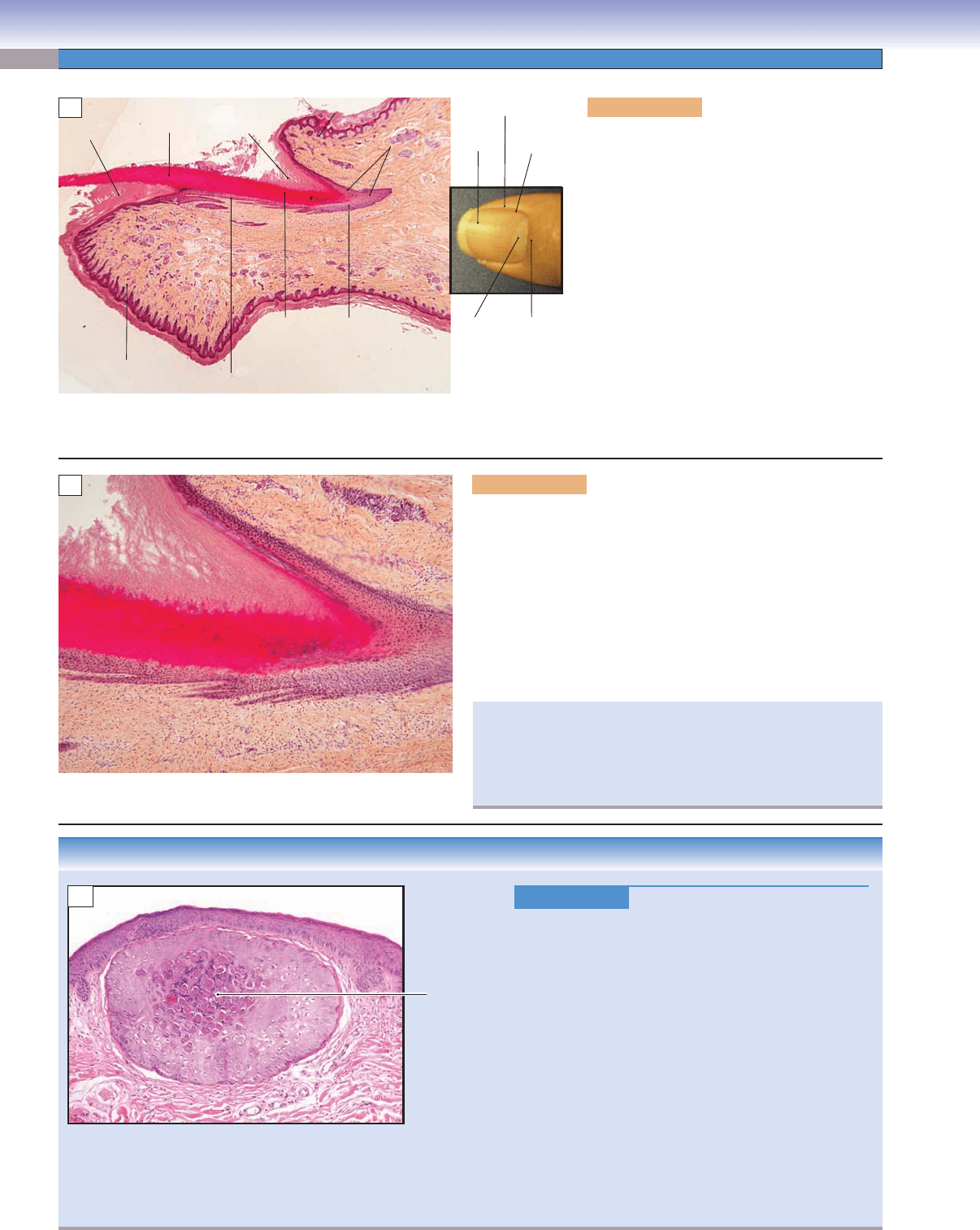
Figure 13-11A. Nail, fi nger. H&E, 17
The nail is a translucent, hard, keratinized
sheet resting on the tip of each digit. It includes
many components: (1) the nail plate, the nail
itself, which is hard keratin; (2) the nail root,
also called the nail matrix, seen as the lunula
in the living state; (3) the nail bed, a layer of
epidermis beneath the nail plate; (4) the epony-
chium, also called the nail cuticle, which is the
junction zone between the skin of the fi nger
and the nail plate and which forms a protec-
tive seal; (5) the perionychium (nail wall), the
skin that surrounds the edge of the nail; and
(6) the hyponychium, the junction seal between
the nail plate and the skin of the fi ngertip. All
of the sealed areas at the edges of the nail plate
protect the delicate nail matrix and nail bed
from dehydration and infection.
Nail
lunula
Nail
lunula
Eponychium
Nail root
(matrix)
Finger tip
(thick skin)
Nail bed
Nail
plate
Hyponychium
Eponychium
(nail cuticle)
Thin skin
Nail
groove
Perionychium
Nail
matrix
Nail
plate
A
Figure 13-11B. Nail root (matrix) and nail bed. H&E, 69
The nail root is a cellular layer and is also called the matrix or
germinal matrix. It contains many layers of epithelial cells, which
are responsible for the production of the nail plate. These cells
proliferate and become fl attened and highly keratinized and are
pushed forward by newly formed cells. As they differentiate, the
cells fi nally lose color and shape and become part of the nail plate.
The nail plate is similar to the hair shaft, but the pattern of kera-
tin formation is different. The nail bed (equivalent to the epider-
mis) rests under the nail plate. The nail bed extends from the nail
matrix to the hyponychium.
Nail
Nail
matrix
matrix
Nail
matrix
Nail plate
Nail plate
Nail plate
Eponychium
Eponychium
Eponychium
Nail bed
Nail bed
Nail bed
B
Normally, the nail bed is smooth and allows for healthy nail
growth and a smooth appearance. If a nail bed is infected by
bacteria or fungus, the nail bed becomes rough, and an accumu-
lation of organic waste materials can react with the nail plate
and cause the nail to become thickened and distorted.
CUI_Chap13.indd 254 6/2/2010 8:21:30 AM