Creagh D., Bradley D. (Eds.) Physical Techniques in the Study of Art, Archaeology and Cultural Heritage. Volume 2
Подождите немного. Документ загружается.

62 D. Creagh
(d)
Ca(SO
4
).2H
2
O (gypsum)
Pb
3
(CO
3
)
2
(OH)
2
(hydrocerussite)
Pb(CO
3
) (cerussite)
SnPb
2
O
4
(tin lead oxide)
C
4
H
6
CuO
4
.H
2
O (copper acetate hydrate)
Cu
2
(OH.Cl)
2
.2H
2
O (calumetite)
Cu
2
CO
3
(OH)
2
Cu
2
CO
3
(OH)
2
(malachite)
Cu
2
Cl(OH)
3
(ataeamite)
Cu
2
Cl(OH)
3
(paratacamita)
CaC
2
O
4
.xH
2
O. x > 2 (weddelite)
+
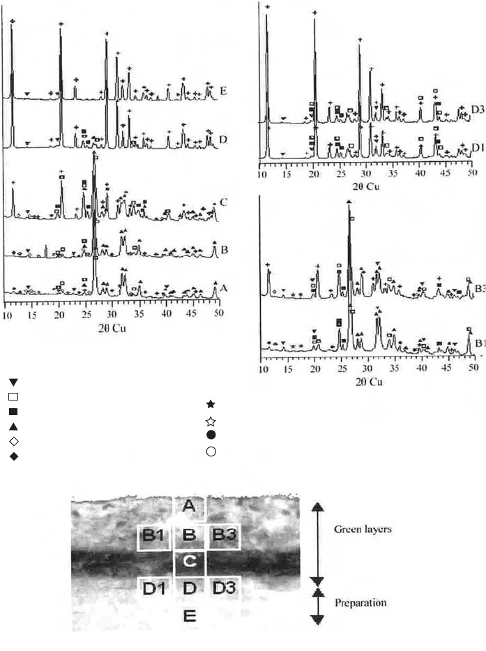
Fig. 9. (d) SRXRD patterns corresponding to a cross section of a sample of the altarpiece
of Constable. The diffraction patterns were taken with a collimator or 100 mm diameter,
using a single-bunch beam from station 9.6 at the SRS Daresbury. The beam current was
20 mA, and the exposure time was 30 s. The location of the diffraction pattern on the pain
chip is indicated by letters. Data used by courtesy of Dr. Manolis Pantos.
With highly focussed, well-monochromated, intense beams available, what experi-
menters can do with the beam is limited only by their imagination. It is possible to study
the diffraction by small crystallites and inclusions in metals, alloys, and minerals using
thin foil or films, and geologically thin sections. The transmitted Laue patterns can be
recorded on imaging plates or on CCD cameras. At the same time, it is possible to meas-
ure the composition of the object of interest. Using the automated stage, it would be possi-
ble to study minerals and organic materials trapped on micropore aerosol or stream filters.
The only constraints are on how best to present the samples to the beam.
In what follows, some applications to material of cultural heritage significance will be given.
5.3.1. Microdiffraction (micro-XRD)
It is possible to study the structure of single fibres of material, such as the cottons, flaxes,
and wools found in ancient tombs and other archaeological sites. An example of this is the
work of Muller et al. (2000), who studied the small-angle scattering and fibre diffraction
produced on single-cellulose fibres, and Muller et al. (2004), who studied the identifica-
tion of ancient textile fibres from Khirbet Qumran caves.
The identification of inclusions in geologically thin sections is possible. It should be
noted that microdiffraction is not the only technique that can be used: Raman microscopy
can be used for mineral-phase identifications. Figure 9(c) shows an optical micrograph of
inclusions in the semi-precious gemstone, rhodonite. Identification of the mineral phases
depends on micro-XRD experiments. Quartz and fluorite may be found in the inclusions
(Milsteed et al., 2005).
Salvado et al. (2002) utilized beamline 9.6 at the SRS Daresbury laboratory, operating
in single-bunch mode (2 GeV, 20 mA). A very small (100 mm diameter) X-ray beam was
used to scan across the cross section of a sample. The sample was a paint chip embedded
in a plastic resin. Transmission geometry was used with the XRD patterns being collected
by a QUANTUM-4 CCD area detector. Figure 9(d) shows SRXRD patterns corresponding
to a cross section of a sample of the altarpiece of Constable.
5.3.2. Microspectroscopy (micro-SRXRF)
As mentioned earlier, the role of the beamline is to deliver a well-conditioned, finely
focussed beam to the specimen. The options for those who seek to undertake microscopy
experiments are many, and are limited largely by the size of the specimen translation stage,
the detectors available, and how they can be located within the experimental hutch. In stud-
ies of corroded ancient bronze objects, De Ryck et al. (2003) used a wide range of exper-
imental techniques to establish which was the most appropriate. In this study, both SRXRD
and SRXRF were performed.
Recently, at BESSY II, Guerra et al. (2005) has continued her research on coinage by
fingerprinting ancient gold by measuring Pt with spatially resolved high-energy SRXRF.
5.4. XAS
A review of XAS has been given by Creagh (2004a). An example of a typical XAS beamline
is shown in Fig. 6(a). Figure 10(a)(i) shows an artist’s impression of the XAS beamline at
Synchrotron Radiation and its Use in Cultural Heritage Studies 63

64 D. Creagh
Bent Flat Collimating Mirror
Double Crystal Monochromator
Adjustable Toroidal Focussing Mirror
a(ii)
a(i)
Fig. 10. (a) (i) Artist’s impression of the XAS beamline at the Australian Synchrotron. This
will be operated in conjunction with a wiggler source (the details of the front end linking to
the storage ring is not shown), which will extend the upper energy significantly. The locations
of the vertical and horizontal focussing mirrors are shown, as is the double-crystal monochro-
mator. (a) (ii) The disposition of optical elements in the beamline shown in Fig. 10(a)(i).
(b)
(c)
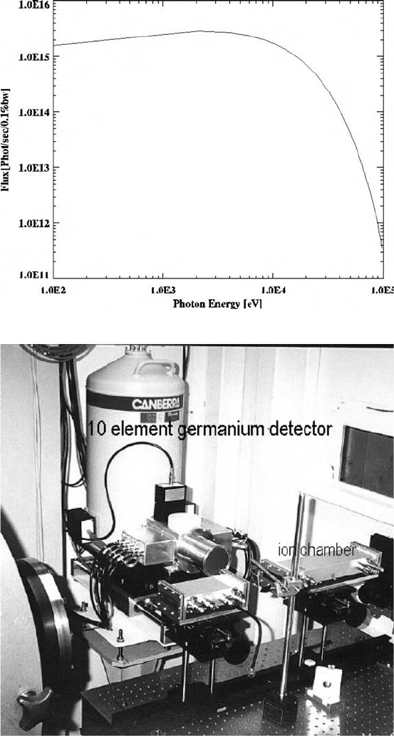
Fig. 10. (b) Calculations of the photon flux of the wiggler as a function of photon energy.
(c) The XAFS station at the Australian National Beamline Facility at the Photon Factory.
Shown are the ionization chambers for transmission XAFS experiments and the 10-element
germanium detector. This has now been superseded by a 30-element detector.

the Australian Synchrotron (Glover et al., 2006). This will be operated in conjunction with
a wiggler source (the details of the front end linking to the storage ring is not shown in this
figure), which will extend the upper energy significantly (Fig. 10(b)). For the W61 wiggler,
the flux is nearly 10
16
photons/cm
2
at 0.1% bandwidth at around 10 keV. The flux remains
above 10
12
, close to 100 keV. The increase in flux and energy range is significant. The
overall range is such that excitation of K-shell electrons up to the uranic elements is possi-
ble. And the flux is very high in the region of 10 keV: close to the K-shell of the transition
metals, and the L-shell emissions of all the elements up to uranium.
The locations of the vertical and horizontal focussing mirrors are shown in Fig. 10(a)(ii),
as is the double-crystal monochromator. The wiggler has magnets of ⬇1.8 T field strength,
K is ⬇20, and there are 18 periods with a period length of 110 mm. Some of the heat load
from the wiggler is taken by the defining aperture. But care has to be taken to cool the bent
flat vertically focussing mirror. Also, the mirror performs a harmonic rejection role, and is
strip coated with silicon, rhodium, and platinum to ensure that harmonic content is a mini-
mum. These have energy cut-offs of 13, 23, 34 keV, respectively. Above 34 keV, the mirror
66 D. Creagh
K
L1, L2, L3
d(i)
Fig. 10. (d) (i) The absorption cross section of molybdenum is plotted as a function of
energy. The contributions of scattering by the photoelectric, Rayleigh, Compton, and pair
production mechanisms are shown. Each element has a different scattering cross section.
In the energy region for which XAFS is a useful technique (4–40 keV), photoelectric scat-
tering is the dominant mode. The circles represent experimental measurements. Note the
discontinuities on the curve for total scattering. These correspond to excitation levels for
atomic shells and subshells, and the discontinuities are referred to absorption edges.
will be moved out of the beam. Further beam definition will be performed using beam-
defining slits.
The double-crystal monochromator has to have good energy resolution (DE/E <10
–4
)
and two cuts of silicon monochromator crystal have to be available: [111] for the range
4–24 keV; [311] for the range 4–50 keV. The first of each crystal pair has to be cryogeni-
cally cooled to reduce the effect that thermal loading has on the reflection of the X-rays.
Because, as we shall see later in this section, the X-ray energy is scanned during an exper-
iment, the exit beam from the monochromator should remain the same, whatever energy is
chosen. Reproducibility of position is also a strong requirement, since, when XANES
spectra are taken, energy differences of 0.1 eV in a measurement of 6.4 keV can be impor-
tant. If time-resolved studies are to be undertaken, the time taken for a scan becomes a
significant parameter in experimental design. The monochromator has to be able to be
operated in the QEXAFS mode (Bornebusch et al., 1999).
Synchrotron Radiation and its Use in Cultural Heritage Studies 67
d(ii)
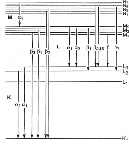
Fig. 10. (d) (ii) A schematic diagram of energy levels in an atom showing how different
photons produce radiations of different energies when transitions occur between levels.
Transitions to the innermost electron shell of an atom (the K-shell) from the next shell (the
L-shell) gives rise to photons with slightly different energies (referred to as K
a1
and K
a2
).
If an electron is removed from the K-shell by the absorption of a photon, the electrons
within the atom rearrange themselves, and a cascade of electronic transitions occur,
causing the emission of photons of various discrete energies (LI, LII, LIII; MI, MII, MIII,
MIV, MV, etc.).
The adjustable toroidal mirror provides both vertical and horizontal focussing. The aim
is to provide a beam of ⬇(0.2 mm ¥ 1 mm) to the sample area. This mirror is rhodium
coated, and it can be rotated about a horizontal axis. At above 23 keV incident beam
energy, this mirror is removed from the system. Since both focussing elements are
removed from the system above 23 keV (just above the K-shell absorption edge of molyb-
denum), the beam size is determined by the slit apertures.
The end station hutch contains the experimental apparatus required for XAS studies. Ion
chambers are used extensively in XAS experiments, both to monitor the beam intensity
and to measure the transmitted X-ray beam. XAS experiments use either transmission
beam measurements or the fluorescence radiation. For the latter, multiple element (up to
100) germanium solid state detectors are used. Figure 10(c) shows the XAFS experimen-
tal station at the Australian National Beamline Facility (ANBF), the Photon Factory,
Tsukuba, Japan. The sample is situated between two ionization chambers. A 30-element
68 D. Creagh
XAFS
Free Atom
Pre-edge
XAFS Region Only
d(iii)
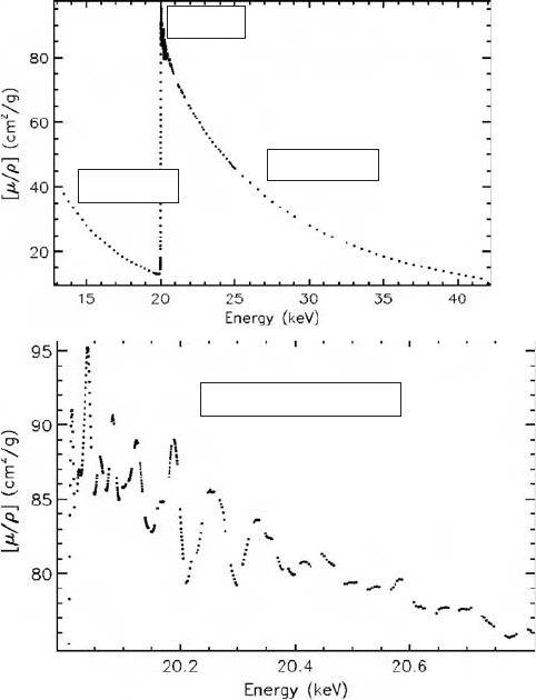
Fig. 10. (d) (iii) XAFS and XANES regions of molybdenum.

germanium detector is located at right angles to the specimen surface for fluorescence
XAFS experiments.
A wide range of sample stages and mounts are used. These may include cooling and
heating stages, and be able to operate with liquids and solids, or at high pressures.
Cryogenic cooling of the sample is desirable to reduce the effect of thermal motion of
atoms in the sample, where this is consistent with experimental constraints. This can some-
times be used to the advantage of the researcher because chemical reactions taking place
outside of the sample cell can be transported into the sample cell, and “stop-frozen” to give
information of the reaction products present.
5.4.1. X-ray absorption fine structure (XAFS)
XAFS useful in probing the environments of selected atomic species in a material. As a
technique, it is used by biologists, chemists, physicists, materials scientists, engineers, and
so on, for monitoring changes in systems, sometimes statically and sometimes dynami-
cally. Since XAFS is a less familiar technique to conservation scientists, I shall give a brief
description of it and how it is used to probe the behaviour of materials. The discussion will
centre on transmission measurements. Fluorescence radiation is related to transmission
radiation through the fluorescence yield, a tabulated function. See http://www.csrri.iit.edu/
periodic-table.html (Krause, 1979).
As its name suggests, XAFS has its origin in the measurement of the transmission
photons through materials. The absorption of atoms has been tabulated by Creagh (2004b)
for all atoms from atomic numbers 1–92. If the intensity of a photon beam is measured
before and after its passage through a material of thickness (t), the relation between the
incident beam intensity (I
o
) and the transmitted intensity (I) is given by
I = I
o
exp (-m
1
t)
Here, m
l
is the linear attenuation coefficient. This is related to the better known mass
absorption coefficient by
A number of different processes contribute to the absorption coefficients: photoelectric
absorption, Rayleigh scattering, Compton scattering, and pair production. In Fig. 10(d)(i) the
absorption cross section of copper is plotted as a function of energy. The contributions of
scattering by the photoelectric, Rayleigh, Compton, and pair production mechanisms are
shown. Each element has a different scattering cross section. In the energy region for
which XAFS is a useful technique (4–40 keV), photoelectric scattering is the dominant
mode. The circles represent experimental measurements. Note the discontinuities on the
curve for total scattering. These correspond to excitation levels for atomic shells and sub-
shells, and the discontinuities are referred to absorption edges.
µ
µ
m
l
=
⎛
⎝
⎜
⎞
⎠
⎟
ρ
Synchrotron Radiation and its Use in Cultural Heritage Studies 69
70 D. Creagh
(a)
(b)
(e)
(a)
(b)
(c)
(f)
(c)
(g)
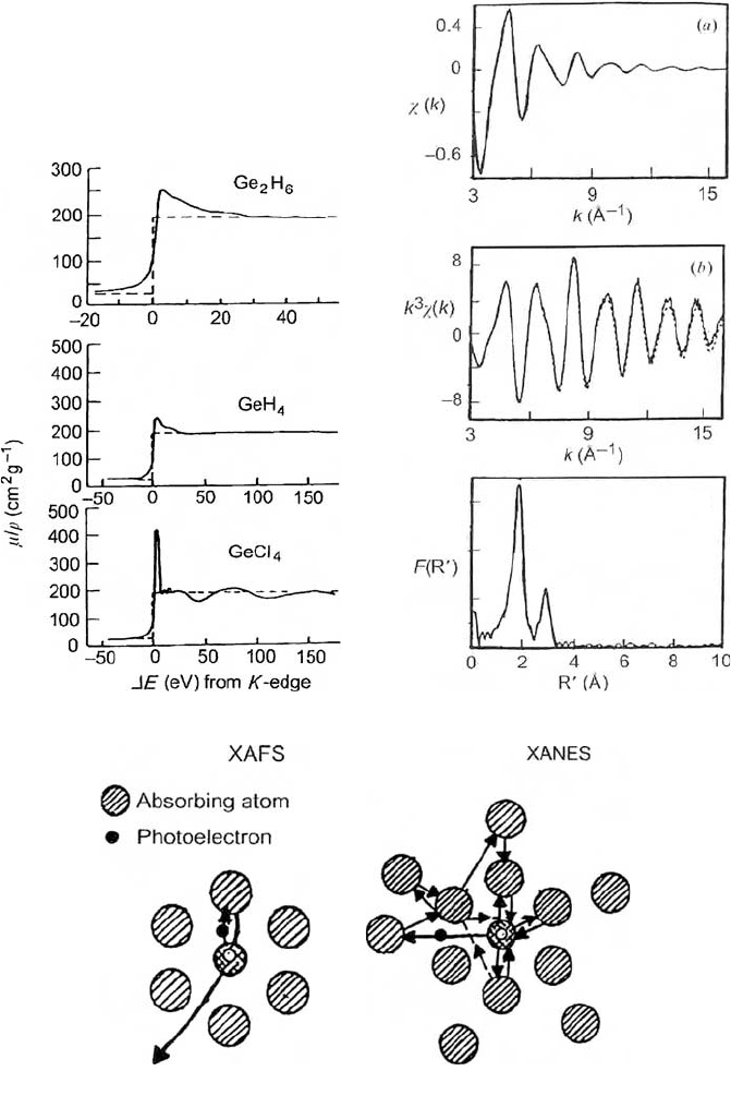
Fig. 10. (e) The effect of molecular structure on the X-ray absorption on gaseous materials
in the region of the K-shall absorption edge: GeCl
4
, GeH
4
, Ge
2
H
6
. Note that these spectra are
XANES spectra since there can be no long-range ordering in the gas phase. (f) Steps in the
XAFS data reduction process. (g) Illustration of the distinction between XAFS and XANES.

Figure 10(d)(ii) is a schematic diagram showing how photons of different energies are
produced when transitions occur between levels. For XAFS studies, absorption by the
K-, L-, and M-shells of atoms are of interest.
The calculations given in tabulations such as Creagh (2004c) are “single atom” calcula-
tions: that is, they are made for a single isolated atom. Using these values to calculate the
absorption of an assemblage of atoms works well at energies remote from an absorption
edge. However, as the absorption edge is approached, deviations from the free atom model
occur. Dramatic effects can occur. Figure 10(d)(iii) shows the mass attenuation coefficient
taken for an energy region that includes the K-shell absorption edge. The upper figure iden-
tifies the pre-edge XAFS and free-atom regions. The lower figure shows just the XAFS data.
Figure 10(e) shows the effect at the edge for three germanium compounds. Evidently the
local environment of the target atomic species, germanium, strongly influences the meas-
ured absorption coefficient.
It is this fact that has lead to the use of XAFS by scientists to probe the local environ-
ments in atoms, and changes that may occur in, say, chemical reactions. The experimental
geometry used in XAFS measurements does not allow absolute measurements to be made.
Readers should peruse the articles of Chantler’s group (2002–2006) to understand the diffi-
culties of making absolute XAFS measurements. Specimen thickness can have a smearing
effect on the XAFS spectra. Thickness should be in the range 2 < ln(I
o
/I) < 4 for best
results.
Nevertheless, XAFS is a valuable research tool, albeit a relative one.
Before giving a brief description of the theory underlying XAFS, the distinction
between XAFS and XANES should be made. XAFS occurs over a considerable energy
range before the edge, 1 keV or more. XANES occur in the energy range, say 100 eV
before and 100 eV after the absorption edge. The origins of the two effects are different,
Synchrotron Radiation and its Use in Cultural Heritage Studies 71
(h)
Fig.10. (h) XANES and pre-edge structures in iron oxides (Foran, 2005). A knowledge of
these structures is, for example, essential for the understanding of the degradation of
historic iron-gall inks on parchment.
