Creagh D., Bradley D. (Eds.) Physical Techniques in the Study of Art, Archaeology and Cultural Heritage. Volume 2
Подождите немного. Документ загружается.

involvement of the SOLEIL synchrotron teams in the setting up of the liaison office. This
work has been partly supported by a grant from the Région
ˆ
Ile-de-France.
REFERENCES
Ando, M., Chen, J., Hyodo, K., Mori, K., Sugiyama, H., Xian, X., 2000. Nondestructive visual search for fossils
in rock using X-ray interferometry imaging. Jap. J. Appl. Phys. 39(2), L1009–L1011.
Bartoll, J., Röhrs, S., Erko, A., Firsov, A., Bjeoumikhov, A., Langhoff, N., 2004. Micro-X-ray absorption near
edge structure spectroscopy investigations of baroque tin-amalgam mirrors at BESSY using a capillary focusing
system. Spectrochim. Acta B 59(10–11), 1587–1592.
Bertholon, R., 2001. Chapter Nettoyage et stabilisation de la corrosion par électrolyse. In La conservation des
métaux (Conservation du patrimoine Tome 5). Le cas des canons provenant de fouilles sous-marines, CNRS
Éditions, Ministère de la Culture et de la Communication, Paris, France: pp. 83–101.
Bertrand, L., 2002. Approche structurale et bioinorganique de la conservation de fibres kératinisées
archéologiques. PhD Thesis, Université Paris 6, Paris.
Bertrand, L., Doucet, J., 2007. Dedicated liaison office for cultural heritage at the SOLEIL synchrotron. Nuovo
Cimento C. 30(01), 35–40.
Bertrand, L., Doucet, J., Dumas, P., Simionovici, A., Tsoucaris, G., Walter, P., 2003. Microbeam synchrotron
imaging of hairs from ancient Egyptian mummies. J. Synchrotron Radiat. 10(5), 387–392.
Bertrand, L., Vantelon, D., Pantos, E., 2006. Novel interface for cultural heritage at SOLEIL. Appl. Phys. A 83(2),
225–228.
Bilderback, D.H., Powers, J., Dimitrova, N., Huang, R., Smilgies, D.-M., Clinton, K., Thorne, R.E., 2005. X-ray
fluorescence recovers writing from ancient inscriptions. Z. Papyrol. Epigr. 152, 221–227.
Brunet, M., Guy, F., Pilbeam, D., Lieberman, D.E., Likius, A., Mackaye, H.T., Ponce de León, M.S.,
Zollikofer, C.P., Vignaud, P., 2005. New material of the earliest hominid from the Upper Miocene of Chad.
Nature 434(7034), 752–755.
Chaimanee, Y., Jolly, D., Benammi, M., Tafforeau, P., Duzer, D., Moussa, L., Jaeger. J.-J., 2003. A Middle
Miocene hominoid from Thailand and orangutan origins. Nature 422, 61–65.
Cotte, M., Walter, P., Tsoucaris, G., Dumas, P., 2005. Studying skin of an Egyptian mummy by infrared
microscopy. Vib. Spectrosc. 38(1–2), 159–167.
Davies, R.J., Burghammer, M., Riekel, C., 2005. Simultaneous microRaman and synchrotron radiation microd-
iffraction: Tools for materials characterization. Appl. Phys. Lett. 87(26), 264105.
De Ryck, I., Adriaens, A., Pantos, E., Adams, F., 2003. A comparison of microbeam techniques for the analysis
of corroded ancient bronze objects. Analyst 128(8), 1104–1109.
Dooryhée, E., Anne, M., Bardiès, I., Hodeau, J.-L., Martinetto, P., Rondot, S., Salomon, J., Vaughan, G.B.M.,
Walter, P., 2005. Non-destructive synchrotron X-ray diffraction mapping of a Roman painting. Appl. Phys.
A 81(4), 663–667.
Fors, Y., Sandström, M., 2006. Sulfur and iron in shipwrecks cause conservation concerns. Chem. Soc. Rev. 35,
399–415.
Gliozzo, E., Kirkman, I.W., Pantos, E. Memmi-Turbanti, I., 2004. Black gloss pottery: production sites and tech-
nology in Northern Etruria, part II: gloss technology. Archaeometry 46(2), 227–246.
Grolimund, D., Senn, M., Trottmann, M., Janousch, M., Bonhoure, I., Scheidegger, A., Marcus, M., 2004.
Shedding new light on historical metal samples using micro-focused synchrotron X-ray fluorescence and
spectroscopy. Spectrochim. Acta B 59, 1627–1635.
Guerra, M.F., Calligaro, T., Radtke, M., Reiche, I., Riesemeier, H., 2005. Fingerprinting ancient gold by measuring
Pt with spatially resolved high energy Sy-XRF. Nucl. Instrum. Methods B 240, 505–511.
Hall, C., Barnes, P., Cockcroft, J.K., Colston, S.L., Hausermann, D., Jacques, S.D.M., Jupe, A.C., Kunz, M., 1998.
Synchrotron radiation energy-dispersive diffraction tomography. Nucl. Instrum. Methods B 140, 253–257.
Hochleitner, B., Schreiner, M., Drakopoulos, M., Snigireva, I., Snigirev, A., 2003. Analysis of paint layers by
light microscopy, scanning electron microscopy and synchrotron induced X-ray micro-diffraction. In Proc.
Conf. Art 2002, Antwerp, Belgium, June 2003.
112 L. Bertrand
Isaure, M.P., Laboudigue, A., Manceau, A., Sarret, G., Tiffreau, C., Trocellier, P., Lamble, G., Hazemann, J.L.,
Chateigner, D., 2002. Quantitative Zn speciation in a contaminated dredged sediment by m-PIXE, m-SXRF,
EXAFS spectroscopy and principal component analysis. Geochim. Cosmochim. Acta 66(9), 1549–1567.
Janssens, K., Proost, K., Deraedt, I., Bulska, E., Wagner, B., Schreiner, M., 2003. Chapter, The use of
focussed X-ray beams for non-destructive characterisation of historical materials. In Molecular and
Structural Archaeology: Cosmetic and Therapeutic Chemicals, Volume 117. Kluwer Academic Publishers,
Dordrecht, The Netherlands: pp. 193–200.
Kanngiesser, B., Malzer, W., Reiche, I., 2003. A new 3D micro X-ray fluorescence analysis set-up – first archaeo-
metric applications. Nucl. Instrum. Methods B 211, 259–264.
Kanngiesser, B., Hahn, O., Wilke, M., Nekat, B., Malzer, W., Erko, A., 2004. Investigation of oxidation and
migration processes of inorganic compounds in ink-corroded manuscripts. Spectrochim. Acta B 59,
1511–1516.
Kempson, I.M., Paul Kirkbride, K., Skinner, W.M., Coumbaros, J., 2005. Applications of synchrotron radiation
in forensic trace evidence analysis. Talanta 67, 286–303.
Kennedy, C.J., Hiller, J.C., Lammie, D., Drakopoulos, M., Vest, M., Cooper, M., Adderley W.P., Wess, T.J., 2004.
Microfocus X-ray diffraction of historical parchment reveals variations in structural features through
parchment cross sections. Nano Lett. 4(8), 1373–1380.
LabsTech European Coordination Network (1999–2002). http://www.chm.unipg.it/chimgen/LabS-TECH.html
Manceau, A., Tamura, N., Marcus, M.A., MacDowell, A.A., Celestre, R.S., Sublett, R.E., Sposito, G.,
Padmore, H.A., 2002. Deciphering Ni sequestration in soil ferromanganese nodules by combining X-ray
fluorescence, absorption, and diffraction at micrometer scales of resolution. Am. Miner. 87(10), 1494–1499.
Marivaux, L., Chaimanee, Y., Tafforeau, P., Jaeger, J.-J., 2006. New Strepsirrhine primate from the late Eocene
of peninsular Thailand (Krabi Basin). Am. J. Phys. Anthrop. 130(4), 425–434.
Nakai, I., Iida, A., 1992. Applications of SR-XRF imaging and micro-XANES to meteorites, archaeological
objects and animal tissues. In Advances in X-ray Analysis, Volume 35. Eds. Barrett, C.S., Gilfrich, J.V.,
Huang, T.C., Jenkins, R., McCarthy, G.J., Predecki, P.K., Ryon, R., Smith, D. Plenum Press: New York, USA,
pp. 1307–1315.
Neff, D., Réguer, S., Bellot-Gurlet, L., Dillmann, P., Bertholon, R., 2004. Structural characterization of corrosion
products on archaeological iron: an integrated analytical approach to establish corrosion forms. J. Raman
Spectrom. 35(8–9), 739–745.
Pantos, E., Tang, C.C., MacLean, E.J., Cheung, K.C., Strange, R.W., Rizkallah, P.J., Papiz, M.Z., Colston, S.L.,
Roberts, M.A., Murphy, B.M., Collins, S.P., Clark, D.T., Tobin, M.J., Zhilin, M., Prag, K., Prag, A.J.N.W.,
2002. Applications of synchrotron radiation to archaeological ceramics. In Modern Trends in Scientific
Studies on Ancient Ceramics, Volume 1011. Eds. Kilikoglou, V., Hein, A., Maniatis, Y. BAR International
Series, Oxford, United-Kingdom: pp. 377–384.
Pickering, I.J., Prince, R.C., Salt, D.E., George, G.N., 2000. Quantitative, chemically specific imaging of selenium
transformation in plants. PNAS 97(20), 10717–10722.
Proost, K., Janssens, K., Wagner, B., Bulska, E., Schreiner, M., 2004. Determination of localized Fe
2+
/Fe
3+
ratios
in inks of historic documents by means of m-XANES. Nucl. Instrum. Methods B 213, 723–728.
Réguer, S., Dillmann, P., Mirambet, F., Bellot-Gurlet, L., 2005. Local and structural characterisation of
chlorinated phases formed on ferrous archaeological artefacts by mXRD and mXANES. Nucl. Instrum.
Methods B 240(1–2), 500–504.
Réguer, S., Dillmann, P., Mirambet, F., Susini, J., Lagarde, P., 2006. Investigation of Cl corrosion products of
iron archaeological artefacts using micro-focused synchrotron X-ray absorption spectroscopy. Appl. Phys.
A 83(2), 189–193.
Salvadó, N., Pradell, T., Pantos, E., Papiz, M.Z., Molera, J., Seco, M., Vendrell-Saz, M., 2002. Identification of
copper-based green pigments in Jaume Huguet’s Gothic altarpieces by Fourier transform infrared microspec-
troscopy and synchrotron radiation X-ray diffraction. J. Synchrotron Radiat. 9(4), 215–222.
Salvo, L., Cloetens, P., Maire, E., Zabler, S., Blandin, J.J., Buffière, J.Y., Ludwig, W., Boller, E., Bellet, D.,
Josserond, C., 2003. X-ray micro-tomography an attractive characterisation technique in materials science.
Nucl. Instrum. Methods B 200, 273–286.
Sandström, M., Jalilehvand, F., Persson, I., Gelius, U., Frank, P., Hall-Roth, I., 2002. Deterioration of the
seventeenth-century warship Vasa by internal formation of sulphuric acid. Nature 415(6874), 893–897.
Synchrotron Imaging for Archaeology, Art History, Conservation, and Palaeontology 113
Sandström, M., Jalilehvand, F., Damian, E., Fors, Y., Gelius, U., Jones, M., Salomé, M., 2005. Sulfur accumula-
tion in the timbers of King Henry VIII’s warship Mary Rose: a pathway in the sulfur cycle of conservation
concern. PNAS 102(40), 14165–14170.
Sciau, P., Goudeau, P., Tamura, N., Dooryhée, E., 2006. Micro scanning X-ray diffraction study of Gallo-Roman
Terra Sigillata ceramics. Appl. Phys. A 83(2), 219–224.
Simionovici, A., Janssens, K., Rindby, A., Snigireva, I., Snigirev, A., 2000. Precision micro-XANES of Mn in
corroded Roman glasses. In X-ray Microscopy: Proceedings of the VI International Conference, Berkeley,
California, USA, 2–6 Aug 1999. Volume 507 of AIP Conference, Eds. Meyer-Ilse, W., Warwick, T. and
Atwood, D. American Institute of Physics: Melville, New York, USA, pp. 279–283.
S
ˇ
mit, Z
ˇ
., Janssens, K., Proost, K., Langus, I., 2004. Confocal m-XRF depth analysis of paint layers. Nucl. Instrum.
Methods B 219–220, 35–40.
Somogyi, A., Drakopoulos, M., Vincze, L., Vekemans, B., Camerani, C., Janssens, K., Snigirev, A., Adams, F.,
2001. ID18F: a new micro-x-ray fluorescence end-station at the European Synchrotron Radiation Facility
(ESRF): preliminary results. X-Ray Spectrom. 30(4), 242–252.
Tafforeau, P., 2004. Aspects phylogénétiques et fonctionnels de la microstructure de l’émail dentaire et de la
structure tridimensionnelle des molaires chez les primates fossiles et actuels: apports de la microtomogra-
phie à rayonnement X synchrotron. PhD Thesis, Université de Montpellier II, France.
Tafforeau, P., Boistel, R., Boller, E., Bravin, A., Brunet, M., Chaimanee, Y., Cloetens, P., Feist, M.,
Hoszowska, J., Jaeger, J.J., Kay, R.F., Lazzari, V., Marivaux, L., Nel, A., Nemoz, C., Thibault, X., Vignaud, P.,
Zabler, S., 2006. Applications of X-ray synchrotron microtomography for non-destructive 3D studies of
paleontological specimens. Appl. Phys. A 83(2), 195–202.
Wess, T.J., Alberts, I., Hiller, J., Drakopoulos, M., Chamberlain, A.T., Collins, M., 2002. Microfocus small angle
X-ray scattering reveals structural features in archaeological bone samples: detection of changes in bone
mineral habit and size. Calcif. Tissue Int. 70(2), 103–110.
114 L. Bertrand
Chapter 3
Holistic Modeling of Gas and Aerosol Deposition
and the Degradation of Cultural Objects
I.S. Cole, D.A. Paterson and D. Lau
CSIRO Manufacturing & Infrastructure Technology, PO Box 56, Highett, Victoria 3190, Australia
Email: Ivan.Cole@csiro.au
ho
.
lis
.
tic (ho
-
-li
ˇ
s’ti
ˇ
k) adj. Emphasizing the importance of the whole and the interdependence of its parts.
Concerned with wholes rather than analysis or separation into parts.
Abstract
This chapter addresses the deposition of gases and aerosols both inside and outside museums and the possible
effects that such deposition may have on cultural objects. This issue is addressed through the concept of holistic
modeling, where all critical factors controlling the deposition and degradation process are defined and linked
together. The types and sizes of particulates both within and exterior to a museum are outlined. The types of gases
found within a dwelling and their relations to exterior pollutants are described. The aerosol and gas deposition
mechanisms and the equations for each mechanism are outlined. In order to define conditions for gas deposition,
the factors controlling condensation and formation of moisture layers are also presented. These principles and
equations are then illustrated by analysis of the generation, transport and deposition of aerosols on cultural
objects in the external environment, followed by a similar analysis for inside buildings. In the case of deposition
inside buildings, the literature is first reviewed, and then three case studies are analyzed that represent significant
cases or highlight unresolved issues in the literature. The case studies clarify the relative importance of each
deposition mechanism. It is evident that the major mechanisms within a building are gravity, vortex shedding and,
in case of significant air flows, momentum-dominated impact. Factors controlling the attachment and detachment
of pollutants both within and outside dwellings are then outlined, as are the common damage forms that result
for some pollutants. Throughout the chapter and especially towards the end, the implications of the findings to
design and maintenance strategies are discussed.
Keywords: Particulates, gases, deposition, condensation, cultural objects, interior spaces, pollutant transport,
degradation, maintenance.
Contents
1. Introduction 116
2. Types and sizes of particles and types of gases 118
2.1. Particulates 118
2.2. Gases 119
3. Deposition mechanisms 121
3.1. Aerosol deposition mechanisms 121
3.2. Gas deposition mechanisms 124
4. Deposition equations 125
4.1. Aerosol deposition equations 125
4.1.1. Gravitational settling 126
Physical Techniques in the Study of Art, Archaeology and Cultural Heritage 115
Edited by D. Creagh and D. Bradley
© 2007 Elsevier B.V. All rights reserved
4.1.2. Momentum-dominated impact 127
4.1.3. Laminar diffusion/Brownian deposition 127
4.1.4. Turbulent diffusion 128
4.1.5. Vortex shedding 128
4.1.6. Filtering 129
4.1.7. Electrostatic attraction 130
4.1.8. Thermophoresis 131
4.1.9. Photophoresis 131
4.2. Pollutant deposition equations 131
4.2.1. Condensation 131
4.2.2. Gas deposition 132
5. Generation, transport and deposition on cultural objects exposed to the external environment 133
5.1. Implication from source transport and deposition models to degradation of objects 136
6. Generation, transport and deposition on cultural objects inside buildings 137
6.1. Literature review 137
6.2. Issue arising from the literature 138
6.3. Case studies 139
6.3.1. Case study 1 – settling on upward-facing surfaces 140
6.3.2. Case study 2 – flow past a vertical surface 140
6.3.3. Case study 3 – deposition on a downward-facing surface 141
6.4. Discussion of case studies 141
6.5. Attachment and detachment 143
6.5.1. Within buildings 143
6.5.2. From exterior surfaces 144
6.6. Factors controlling gaseous pollutant levels within museums 145
6.6.1. Implications to cultural practice of pollutant sources and deposition 147
7. Surface forms and degradation 148
7.1. Oxide products 148
7.2. Implications of pollutants to object degradation 149
8. Implications for design and maintenance strategies 151
9. Conclusions 152
References 152
1. INTRODUCTION
This chapter addresses the deposition of liquid and solid particulates and gases on cultural
objects that may be open to the external environment or protected inside buildings, and the
effects of such pollutants and condensates.
In an external environment, the major pollutants are marine aerosols and acidic partic-
ulates, which may promote corrosion (oxidation), lead to damage from crystallization and
promote condensation, providing a culture medium for biodegradation. Within buildings
(museums or historic structures), there may be a wide range of particulates and pollutants
(see Section 2). These may be damaging to collections in a number of ways. They may:
∑ act as a carrier for more degradation species;
∑ lead to visual but not chemical degradation;
∑ chemically degrade artwork; and
∑ act as a catalyst for chemical degradation.
Where benign particulates deposit and do not chemically interact, damage may be
induced secondarily when treatments for removal lead to the ingress of deposited material
116 I.S. Cole
et al.

Holistic Modeling of Gas and Aerosol Deposition and Degradation 117
into the surface structure of an object. This may occur with dry-surface-cleaning treatments
involving brushing or rubbing, or wet surface swabbing or washing.
A further consideration is the susceptibility of the object to the deposited particulate or
gas. Inorganic materials may be relatively unaffected by microbial deposits, and acidic
deposits are generally considered aggressive for most materials, while co-deposited gases
or particulates may act to neutralize an acidic or alkaline pollutant.
Baer and Banks (1985) indicate that soot and organic compounds may absorb damaging
gases such as SO
2
. Soot may also lead to visual degradation of paintings. (NH
4
)
2
SO
4
can
induce bloom on varnish, while other S-rich materials can be responsible for discoloring
of the pigments by oxidation to H
2
SO
4
(De Santis et al., 1992). This process can be
catalyzed by Fe-rich particles.
There have been a range of studies looking at pollutant sources, transport and deposi-
tion, both in the exterior and interior environments, and these are reviewed in Sections 4.1,
5 and 6.1. One limitation of many of these studies is that they tend to look at a particular
component of the problem of degradation, rather than study the series of processes that
lead to it. Degradation is an interconnected process involving pollutant generation, trans-
port and deposition, followed by attack on the surface and physico-chemical modification
of the surface. The current authors have developed a holistic model of degradation that
links and models the processes controlling atmospheric corrosion on a range of scales,
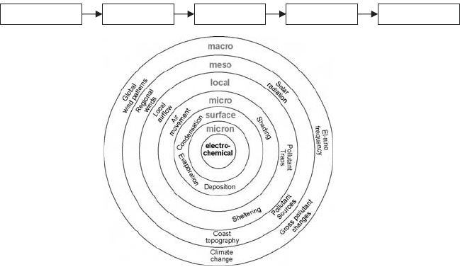
from macro through meso to local, micro and lastly micron (Cole et al., 2003). A schematic
presentation of the holistic model as defined by the event sequence and scale diagram is
given in Fig. 1.
These scales are defined in line with EOTA (1997), so that “macro” refers to gross mete-
orological conditions (polar, subtropical, etc.), “meso” refers to regions with dimensions up
to 100 km, “local” is in the immediate vicinity of a building, while “micro” refers to the
absolute proximity of a material surface. As indicated above, the holistic model links
Generation Transport Deposition Attack
Degradation
Fig. 1. Framework for a holistic model. A holistic view of the time sequence, of the environ-
ment and of degradation mechanisms.
processes controlling degradation across a range of scales, so that the formation of marine
aerosols is modeled on the meso-scale from wave activity in the oceans, while oxide
growth is modeled on the micron scale. The emphasis of the model shifts when modeling
exterior or interior conditions.
In the case of the exterior of a building, the emphasis of the research is on determining
the aerosol concentration adjacent to a dwelling (which involves significant source and
transport modeling). Deposition onto a dwelling is presented, but can be modeled rela-
tively simply as it is primarily controlled by turbulent diffusion (Cole and Paterson, 2004).
In contrast, deposition within a building can occur by a variety of mechanisms (Camuffo,
1998), of which turbulent diffusion is one of the least important. The pollutant concentra-
tion within a building is controlled by exterior pollutant levels and by ad hoc factors within
a building. Thus, in this chapter, the discussion of internal deposition will focus on the
mechanism of pollutant deposition.
This chapter will address the types of particulates and gases, deposition mechanisms,
deposition equations, generic case studies for cultural objects outside and inside buildings,
attachment and detachment issues, surface forms of corrosion and degradation, and impli-
cations for design and maintenance.
2. TYPES AND SIZES OF PARTICLES AND TYPES OF GASES
2.1. Particulates
In this chapter, the word “particle” is used for both solid and liquid aerosols, but not for
gaseous pollutants. Common dangerous classes of particulate deposits are (Hill and
Bouwmeester, 1994):
∑ acidic substances,
∑ oxidizing substances,
∑ soot and tarry particles from burning fuel,
∑ large abrasive particles,
∑ hygroscopic materials,
∑ particles containing traces of metals (that act as catalysts),
∑ wet and oily particles from food preparation,
∑ alkaline particles from new concrete,
∑ salt crystals and dissolved salts (corrosion and microorganisms),
∑ textile fibers and skin fragments as food for insects.
Particulates may arise from exterior sources, either natural (sea salt aerosol, bushfires,
windblown dust, pollen and other plant products and insect, arachnid and bird products), agri-
cultural (agricultural sprays), transport (oil and soot) or industrial (organics and sulfuric acid).
Particles may also arise from within, either human-related (lint, dirt, hair, skin flakes,
droplets and food), microorganism-related (mites, mold and other microorganisms),
combustion-related (cigarette smoke, soot and ash from candles, incense, smoke from the
kitchen range and frying oil), renovation-related (brick, plaster and asbestos dust and clean-
ing products) or resuspended (from vacuuming, walking, dusting and sitting on furniture).
118 I.S. Cole
et al.

In a study of particulate soiling in museums and historic houses (Yoon and Brimblecombe,
2001), sticky samplers were used to collect particulates. The main components were soil
dust, soot and fibers with lesser amounts of human hair and skin, plant and paint fragments
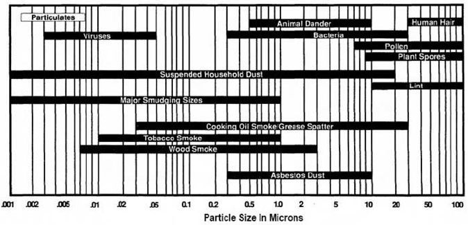
and insect parts. Some typical particle size ranges are given in Fig. 2 (Annis, 1991).
2.2. Gases
In the external environment of urban regions, the major acidic and oxidizing gaseous
pollutants occur as a result of industrial activity. Sulfur dioxide and nitrogen dioxide are
produced from the combustion of fossil fuels (coal, gas and oil) used in energy production
and transport. While natural sources may be the primary contributors for these pollutants
in remote areas, human activity is by far the major source in the modern built environment.
Although naturally occurring in the stratosphere, ozone, an oxidizing pollutant, is found at
ground levels and is most often a result of photochemical smog. Levels of ozone can be
directly related to those of SO
x
and NO
x
.
The consequences of outdoor materials are (1) acidic and (2) oxidizing. Specific impli-
cations of acidic attack exist for calcareous materials, cellulosic materials and particularly
ferrous but most metals. Buildings and monuments made of limestone, marble and other
alkaline stones are literally dissolved through the neutralizing reaction with acids.
Oxidizing attack occurs through destructive oxidation of chemical bonds within organic
materials, e.g. synthetic polymers, natural polymers (cellulose and protein) and dyes.
However, the short half-life of ozone somewhat modifies its powerful oxidizing action.
Acidic activity of any particular acid is indicated by its dissociation constant pKa
(see Table 1). The overall acidic effect experienced by a surface is contributed to by the sum
of all the atmospheric acids present, including carbonic (from CO
2
) and organic acids, but
is influenced by pKa. Therefore, considering that nitric and sulfuric acids have a pKa much
Holistic Modeling of Gas and Aerosol Deposition and Degradation 119
Fig. 2. Size ranges of indoor pollution particles.

lower than carbonic acid, in a solution where the concentration of nitric and sulfuric acids may
be much lower, these highly acidic species will contribute much more to the total acidity.
While particulates are generally considered to have adverse effects for both humans and
objects, the definition of pollutant gases for collections remains more open to interpreta-
tion and reliant on the particular situation. Gases that have measurable health risks for
humans are not always dangerous for collections, and the reverse also applies. Those most
often associated with harm to collections are SO
x
, NO
x
, carboxylic acids (acetic, formic
and fatty acids), ozone and some amines. Volatile organic compounds (VOCs) are included
as they are often present in association with the short-chain carboxylic acids and their
precursors, although the specific effects of most VOCs are still unknown. Larger molecules
may photodissociate to produce active smaller molecules or be involved in inter- or intra-
molecular chemical reactions. For example, VOCs with primary alcohol groups may react
with oxygen to become aldehydes, and then carboxylic acids. Gases can also interact and
influence the particulate composition of an environment. Ozone and terpenes have been
shown to increase the number and mass concentrations of sub-micron particles, resulting
in an undocumented source of indoor particulates (Weschler and Shields, 1999).
It is also worth noting that in studies of concentrations of indoor air VOCs, total VOC
(TVOC) concentrations are considerably higher than individually measured VOCs (Brown
et al., 1994). This suggests that there are a large number of chemical compounds present,
but only a select few are evaluated using current monitoring methodologies.
120 I.S. Cole
et al.
Table 1. pKa values for acids at 25∞C in water (CRC
Handbook of Chemistry and Physics, Edition 76, 1995–1996)
Acid Formula pKa
Perchloric HClO
4
~-7
Hydrochloric HCl ~-7
Chloric HClO
3
~-3
Sulfuric (1) H
2
SO
4
~-2
Nitric HNO
3
~-1.3
Oxalic (1) H
2
C
2
O
4
1.23
Sulfuric (2) 1.92
Chlorous HClO
2
1.96
Phosphoric (1) H
3
PO
4
2.12
Nitrous HNO
2
3.34
Formic HCOOH 3.75
Oxalic (2) 4.19
Acetic CH
3
COOH 4.75
Carbonic (1) H
2
CO
3
6.37
Hydrosulfuric H
2
S 7.04
Ammonium ion 9.25
Hydrogen peroxide H
2
O
2
11.62
(1) and (2) refer to the first and second deprotonation stages of the acid.
NH
4
+
H
CO
24
−
HSO
4
−
In the inside-museum environment, gaseous pollutants arise from both internal and
external sources. Internally generated VOCs are generally recognized to occur in far
greater concentration indoors compared with the external environment, in the ratio 8:1 for
established buildings and greater than 200:1 for new buildings (Brown, 2003). Conversely,
pollutants generated outdoors are found to be in much lower concentration indoors, as
exemplified by the measurements of SO
2
, O
3
, NO
x
and NO given in Table 2.
Gaseous emissions may occur from the artworks themselves, or from the materials and
coatings used in storage, transport and display. Processed wood products are the most often
cited sources of VOCs such as acetate and formate. Newer building materials are expected
to reduce emissions to an acceptable level with time; however, the persistence of emissions
is demonstrated in older wood in a study by Rhyl-Svendsen and Glastrup (2002). It was
found that a 15-year-old oak plank had an acetic acid emission of 55.7 (±5.6) mg/m
2
h and
a formic acid emission of 11.1 (±2.5) mg/m
2
h; and a 12-year-old coin collection drawer
with a Masonite board base and maple wood edges had an acetic acid emission of 172.5
(±6.9) mg/m
2
h and a formic acid emission of 94.1 (±7.2) mg/m
2
h.
3. DEPOSITION MECHANISMS
3.1. Aerosol deposition mechanisms
There are a number of mechanisms that can lead to the deposition of particulates (Camuffo,
1998; Van Greiken et al., 1998). In this chapter, the most important of these are restated,
some with new or improved equations, and a few more possible deposition mechanisms are
introduced.
The main deposition mechanisms for aerosol particles are:
∑ gravitational settling,
∑ turbulent diffusion,
∑ laminar diffusion (also called Brownian deposition and diffusiophoresis),
∑ thermophoresis (migration from high temperatures to low),
∑ electrostatic attraction,
∑ momentum-dominated impact,
∑ vortex shedding (transport by transient laminar flows),
∑ filtering (flow past or through raised fabrics) and
∑ photophoresis (motion generated by an intense beam of light).
These are illustrated in Fig. 3.
Different particle sizes are affected by different mechanisms. Gravity and
momentum-dominated impact are most significant for the largest particles. Turbulent
diffusion and vortex shedding are independent of particle size. Electrostatic attraction and
laminar diffusion are strongest for the smallest particles. Thermophoresis and filtering
display a weak dependence on particle size, with smaller particles being favored in
thermophoresis.
Because the effect of vortex shedding on deposition is not widely known, it is described
here in a bit more detail than the others. It is similar to turbulent diffusion, but occurs at much
Holistic Modeling of Gas and Aerosol Deposition and Degradation 121
