Boros Mihaly. Surgical Techniques
Подождите немного. Документ загружается.

VIII. Hemorrhagic shock
“Igitur corde percusso sanguis multus fertur, ve-
nae elanguescunt, color pallidissimus, sudores frigidi
malique odoris tamquam inrorato corpore oriuntur, ex-
tremisque partibus frigidis matura mors sequitur.”
Aulus Cornelius Celsus: De Medica (1478) Liber V. 28.8.
1. General remarks
Hemorrhagic shock was well characterized by Cel-
sus (1st century Roman savant): “When the heart is
injured, much blood is lost, the pulse becomes feeble,
the skin becomes extremely pale, the body is covered
with a malodorous sweat, the extremities are frigid,
and death occurs speedily”.
Shock is a condition leading to death - according to
John C. Warren (1895): “a momentary pause in the
act of death”. e treatment depends on the ear-
ly recognition. Shock may be presumed suspected
from the anamnesis and the risk factors.
Shock is not equal to hypotension (shock is not al-
ways accompanied by hypotension due to the com-
pensation mechanisms in the circulation). Not every
hypotensive state indicates a shock circulation, and
not every shock state is accompanied by low BP.
e main types of shock, depending on the cause of
the syndrome: hypovolemic, cardiogenic, distribu-
tive, and others, such as obstructive. ere are many
other shock states; the clinical shock types are not
strict categories.
Independently of the category, the key factor of
shock is inadequate tissue perfusion (CO may be low
or high), i.e. independently of the main cause (trig-
ger), the main pathogenetic factor is an imbalance
between the O₂ delivery and demand, resulting in a
disordered cell function.
2. Types of shock
Hypovolemic Distributive Cardiogenic Others
Dehydration,
starvation
Anaphylactic
Congenital
cardio-
myopathy
Heat
Gastro-
enteritis
Neurogenic Ischemic
Pulmonary
emboli
Burns
Drugs
(toxicity)
Anoxic Pancreatitis
Hemorrhage Septic Tamponade Obstructive
Shock may be comprised of components of different
subtypes (e.g. distributive and cardiogenic): the types
are not obligate!
3. e essential patterns of
circulatory shock
4. Anamnesis of shock
Signs upon physical examination:
depressed central nervous system activity,
an abnormal mucosal color,
a decreased urine output (a sign of systemic tissue hypo-
perfusion),
tachypnea, tachycardia,
decreased capillary refill.
Biochemical changes:
arterial blood gases: acidosis with a base deficit (a
sign of tissue hypoperfusion),
venous blood gases: decreased venous O₂ saturation,
an electrolyte imbalance.
5. Compensatory mechanisms
after blood loss
5.1. Baroreceptor reflex
is responds to small changes in vascular tone/pres-
sure. It leads to a decreased vagal tone, which increas-
es the HR, and decreases the coronary resistance (im-
proves the myocardial O₂ supply). e sympathetic tone
is increased, which causes venoconstriction, constric-
tion of blood reservoirs (increasing circulating blood
volume), and decreased perfusion in the skin and skel-
etal muscle.
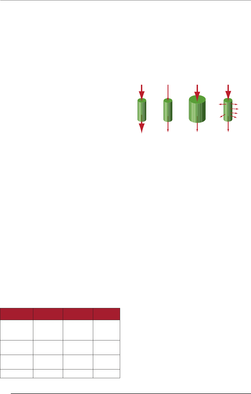
Normal CO
Normal perfusion
Normal circulation
Reduced perfusion
� Cardiogenic shock
– Inadequate MC
– Inadequate HR
– Excessive afterload
Reduced perfusion
� Sepsis, anaphylaxis
– Dilated container
without change in
fluid volume
Reduced perfusion
� Hypovolemic shock
– Abnormally low
circulating blood volume
– Leak in container
Normal COReduced CO
Normal /
elevated CO
SURGICAL TECHNIQUES
70
VIII. HEMORRHAGIC SHOCK
5.2. Chemoreceptors
ese are sensitive to O₂ and CO₂ and sense hypoxia
(due to inadequate blood flow in the peripheral tissues
and a MAP of ~ 60 mmHg). Important chemoreceptors
are the carotid and aortic bodies. Reflexes that regulate
BP are integrated in the medulla. e results are further
vasoconstriction and an improved venous return (to the
pump).
5.3. Endogenous vasoconstrictors
e adrenal medullary hormones norepinephrine and
epinephrine cause vasoconstriction and an increased
CO. Vasopressin (anti diuretic hormone - ADH) re-
leased from the posterior pituitary causes intense va-
soconstriction in cases of extremely low MAP. Renin
(from a decreased renal perfusion) leads to angiotensin-
ogen and angiotensin II production. e endothelium-
derived factors endothelin-1 and prostaglandin-derived
growth factor are both potent vasoconstrictors.
5.4. Brain ischemia
Low MAP (60 mmHg) causes a decreased cerebral per-
fusion pressure and an increased sympathoadrenal ac-
tivity (which is higher than that induced by barorecep-
tors), involving the increased release of catecholamines
from the adrenal glands and sympathetic nerves (lead-
ing to vagus nerve stimulation, which has opposite ef-
fects).
5.5. Changes in renal water
metabolism
e aldosterone release stimulated by vasopressin re-
sults in Na
+
reabsorption in the distal tubules of the kid-
neys; water follows the Na
+
.
5.6. Reabsorption of tissue fluids
(“fluid shift”)
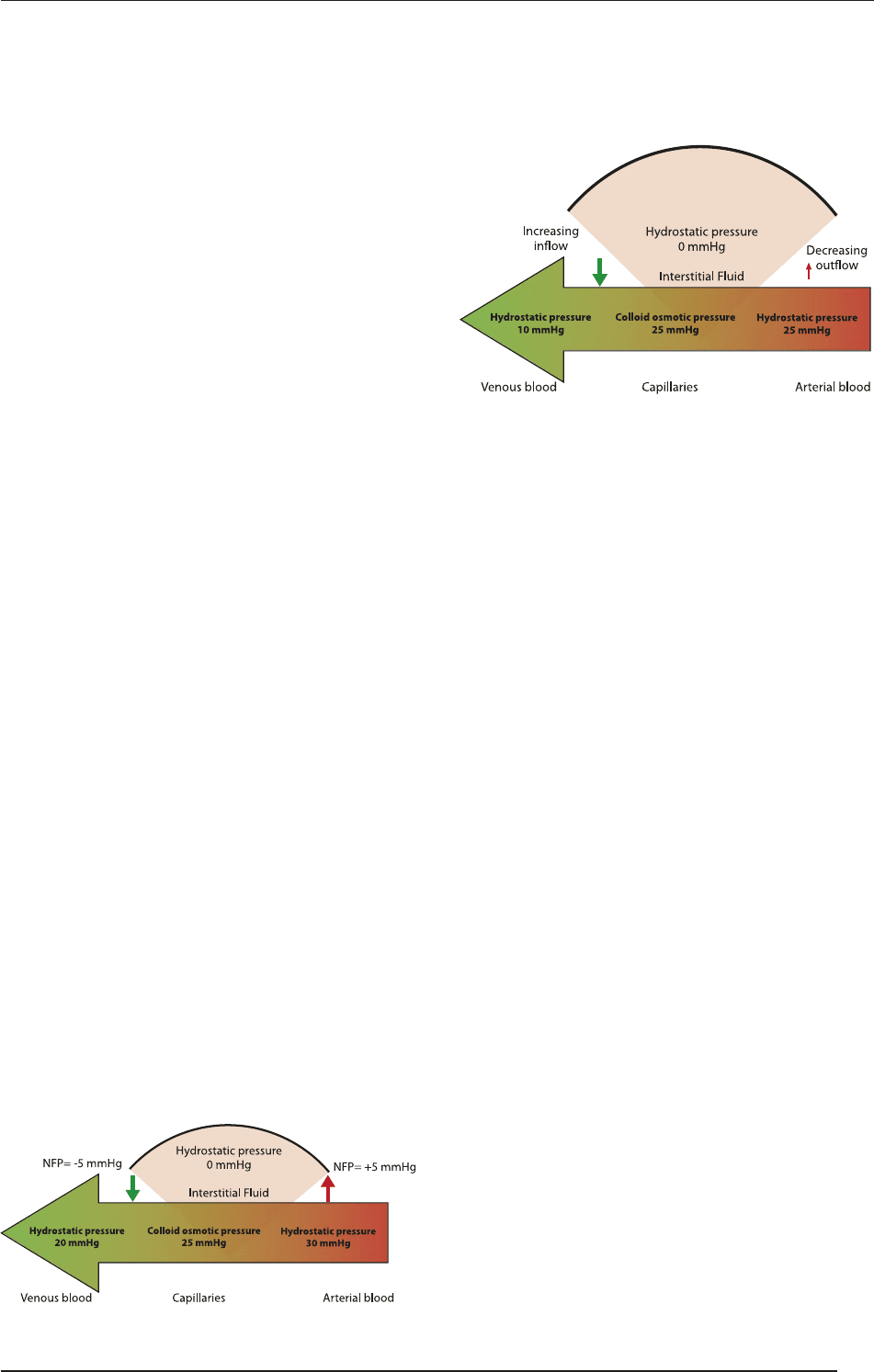
At the arterial end, the hydrostatic pressure dominates,
and fluid moves out of the circulation with +5 mmHg. At
the venous end, the oncotic pressure predominates and flu-
id moves into the bloodstream with an NFP of -5 mmHg.
Decreased MAP and arteriolar constriction lead to a de-
creased hydrostatic pressure and a decreased venous pres-
sure. e oncotic pressure is constant, so the fluid exchange
from the capillaries to the extracellular space decreases, and
the fluid return from the extracellular space to the capillar-
ies increases. is “fluid shi system” increases the blood
volume, which increases MAP and helps compensate for
shock (the fluid shi in adults is 1 ℓ/h). Two remarks:
1. “One great consequence of blood loss is the intense vaso-
constriction, the shrinkage of the capacity of the vascular
bed to accommodate the decreased blood volume...adjust-
ments for blood loss take place...the entry of fluid into the
blood vessels in a compensatory attempt. e greatest ex-
travascular store of readily available fluid in the body is...
in the extracellular space.” (Beecher et al. Recent Advanc-
es in Surgery I. e internal state of the severely wounded
man on entry to the most forward hospital. Surgery, 1947).
2. Caveat: “Possibly, too much attention has been given
to the fact that on many occasions [patients in shock may
have a normal blood pressure]. ...this has led to a tendency
to dismiss the blood pressure as a helpful sign even when it
is low - a fatal error, on some occasions. More helpful than
the level of the blood pressure is the direction of its swing
- a falling blood pressure, a rising pulse rate, are in most
cases an urgent indication of the need for blood.” (Beecher,
LTC and Henry K: Annals of Surgery, 1945).
6. Decompensatory mechanisms
after blood loss
6.1. Cardiac failure
is has many potential etiologies (i.e. the actual etiol-
ogy is controversial). e myocardial strength may de-
crease from ischemia secondary to a reduction of cir-
SURGICAL TECHNIQUES
71
VIII. HEMORRHAGIC SHOCK
culating red blood cells, lower oxygen saturation, or
decreased coronary perfusion secondary to hypotension
(especially diastolic hypotension).
6.2. Acidosis
Hypoperfusion leads to an anaerobic metabolism and
lactic acidosis. As a depressant of the myocardial func-
tion, there is a decreased response to catecholamines in
both the myocardium and the peripheral vasculature.
Caveat: “Although this is a time-honored concept, recent
data do not find evidence of this phenomenon. Metabol-
ic acidosis is a sign of underlying lack of adequate oxy-
gen delivery or consumption and should be treated with
more aggressive resuscitation, not exogenous bicarbon-
ate” (John P. Pryor: Hemorrhagic Shock, 2004).
6.3. Central nervous system
depression
is is due to opioid release (enkephalins and beta-
endorphin). Naloxone has been used as treatment in
shock, with some success.
6.4. Disseminated intravascular
coagulation (DIC)
Abnormalities of the clotting system develop as a result
of attempts to control hemorrhage, but also dilution/
loss of clotting factors. Gastrointestinal hemorrhage is
seen as a complication of acute hemorrhage, hours aer
the initial event.
6.5. Reticuloendothelial system
dysfunction
Loss of the antibacterial function can lead to endotox-
in release from native bacteria, aggravating an already
compromised situation.
7. Stages of hemorrhagic shock
7.1. Compensated shock
is entails some decrease in tissue perfusion, but the
body’s compensatory responses are sufficient to over-
come the decrease in available fluid.
7.2. Decompensated shock
Blood moves to more vital organs. e decreased venous
return results in a fall in CO. Viscera (lung, liver, kid-
neys and gastrointestinal mucosa): ese are congest-
ed due to the stagnant blood flow. Respiratory system:
Attempts are made to compensate for the acidosis by
increasing respiratory rate and producing a partially
compensated metabolic acidosis. Activation of clotting
mechanisms leads to hypercoagulability (DIC).
7.2.1. Main microcirculatory phases
during decompensation:
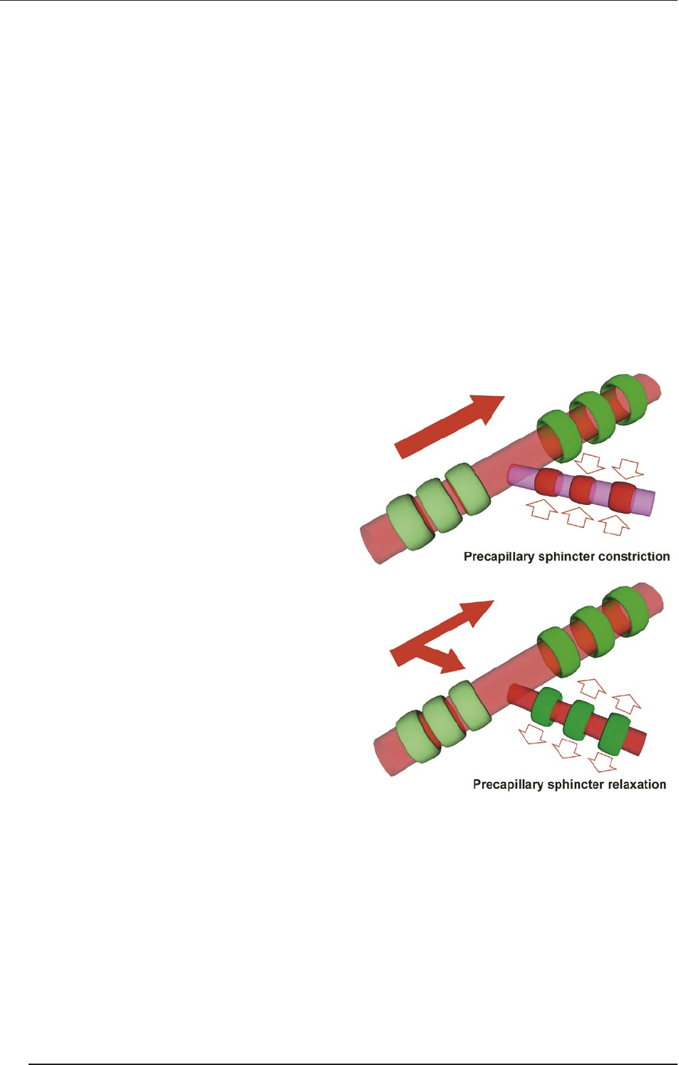
a. e precapillary sphincters relax due to shock-relat-
ed stimuli.
b. e postcapillary sphincters resist local effects and
remain closed => pooling/capillary stasis, capillary
engorgement.
c. Increasing hypoxemia and acidosis lead to the open-
ing of additional capillaries, and the vascular space
expands greatly.
e degree of change is so great that even the nor-
mal blood volume can not fill the available space.
e circulatory blood volume can not fill the vena cava.
Decompensated shock progresses to irreversible shock
if fluid resuscitation is inadequate or delayed.
SURGICAL TECHNIQUES
72
VIII. HEMORRHAGIC SHOCK

7.3. Irreversible shock
e body is no longer able to maintain the systolic BP,
and both the systolic and diastolic BP begin to drop. e
pulse pressure may be narrowed to such an extent that
it is not detectable with a BP cuff. e loss of arterial BP
causes damage from which ultimate recovery is not pos-
sible despite temporary restoration of the MAP. Multiple
organ system failure and organ damage (MOF and MOD)
occur, and even with treatment death is the result.
8. Signs of progressing shock
Bradycardia
Serious arrhythmias
Serious hypotension
MOF
A pale, cold and clammy skin
Prolonged capillary filling/stagnation
Cardiopulmonary failure.
9. Ischemia-reperfusion injury
A complex cascade mechanism occurs in two steps, an
inflammatory (a local and then a systemic) reaction, lead-
ing to MOD and MOF. e target organs of ischemia-re-
perfusion injury are the heart, lung, skeletal muscle and
gastrointestinal tract. During ischemia, an anaerobic me-
tabolism and cellular function disorders are initiated. In-
jury and cell death (necrosis and apoptosis) are caused
by reductive stress. During reperfusion, the production
of reactive free radicals (oxygen and nitrogen) is started
with the activation of leukocyte-endothelial cell interac-
tions and enzymes, etc. Injury/cell death (apoptosis and
necrosis) are caused by oxidative stress.
10. Intestinal mucosa injury
Splanchnic redistribution is one of the compensato-
ry mechanisms.
ere are serious consequences if shock process-
es are prolonged (the mucosa is sensitive to hypoxia
and ischemia-reperfusion).
Injury to the intestinal epithelium leads to bacterial
translocation (intestinal flora, toxins, etc. are translocat-
ed into the circulation); the consequences are systemic
inflammatory response syndrome, MOD and MOF.
11. Shock diagnosis
e first thing to note is that this is a clinical diagnosis!
In most cases, the diagnostic tools are only available af-
ter initiation the therapy.
Decreasing Hbg, hematocrit (responds only later due
to the fluid shi).
Decreasing CVP (1–9 cmH₂O).
Decreasing pulmonary capillary wedge pressure:
5–12 mmHg.
Decreasing CO (4–6 ℓ/min).
Decreasing mixed (venous) O₂ saturation (the nor-
mal value is ~ 75%).
Increasing total peripheral vascular resistance (the
normal values are ~ 800–1400 dyne s/cm⁵).
12. Relationship between
mortality and time elapsed
from injury to therapy
Interval
[h]
Mortality
[%]
World War I 12–18 8.5
World War II 6–12 5.8
Korean War 2–4 2.4
Vietnam War 1–4 1.7
Approximately 53% the cases of traumatic death oc-
cur onsite, 7.5% in the emergency room, and39.5% in
hospitals. In the hospital 50% of the deaths are caused
by central nervous system-related problems, 31% by
hemorrhage and 18% by sepsis (Trunkey DD, Holcro
JW. Trauma: general survey and synopsis of manage-
ment of specific injuries. In: Hardy’s Textbook of Sur-
gery, 1988).
13. Treatment of hemorrhagic
shock
1. Evaluation: Internal or external hemorrhage? Are
there underlying cardiac problems? Amount of
blood lost? Duration of bleeding? And stop the
bleeding!
2. Level of consciousness (motto: “Report and record”):
alert,
verbal response to stimuli,
pain response to stimuli,
no response to stimuli.
3. Determination of aims (to determine and increase
tissue perfusion and oxygenation, and to eliminate
and treat the triggering cause. e cause, and not the
shock, is what must be treated!).
SURGICAL TECHNIQUES
73
VIII. HEMORRHAGIC SHOCK

4. e first steps involve positioning, the ABC ap-
proach, keeping the patient at normal temperature
to prevent hypothermia. Ongoing assessment (every
10–15 min) is mandatory.
A = Airways
Depends on etiology: from minimal to com-
plex therapy (intubation, and mechanical
ventilation).
If the patient can say his/her name: the air-
ways are free (!)
B = Positive-pressure ventilation
Breathing: Patients need respiratory support
(intubation or other respiratory support) and
monitoring. In general, respiratory support
and monitoring are needed even in the case
of adequate O₂ saturation to help compensa-
tion for metabolic acidosis.
C = Circulation
What is to be given? “Volume” (!)
How to give it? e flow in the catheter is inverse-
ly proportional to the length of the tube and directly
proportional to the cross-section: a short, wide and
peripheral iv. infusion set must be used!
Where to give it? Into peripheral (antecubital) vis-
ible/palpable veins. Intraosseal colloid therapy is
probable in children, if an iv. route cannot be se-
cured quickly. A central vein can be used only aer a
routine has been attained!
What type of fluid? Isotonic (in the ideal case iso-
oncotic) fluid should be given. Physiological salt so-
lution is available everywhere; first, 2 ℓ of salt solu-
tion or lactated Ringer, but in most cases much more
(20 mℓ/kg!) is given. 3 units of crystalloid is ℓ unit of
intravascular fluid (!), colloid solutions supplement
the volume in a 1:1 ratio (see above).
2 U of red blood cells are given if the circulation
is unstable aer the administration of 2000 mℓ of
crystalloid. More blood may be needed during ac-
tive bleeding (warming, taking a blood sample be-
fore transfusion, cross-reaction!).
Fresh frozen plasma and a thrombocyte suspension
are indicated for the treatment of the symptoms of
coagulopathy (usually aer giving 6-8 U of blood).
D = Definitive therapy / Drugs:
e goals are to 1. increase the preload, 2. to increase
the contractility, and 3. to decrease the aerload.
Correction of acidosis
Background: A pH < than 7.25 will interfere with
the effects of catecholamines and inotropic resis-
tant hypotension evolves.
Method (see above): Na-bicarbonate is given if the
deficit > 6 meq/ℓ.
A useful formula: 0.3 × kg bw × base deficit = meq
NaHCO
3
will compensate for half of the loss. It
should be given slowly in a 1–2 meq/kg bolus; 10–
20 meq/kg could be needed, which means a large
Na
+
load and hyperosmolarity.
Treatment with pressors
β effects: Increase inotropy and chronotropy - in-
creasing CO (beta-1), and also some pulmonary and
peripheral vasodilation (beta-2).
α effects: Increase the systemic vascular resistance -
maintaining the BP
Vasodilators: Decrease the systemic vascular resis-
tance, and decrease the aerload, potentially im-
proving the cardiac function, but also dramatically
reducing the MAP in hypovolemic patients.
Alpha
Peripheral
Beta 1
Cardiac
Beta 2
Peripheral
Norepinephrine
alpha and beta,
more alpha
++++ ++++ 0
Epinephrine
beta and alpha,
stronger beta
++++ ++++ ++
Dopamine ++++ ++++ ++
Isoproterenol 0 ++++ ++++
Dobutamine
beta-1 alone
+/0 ++++ +
(Source: NEJM, 300:18, 1979)
5. Further important interventions – correction of
electrolyte abnormalities.
e Na
+
level can be markedly abnormal as a result
of the underlying disease (hypo/hypernatremic de-
hydration); it can become elevated during the pro-
cess of correcting a base deficit. e goal should be
to normalize the Na
+
level- slowly! Hyperchloremia
may aggravate acidosis.
e K
+
level can be elevated to the point of cardiac
dysrhythmias. As the correction of acidosis occurs,
K
+
can be driven back into the cells, severe hypoka-
lemia developing in some cases.
Ca
++
can be chelated in the treatment of a base deficit
and dramatically decrease, leading to problems from
seizures, hypotension and a myocardial dysfunction.
SURGICAL TECHNIQUES
74
VIII. HEMORRHAGIC SHOCK

Glucose: As part of the response to compensato-
ry mechanisms (epinephrine and corticosteroids),
hyperglycemia is a common occurrence in stressed
children. is can cause problems from osmotic di-
uresis and glucose intolerance. Care should be made
not to overload the glucose management system in
the body (i.e. no dextrose in flush solutions).
6. Blood gases: It is important to maintain good DO
2
so as to minimize the anaerobic metabolism and ac-
idosis. Venous blood gases are also of benefit since
mixed venous O
2
saturation is a measure of tissue
perfusion and CO.
7. Hemodynamics: MAP and ECG: A decreasing MAP
may be a sign of decompensation. Monitoring the
CVP may be indicative of tissue hydration and the
preload (see above).
8. Coagulation status: DIC is a common complication
even early in shock.
9. Urinary output: is is representative of the organ
perfusion. An improving urinary output can be a
sign of an improving volume status, while a wors-
ening output suggests the need for more aggressive
therapy.
10. Neurologic status: Indicative of brain perfusion.
14. Signs of cardiovascular
stabilization
MAP is stable.
HR is decreasing.
Consciousness, and decreased anxiety.
Increasing capillary refilling, improved color of mu-
cous membranes.
e urinary output exceeds 30 mℓ/h.
15. Medical – legal pitfalls
Unrecognized occult bleeding.
Hypotension aer head trauma (hypotension and
other causes!).
Omission of rectal finger examination.
Undiagnosed bleeding source.
Inadequate resuscitation (immediate, correct, sus-
tained therapy)
16. Variations in physiological
responses to hemorrhagic
shock
It is important that there are significant differences in
relation to the following parameters:
Age and relative health
General physical condition
Preexisting diseases
Ability to activate compensatory mechanisms
Older adults are less able to compensate (they devel-
op hypotension early)
Children compensate longer and deteriorate faster
Medication may interfere with compensatory mech-
anisms.
SURGICAL TECHNIQUES
75
VIII. HEMORRHAGIC SHOCK
IX. Wounds
“Notae vero inflammationis sunt quattuor: rubor et
tumor cum calore et dolore”.
Aulus Cornelius Celsus (BC 25-50 – 45-50 AD)
De Medica (1478) Liber III. 10.
e etymology of the word wound is the Old English
“wund” and the Old Norse “und”. A wound is a disrup-
tion of the continuity of tissues produced by an exter-
nal mechanical force, a cut or break in the continuity of
any tissue, caused by an injury or operation. As a con-
sequence, the circulating blood volume is lost, while the
breakdown of the defense mechanisms of the skin leads
to the entrance of pathogens and foreign materials into
the body. e exposure of body cavities and internal or-
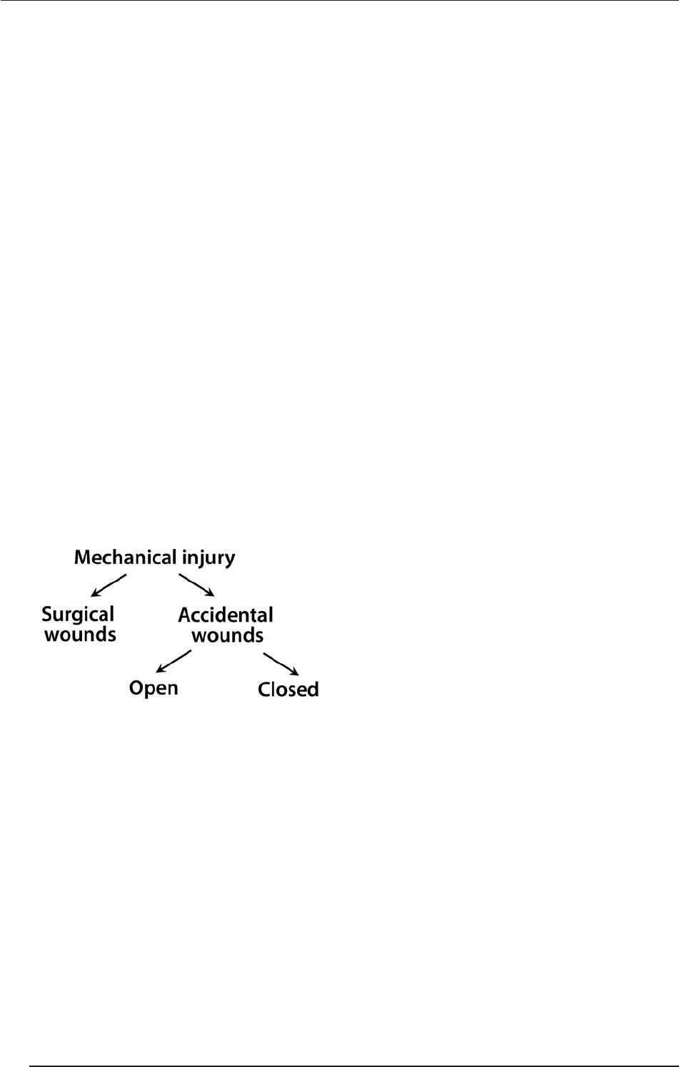
gans means a further risk. Surgical wounds are usual-
ly made under sterile circumstances and are closed layer
by layer upon completion of the surgical intervention.
Accidental wounds, however, are caused by mechanical
trauma and can be either open or closed. Wounds can
result from mechanical, thermal or chemical forces and
irradiation, but the focus here will be on the character-
istics and management of wounds caused by mechani-
cal injury. e term injury is used synonymously with
wound, but can have a wider meaning; wound (in sur-
gery) = mechanical injury.
1. Classification of accidental
wounds
1.1. Morphology / classification
depending on the penetration
route
A puncture wound (vulnus punctum) is caused by a
sharp pointed tool, wood splinters, pins, nails, glass,
scissors or knives and usually, misleadingly, seems to
be negligible. Some punctures are merely on the sur-
face, whereas others can be very deep, depending on the
source and cause.
Treatment: Such wounds usually close quickly on their
own. Treatment may be necessary to prevent infection,
as the object that caused the wound may carry bacteria
or tetanus spores into the skin and tissue.
An incised wound (vulnus scissum) is caused by sharp
objects; it involves a linear cut in the skin, which is usu-
ally superficial, but may involve deep structures (surgical
incisions). is type of wound exhibits the best healing.
e extent of opening of the wound depends on the tissue
flexibility and the directions of the Langer lines. ese
wounds are accompanied by considerable bleeding.
A cut wound (vulnus caesum) is similar to an incision,
but with an additional direct, perpendicular force. e
impact bursts the tissues open (e.g. an axe injury).
A crush wound (vulnus contusum) is caused by a blunt
force and can be either open or closed. Heavy objects
split the skin and shatter or tear the underlying struc-
tures. Fingers and toes are commonly involved. is is
a painful injury with much swelling. e wound edg-
es are usually uneven and torn. e bleeding is negligi-
ble, but the pain is proportionately greater than would
be expected from the size of the injury (termed wound
stupor).
A torn wound (vulnus lacerum) is caused by great tear-
ing or pulling forces and can result in the incomplete
amputation of certain body parts.
A shot wound (vulnus sclopetarium) consists of an ap-
erture, a slot tunnel and an output. A shot from close
range is usually accompanied by some degree of burn
injury at the aperture. Other characteristic features are
the incorporated foreign materials: textile fibers, bullets
and the various types of tissues penetrated.
A bite wound (vulnus morsum) is a ragged wound with
crushed tissue characterized by the shape of the biting
teeth and the force of the bite. It is also accompanied by
the features of torn wounds. ere is a high risk of infec-
tion (transmission of malaria, rabies, etc.; human bites
carry a considerable risk of infection through the trans-
mission of HIV or hepatitis B). Such wounds should not
be sutured.
1.2. Classification according
to “cleanliness” – bacterial
contamination
Clean wounds (operation or sterile conditions; only the
normally present skin bacteria are detectable) with no
signs of inflammation.
SURGICAL TECHNIQUES
76
IX. WOUNDS
Clean-contaminated wounds (the contamination of
clean wounds is endogenous or comes from the envi-
ronment, the surgical team, or the patient’s skin sur-
rounding the wound). ey include opening of the di-
gestive, respiratory or urogenital tract.
Contaminated wounds (large contaminates infect the
wound) arise when an incision is performed in a puru-
lent area or in cases of a leakage from the gastrointesti-
nal tract.
Dirty wounds (the contamination comes from the es-
tablished infection), in which there are residual nonvia-
ble tissues and chronic traumatic wounds.
1.3. Classification depending on the
time since the trauma
Acute wounds (mechanical and other injuries):
Fresh wound: treatment within 8 h.
Old wound: ≥8 h aer trauma/discontinuity of the skin.
Chronic wounds (venous, arterial, diabetic and other
ulcers, and skin or so tissue defects):
ey do not heal within 4 weeks aer the beginning
of wound management.
ey do not heal within 8 weeks.
With chronic wounds, the normal process of healing is
disrupted at one or more points (in most cases, the heal-
ing process is 'stuck' in the inflammatory or prolifera-
tive phase; see later).
1.4. Classification depending on the
number of skin layers involved
Grade 1: Non-blanchable erythema of intact skin.
Discoloration of the skin, warmth, edema, indura-
tion or hardness may also be used as indicators in
people with dark skin.
Grade 2: Partial-thickness skin loss involving the
epidermis, dermis or both. e ulcer is superficial
and presents clinically as an abrasion or blister.
Grade 3: Full-thickness skin loss involving damage
to or necrosis of subcutaneous tissue, which may ex-
tend down to, but not through the underlying fascia.
Grade 4: Deep wounds or complex wounds (e.g. lac-
erations, or vessel or nerve injuries), or wounds of
the bone or supporting structures, the opening of
body cavities, or penetrating injuries of organs.
1.5. Classification depending on the
factors affecting wound healing
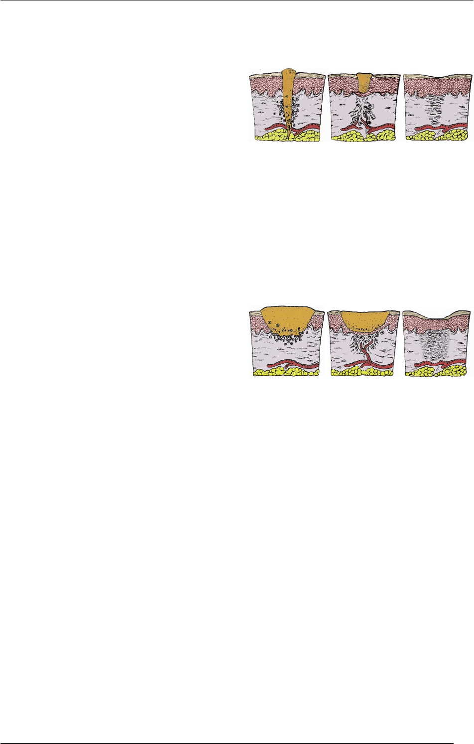
The scheme of sanatio per primam intentionem (“p.p. healing”). Ac-
cording to Galen (Galenus, BC 129-199): “the major aim” of a doc-
tor is the gap-free healing of wounds. Wounds are closed surgical-
ly by reconstruction of the skin continuity (a) by simple suturing,
(b) by the movement (relocation) of skin fragments from the sur-
rounding area (flaps), or (c) by the transplantation of free skin el-
ements (grafts) of different thicknesses (e.g. split- or full-thickness
grafts). Primary healing is usually the case in all wounds in which
the anatomical location and the size allow the skin continuity to be
restored (no significant degree of tissue loss); healing takes place
from the internal layers outward.
The scheme of sanatio per secundam intentionem. Secondary heal-
ing is the mode of healing of abrasions or split-thickness graft donor
sites. The tissue loss is compensated by a granulation tissue “accord-
ing to the second potential goal of the doctor”. (Due to the abacteri-
al or purulent inflammation, the wound is filled with connective tis-
sue which transforms into scar tissue. If there is significant tissue loss
in the formation of the wound, healing will begin by the production
of the granulation tissue wound base and walls. After wound de-
bridement and preparation, the wound is left open to achieve suffi-
cient granulation for spontaneous closure (re-epithelialization from
the remaining dermal elements or from the wound borders).
Tertiary (delayed primary) healing occurs in pri-
mary contaminated wounds or mixed tissue trauma
wounds (e.g. aer the reconstruction of hard tissue).
Factors influencing wound repair
Drugs: Glycocorticoids inhibit fibroblast activity,
protein synthesis and immune responses. Some an-
tibiotics inhibit collagen biosynthesis. Cytostatic
agents slow down metabolic processes. Anti-inflam-
matory agents reduce hyperemia and the blood sup-
ply to the wound (they may slow down the healing
process if they are taken aer the first several days of
healing, following which anti-inflammatory drugs
should not have an effect on the healing process).
SURGICAL TECHNIQUES
77
IX. WOUNDS
General condition, nutrition, protein level, vitamins
B, C and K, and trace elements (Zn and Mg) (malnu-
trition slows down the healing process).
Diabetes mellitus: ere is a risk of infection, dys-
function of the micro- and macrocirculation, and
hyperglycemia = chronic wounds.
Icterus and anemia.
Bacterial/other infections: Bacterial contamination
slows down the healing process.
Age: e older the patient, the slower the wound
heals.
e location of the wound: Poorly vascularized ar-
eas or areas under tension heal more slowly than ar-
eas that are highly vascularized.
1.6. Classification depending on
wound closure
Primary wound management was pioneered by Paul
Leopold Friedrich (1898).
Primary suture: Immediate surgical wound closure can
be performed within 12 h aer the injury if no signs of
inflammation or contamination can be detected (see be-
low).
Delayed primary wound closure: Aer 3–8 days of
open wound management, surgical wound closure is
performed (see below).
Early secondary surgical wound closure: Aer 2 weeks
of open wound management, surgical wound closure is
performed (see below).
Late secondary surgical wound closure: Aer 4–6
weeks of open wound management, surgical wound clo-
sure is performed (see below).
2. Surgical wounds
2.1. Determinants of healing of
surgical wounds
Preparation of the operating site, hygiene, shaving,
disinfection and isolation.
e incision should be parallel to the Langer lines.
e skin is stretched, the scalpel is held in a verti-
cal position and the incision is performed until the
sc. layer is reached.
It is important to be aware of the anatomical aspects
of the involved area. e muscle is separated along its
fascia, the handling of bleeding is of importance, etc.
2.2. Skin incision
A skin incision is made on a prepared (cleansed,
draped) operative field.
During the incision, the surgeon and the assistant
stretch the skin with sterile towels on both sides of
the operative field.
Usually a scalpel (e.g. #20 blade, #4 handle) is used.
e type of the scalpel depends on the site of the in-
cision.
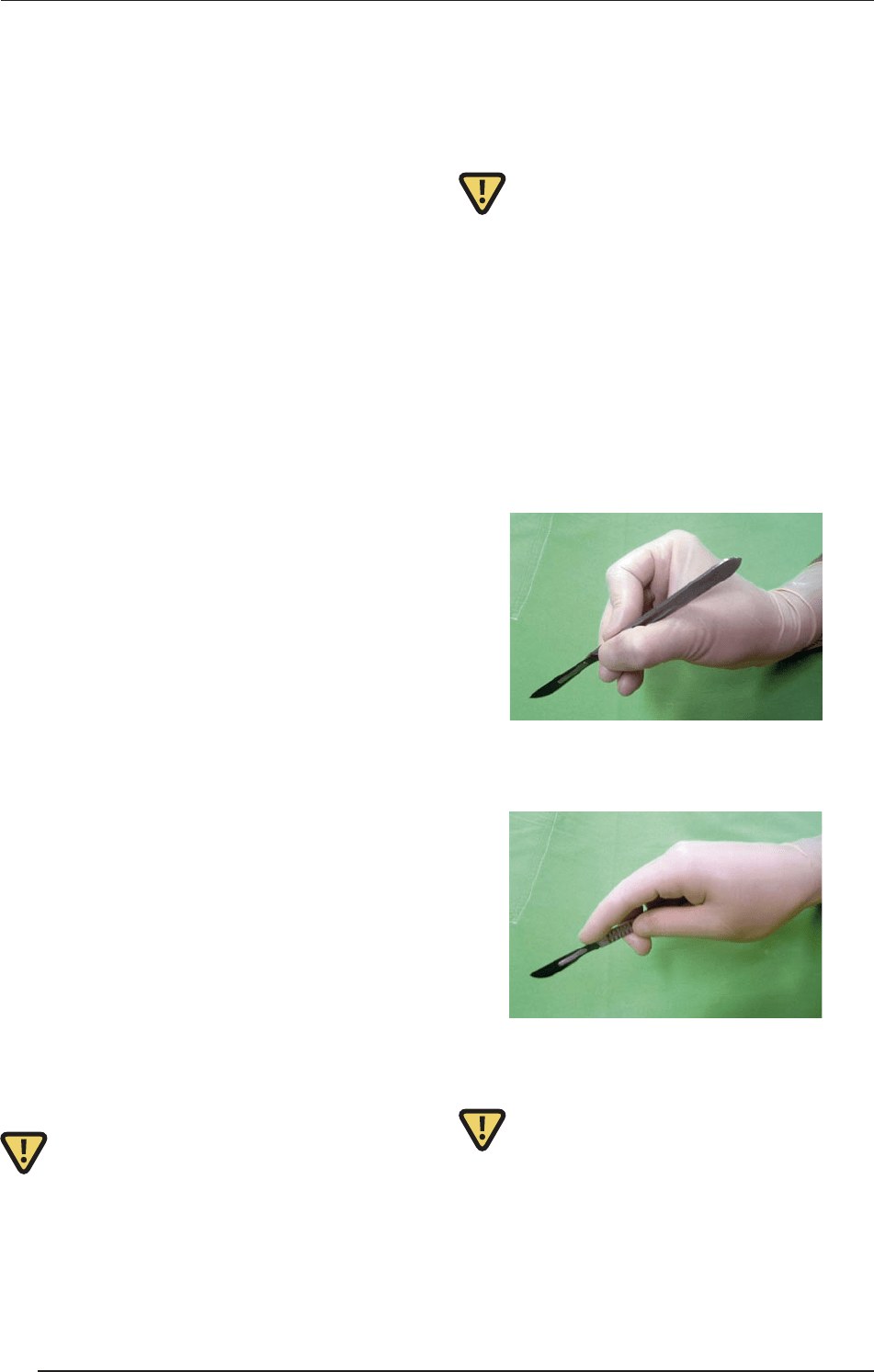
e manner of holding the scalpel varies according
to the use.
For the delicate, curved incision of fine structures,
the scalpel is held like a pen.
For a long straight incision, the scalpel is held like
a fiddle bow.
2.3. e requirements of skin incision
e length of the incision should be appropriate for
safe surgery.
Vessels and nerves should not be damaged.
e skin edges should be smooth.
e incision is made perpendicularly to the skin
with a single definite cut (failed attempts result in
ragged edges and prevent wound healing).
SURGICAL TECHNIQUES
78
IX. WOUNDS

e direction of the incision depends on the location
of the organ being operated on.
e skin is incised parallel to the Langer lines (bet-
ter wound healing and less scar formation), usually
toward the operator, and from le to right.
The depth of the incision must be the same
throughout the whole length. At the beginning,
the tip of the scalpel is inserted perpendicularly
into the skin, the cut is made an angle of 45° with
the blade of the scalpel (not with the tip!), and the
incision is completed with the scalpel held per-
pendicularly.
e skin scalpel is discarded into the container aer
the skin incision. In the deeper layers, another scal-
pel is used.
2.4. Main types of skin incisions
(See details later, on page 93)
Kocher’s transverse incision at the base of the neck
(thyroid gland), sternotomy, thoracotomy.
Subcostal (gallbladder or spleen), median/parame-
dian laparotomy (this may be upper or lower relative
to the umbilicus).
Transrectal/pararectal/transversal laparotomies.
Pfannenstiel suprapubic incision (bladder, uterus or
ovaries).
McBurney incision (appendectomy).
Inguinal incisions (hernia).
2.5. Closure of surgical wounds
Fascia and subcutaneous layer: Interrupted stitches.
e fat must not be sutured (fat necrosis).
Skin: Tissue-sparing technique, with accurate ap-
proximation of the skin edges. Tension and ischemia
of the skin edges are to be avoided. A simple inter-
rupted stitch is the most fundamental type in cu-
taneous surgery (other possibilities: Donati vertical
mattress suture, Allgöwer, continuous intracutane-
ous, etc.; Steri-Strips, clamps and tissue glues may be
applied).
Dressing: Sterile, moist, antibiotic and non-adhe-
sive dressings. Gauze placed directly on the wound
makes dressing removal difficult and painful: tear-
ing of the closure is possible.
Holding the dressing: Stretchable adhesive tape,
such as Hypafix.
e dressing is removed on the 2nd postoperative
day, and daily in cases of infection.
Sutures are usually removed aer 4–6 days. In areas of
good blood supply, such as the face, it is aer 5–7 days,
and in the trunk and extremities aer 10–14 days.
3. Early complications of wound
closure
(See also sections I.4 and V.7.2.2.2)
Hematoma
Seroma
Wound infection (see also SSI). erapy in general:
e type of surgery (clean, clean/contaminated,
contaminated or dirty) will determine the level
of the risk of infection and the likely spectrum of
pathogens. Empirical antibiotic therapy should be
primarily directed against Staphylococcus aureus.
Swabs are commonly sent for culture; pus (if
available) is a better sample. Other fluids or tissue
biopsy samples may also be cultured. Blood cul-
turing is recommended in febrile patients.
If wounds are not grossly infected, they may re-
spond to local measures such as the removal of
sutures. Frequent saline bathing should be un-
dertaken and the wound requires a drain to al-
low healing. Deep-seated infection may require
broad-spectrum antibiotics and possible surgical
intervention.
Superficial SSI
1. Diffuse and superficial (e.g. erysipelas). Streptococ-
cus haemolyticus-induced lymphangitis, linear, dif-
fuse subcutaneous inflammation. Treatment: Rest,
antibiotics and dermatology consultation.
2. Localized (e.g. abscess, stitch abscess, filum suppu-
ratio). is can occur anywhere: under the skin, be-
tween the muscles, subfascially, in the chest, brain
or liver. erapy: Radical surgery and drainage. In
the presence of dead tissue, the most critical aspect
of treatment is the surgical removal of pus (Motto:
“cut out the rubbish”). Antibiotics have a support-
ive role.
3. Foreign material (corpus alienum) could be present
even years later (importance of X-ray examination!).
Deep SSI
1. Diffuse (e.g. anaerobic necrosis).
2. Localized (e.g. empyema) in body cavities (chest and
joints). erapy: Surgical exploration and drainage
(Staphylococcus aureus!)
Mixed SSI
1. Gangrene: Necrotic tissues with putrid and anaerobic
infection; this is a highly lethal, severe state. e terms
gas gangrene and clostridial myonecrosis are used in-
terchangeably and refer to the infection of muscle tis-
sue by toxin-producing clostridia. erapy: A combi-
nation of aggressive surgical debridement and effective
antibiotic therapy is the determining factor.
2. Generalized reaction: Bacteremia, pyemia and sepsis.
SURGICAL TECHNIQUES
79
IX. WOUNDS
