Boros Mihaly. Surgical Techniques
Подождите немного. Документ загружается.


e appendix is clamped with a Kocher clamp dis-
tal to the crushed line and cut above the base tie,
just below the Kocher clamp (the scalpel and the
appendix should be thrown into the kick bucket).
e stump of the appendix is disinfected with po-
vidone-iodine and cauterized (to prevent the later
secretion of mucus).
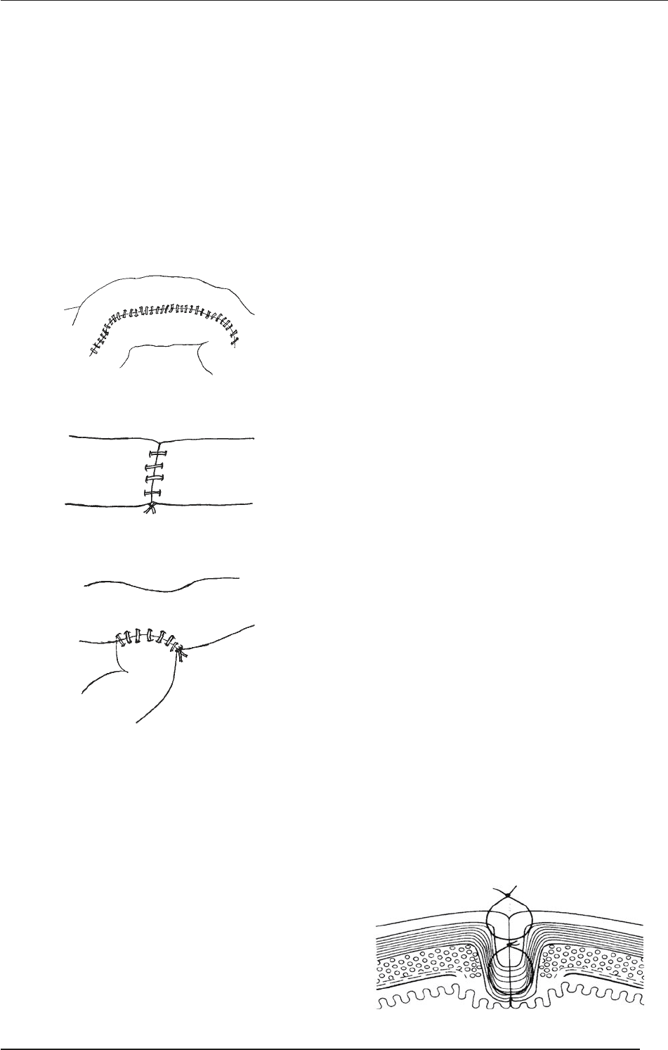
e stump of the appendix is buried (the stump
will be inverted in the lumen of the intestine), and
the purse-string suture is then tied. e buried ap-
pendix stump is covered with a serosa layer with a
“Z” stitch, i.e. with a zed-like serosa stitch; thin lin-
en thread and taper needle are used (this step is not
obligatory in humans).
e cecum and appendiceal stump are then placed
back into the abdomen. If free perforation is en-
countered, thorough irrigation of the abdomen with
warm saline solution and drainage of any obvious
cavity and well-developed abscesses is required.
e peritoneum is identified, and closed with a
continuous 2 or 3–0 suture. e inferior oblique
muscles are re-approximated with a figure-of-eight
interrupted absorbable 0 to 3–0 suture, and the ex-
ternal oblique fascia is closed with an interrupted
2–0 PG suture. e skin may be closed with staples
or sc. sutures.
In cases of a perforated appendicitis, the skin should
be le open, with delayed primary closure on post-
operative day 4 or 5.
ADVANCED MEDICAL SKILLS
100
IV. BASIC SURGICAL PROCEDURES ON THE INTESTINES. APPENDECTOMY
V. Anastomoses
e origin of the word is late Latin (by Galen) and Greek
(anastomoun = to provide with a mouth; ana + stoma
= mouth, orifice). e basic types are side-to-side, end-
to-end and end-to-side anastomoses. Anastomoses are
applied not only in gastrointestinal surgery, but also in
urology and vascular (etc.) surgery. However, the basis
of surgical techniques can be best practised in the case
of the small intestine. An important general principle
is that the techniques (e.g. restoration of the anatomy)
serve to restore function (!).
1. Healing of the anastomosis
e most important factors influencing the healing of
the anastomosis are the good blood supply of the tis-
sues, the lack of tension, and an adequate surgical tech-
nique, securing the appropriate approximation for the
beginning of collagen formation:
Early phase (days 0–4): ere is an acute inflamma-
tory response, but no intrinsic cohesion.
Fibroplasia (days 3–14): Fibroblast proliferation oc-
curs with collagen formation.
Maturation stage (>10 days): is is the period of
collagen remodeling, when the stability and strength
of the anastomosis increase.
2. Causes of anastomosis
insufficiency
Distal obstruction of the lumen
Perianastomotic hematoma, infection or sepsis
Hypotension or hypoxia
Icterus, uremia or diabetes
Corticosteroids
3. e characteristics of
a good technique
e precise joining of cut tissues results in primary
wound healing (per primam intentionem, p.p.).
Placing the lowest possible amount of foreign mate-
rial (suture) into the tissues causes the least disrup-
tion of the local circulation.
4. Complications
Suture insufficiency
Stricture
5. Anastomosis techniques
Traditional methods
Suturing by hand (there is no evidence that suturing
by hand is better than stapling with staplers)
Staplers or clips (the Hungarian surgeon Aladár Petz
(1888–1956) invented the gastric stapler and pio-
neered the technique).
New methods
Compression (biodegrading) rings
Tissue adhesives
5.1. Two-layered anastomosis technique
is is the traditional method for anastomoses of the
gastrointestinal tract
An inner continuous catgut (absorbable) suture, with
stitching of all layers
An outer, seromuscular, interrupted silk (nonabsorb-
able) suture
Serosa apposition and mucosa inversion; the inner
layer has a hemostatic effect (there is no significant
bleeding), but the mucosa is strangulated.
ADVANCED MEDICAL SKILLS
101
V. ANASTOMOSES
5.2. Single-layered technique
is is a newer, more up-to-date technique of gastro-
intestinal anastomosis
An interrrupted seromuscular suture, with absorbable
(e.g. 3/0 Vicryl) thread. e submucosal layer is strong
and the blood supply is only minimally damaged.
5.3. Stapler-made anastomosis
is can be a side-to-side anastomosis with a straight
sewing machine (e.g. GIA = gastrointestinal anasto-
mosis staplers).
It can be an end-to-end anastomosis with a circular
machine (e.g. CEEA = circular end-to-end anasto-
mosis stapler).
e stapler decreases the frequency of radiologically
demonstrated anastomosis insufficiency, but the in-
cidence of anastomosis stricture is increased.
6. Surgical techniques of
intestinal anastomoses
Requirements include a supine position, general an-
esthesia, a midline laparotomy and a good exposure;
the affected bowel must be mobilized (freed).
e gastrointestinal tract should always be considered
infected when the intestinal lumen has been closed;
new, sterile instruments and draping are necessary.
e pathological tissue must always be excised with
a normal intact margin (!); the blood supply of the
remaining intestinal tissue is critical.
Relatively equal diameter segments of bowel should
be sewn together. e anastomosis should be ten-
sion-free and leak-proof.
e mesenteric defect is closed (prevention of inter-
nal hernia formation).
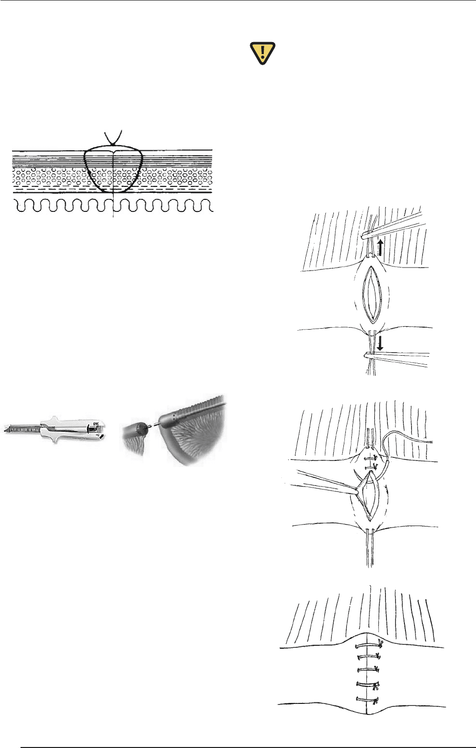
7. Closure of enterotomy
Aer laparotomy, the injured bowel segment is
identified and isolated. e borders are temporar-
ily closed (Klammer intestinal clamps) and the de-
fect is enlarged/incised, i.e. converted to a surgical
incision.
A horizontal suture or end-to-end anastomosis is
performed.
Irrigation, handling of bleeding and closure in layers.
ADVANCED MEDICAL SKILLS
102
V. ANASTOMOSES
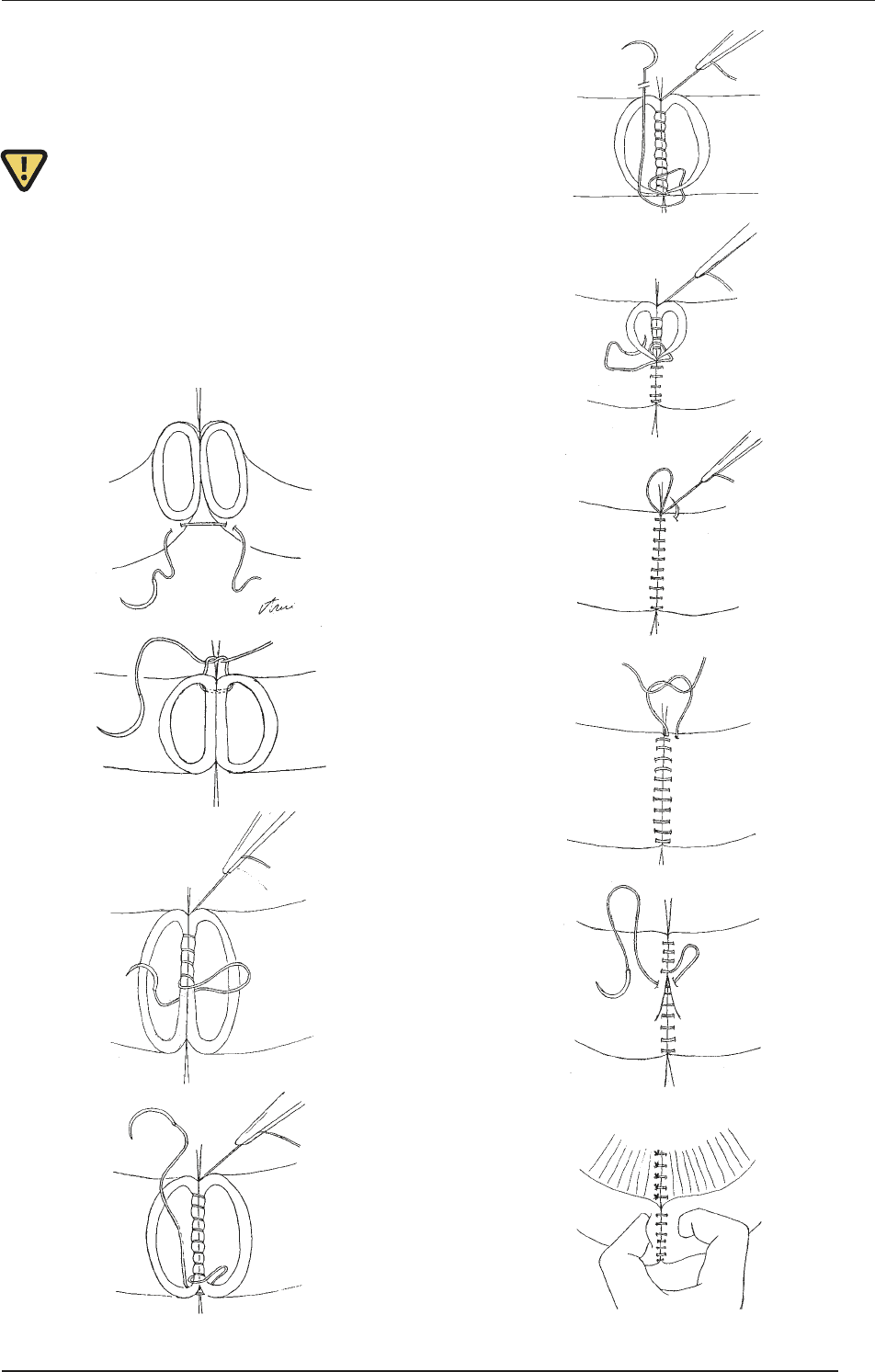
8. Surgical unification of bowel
segments by end-to-end
anastomosis
e two bowel ends are put in close approximation
and two interrupted, holding sutures are placed. A
continuous running suture is applied to close the
back, and then the front part of the intestinal wall.
Aer closure of the deeper layer, the serosa (second
layer) is closed.
e passage of the anastomosis is checked by exami-
nation with the fingers.
1
2
3
4
5
6
7
8
9
10
ADVANCED MEDICAL SKILLS
103
V. ANASTOMOSES
VI. Abdominal drainage
e most frequent causes of surgical diseases of the
small intestine are mechanical causes (obstruction,
strangulation/adhesion, volvulus, intussusception or fe-
cal impaction), vascular causes (ischemic colitis, occlu-
sion/infarct, or arteriovenous malformations), inflam-
mation (diverticulosis/diverticulitis, ulcerative colitis,
Crohn’s disease or appendicitis) or traumas (blunt/pen-
etrating injuries). Invasive abdominal diagnostic inter-
ventions may be needed primarily in these latter cases.
1. Historical background
of invasive diagnostic
procedures
1950 Four quadrant needle paracentesis.
1965 Diagnostic peritoneal lavage (DPL – the term
was coined by Root HD et al. Diagnostic peri-
toneal lavage. Surgery. 1965; 57:633–637). e
sensitivity is 98%, but the specificity is only
80% (no information is provided on the retro-
peritoneum).
1990s Laparoscopy became widespread. It has the ad-
vantage of good visualization of the intraab-
dominal organs, whereas it is disadvantageous
that no information is available on the retro-
peritoneum, and the closure is ‘complicated’ as
compared with punctures.
2. Indication of diagnostic
peritoneal lavage
An equivocal clinical examination and difficulty in
assessing a patient.
Persistent hypotension, despite adequate resuscitation.
Multiple injuries, or stab wounds where the perito-
neum has been breached.
Lack of alternative diagnostic methods (US or CT).
2.1. Open system
Aer insertion of a urinary catheter and a nasogastric
tube, local anesthesia is started. A vertical, ~ 2-cm sub-
umbilical incision is made, and the linea alba is divided.
An incision is made in the peritoneum, a peritoneal
dialysis catheter is inserted, the free blood or gastric
content is aspirated, etc.
If no blood is seen, 1 ℓ of normal saline is infused,
a period of 3 min being allowed for equilibration.
e drainage bag is placed on the floor and drainage
proceeds (motto: “Gravity is our friend”).
A 20-mℓ sample should be sent to the laboratory for
the measurement of red blood cells, white blood cells
and microbiological examination (DPL is positive if
the red cell count is > 100,000 / mm
3
, the white cell
count is > 500 / mm
3
, or bile, bacteria or fecal mate-
rial is present).
In the event of positive results, DPL is continued un-
til surgical exposure (laparotomy), and the demon-
stration and treatment of the causes.
e peritoneum is closed with a purse-string suture,
and the skin and sc. layers are then closed with an
interrupted suture.
2.2. Closed system
Aer insertion of a urinary catheter and nasogas-
tric tube, local anesthesia is initiated, aer which a
catheter is introduced with the aid of a guide wire (a
blind technique; the morbidity of 9%, is mostly due
to vessel injury).
e routine is modified in obese patients (special in-
dication for closed DPL):
Computer tomography is impossible (weight, diam-
eter limits, poor image, higher radiation).
Open DPL is contraindicated as the depth of the
puncture (peritoneum) can not be judged, and
hence the complication rate of the closed technique
is much higher. e half-closed/blind Seldinger or
modified Seldinger technique is possible.
3. erapeutic (chronic) lavage:
peritoneal dialysis
Dialysate is injected into the peritoneal space
through a two-way Tenckhoff catheter, which re-
mains permanently in place. e peritoneal dialy-
sate, composed mostly of salts and sugar (glucose),
ADVANCED MEDICAL SKILLS
104
VI. ABDOMINAL DRAINAGE
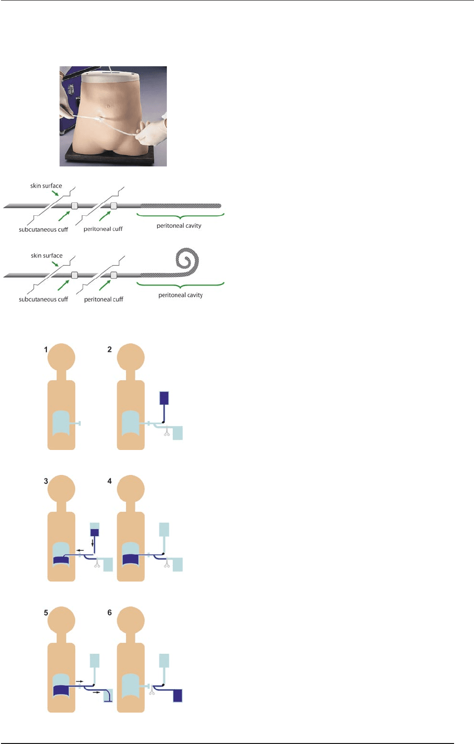
encourages ultrafiltration. e peritoneum allows
waste and fluid to pass from the blood into the dial-
ysate, which is pumped out.
e catheter exits the skin laterally to the midline.
A 20–30 cm long connecting tube (transfer set) can
be fastened to this with a screw thread, with the help
of which the sacks containing the dialysing solution
can be attached. e transfer tube can be closed with
a roller-wheel or with a sterile screw stopper.
4. erapeutic (postoperative)
rinsing drainage
(see the basics in section IV.10)
e main indication of continuous postoperative
drainage was earlier severe sepsis. Today it is used
mostly for intraabdominal abscesses and inflam-
matory processes. It is simple and cheap and can be
life-saving (see details in Csaba Gaál: Alapvető Se-
bésztechnika, Medicina, 1998).
Principle: If a cavity is present, it must be open; pri-
mary closure is forbidden.
Problems: Clotting, fibrin plug and cavity compart-
ment formation resulting from adherence, which in-
creases the risk of bacterial infections.
Main types: Rubber tubes and suction tubes (see sec-
tion IV.10).
ADVANCED MEDICAL SKILLS
105
VI. ABDOMINAL DRAINAGE
VII. Basic thoracic
surgical practicals
A thoracic trauma is generally sudden and dramatic;
it plays a role in 25% of the cases of mortality caused
by traumas overall. Two-thirds of the deaths occur af-
ter admission to hospital. It has serious complications:
hypoxia, hypovolemia, and respiratory and circulato-
ry insufficiency are frequent. It can be blunt and non-
penetrating (a traffic accident, a direct blow, a fall, or
a deceleration and compression injury) or penetrating
(shot and stabbed wounds, in which primarily the pe-
ripheral lung is affected). e chest wall, pleura, lung
parenchyma, upper airways/mediastinum or heart may
be injured. If the state of the patient is unstable, tension
pneumothorax, pericardial tamponade and massive he-
mothorax may be suspected (the route of the trauma
may be an indicator). Complementary examinations are
oen needed (echocardiography, bronchoscopy, esoph-
agography, esophagoscopy and aortography). Iatrogen-
ic traumas are frequently caused by the introduction of
nasogastric tubes (endobronchial introduction), chest
tubes (sc., intraparenchymal or intrafissural introduc-
tion) or central venous catheters.
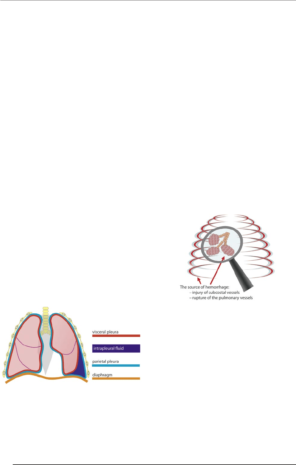
1. Types of pleural effusion
Transudate (protein content < 3.0 g/mℓ), serous fluid
(e.g. malignancies)
Exudate (protein content > 3.0 g/mℓ) caused by in-
flammation
Hemothorax (blood in pleural sac)
Empyema (pus in pleural sac), fibropurulent exudate
Chylothorax (lymph)
2.1. Mechanism/causes of thoracic
effusion formation
Increased hydrostatic pressure – in chronic heart failure
Increased capillary permeability – inflammation
Decreased colloidal oncotic pressure – nephrotic
syndrome, liver disease
Decreased lymphatic drainage – metastatic obstruc-
tion
2.2. General principles of treatment
Find the cause
Analgetics for pleurisy
oracocentesis/thoracic tube
3. Hemothorax
Definition: ere is blood in the pleural sac, usual-
ly caused by a penetrating trauma. e source of the
bleeding can be the alveoli, bronchi or thoracic vessels.
e accumulating blood compresses the heart and the
thoracic vessels (one half of the lung may contain ~ 1.5 ℓ
of blood). Respiratory disorders can occur, but circula-
tory disorders are more common. Signs: Tachycardia, a
weak pulse and shock-like symptoms. e diagnosis can
be established via a chest X-ray (pleura infiltration) and
diagnostic thoracocentesis.
3.1. Treatment of hemothorax
Fluid, volume and oxygen therapy
Small (300–500 mℓ). is may be le alone; it will be
reabsorbed.
Moderate (500–1000 mℓ): is requires computer
tomography (CT) and drainage
Large (> 1000 mℓ): CT, drainage and surgery (con-
trol of arterial bleeding) are needed.
4. Pneumothorax (PTX)
Definition: is is a condition in which air or gas is present
in the pleural space. is leads to an increased intrapleural
pressure, which causes partial or total collapse of the lung.
ADVANCED MEDICAL SKILLS
106
VII. BASIC THORACIC SURGICAL PRACTICALS
4.1. Etiology of PTX
Spontaneous, primary PTX
Blunt chest traumas (motor vehicle accidents and
falls)
Penetrating traumas (gunshot and knife injuries),
rib fractures and flail chest
Rib rupture and unstable chest
4.2. Clinical signs of PTX
Tachypnea and tachycardia. Questions: Are there
breathing difficulties, or pleurisy? Show its location.
Cyanosis
Diminished breath sounds; hyper-resonance on the
affected side
Neck vein engorgement
Paradoxical movement of the unstable chest; deviat-
ed trachea
Cardiogenic shock
4.3. Types of PTX
Closed or open
Traumatic or spontaneous
Simple or tension
Primary or secondary
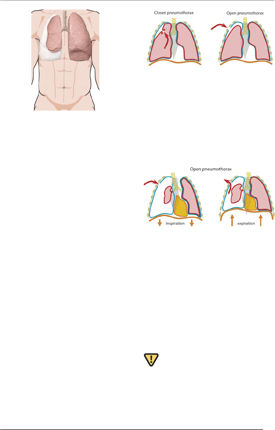
4.4. Closed PTX
is can occur spontaneously, or it may be a conse-
quence of a blunt trauma or an abrupt pressure rise (a
blast or diving). It is particularly frequent in thin, 20–
40-year-old male smokers. Most cases resolve aer 1–2
days. Chest tubes and surgical repair are rarely required
(< 10%). Treatment: Oxygen, iv. fluid, circulatory moni-
toring and a chest tube if needed.
4.5. Open PTX
Definition: A hole in the chest wall allows atmospher-
ic air to flow into the pleural space, leading to an in-
creased intrapleural pressure, resulting in partial or
total collapse of the lung. is can be caused by a pen-
etrating injury or be a side-effect of a therapeutic pro-
cedure, e.g. the insertion of a central venous or pulmo-
nary artery catheter.
4.5.1. Signs of open PTX
A sucking or hissing sound is audible on inspiration
as the chest wall rises
Blood, foam and blood clots are coughed up
Shortness of breath / difficulty in breathing
Pain in the shoulder or chest that increases with
breathing
4.5.2. Treatment of open PTX
Wound toilette: Larger wounds should be treated first.
Check for entry and exit wounds (look and feel). An air-
tight, complete cover should be provided at least 5 cm
beyond the edges of the wound. ree edges of airtight
material (the top edge and two sides) are taped down so
as to create a “flutter valve” effect that allows air to es-
ADVANCED MEDICAL SKILLS
107
VII. BASIC THORACIC SURGICAL PRACTICALS
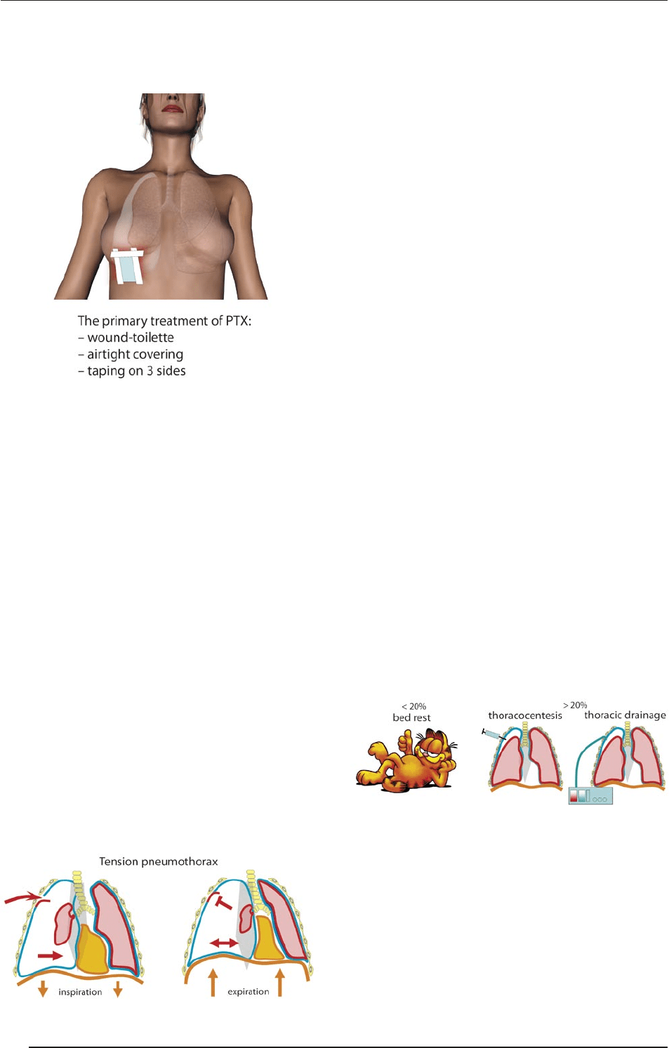
cape from, but not enter the chest cavity (in the USA,
“Petroleum Gauze” or “Asherman Chest Seal” (flutter-
valve seal) can be used).
4.6. Tension PTX
Iatrogenic or traumatic lesions of the visceral or pari-
etal pleura (oen associated with rib fracture) is respon-
sible for one-third of preventable thoracic deaths(!); in
these cases, the rupture of the pleura behaves as a one-
way valve. Mechanism: A one-way valve allows air to en-
ter the pleural space and prevents the air from escaping
naturally. e increased thoracic pressure leads to col-
lapse of the ipsilateral lung, and pushes the heart, vena
cava and aorta out of position (mediastinum shi), lead-
ing to a poor venous return to the heart, a decreased CO
and hypoxia. Etiology:
barotraumas
secondary to positive-pressure ventilation (PEEP)
complication of enteral venous catheter placement,
usually subclavian or internal jugular
conversion of idiopathic, spontaneous, simple PTX
to tension PTX (an occlusive dressing functions as a
one-way valve)
chest compressions during cardiopulmonary resus-
citation
fiberoptic bronchoscopy with closed-lung biopsy
markedly displaced thoracic spine fractures.
4.7. Signs and symptoms of tension
PTX
Early findings
Chest pain and anxiety
Dyspnea, tachypnea and tachycardia
Hyper-resonance of the chest wall on the affect-
ed side; diminished breath sounds on the affected
side
Late findings
A decreased level of consciousness
A tracheal deviation toward the contralateral side
Hypotension and cyanosis
Distension of the neck veins (this may not be present
if the hypotension is severe) and increased CVP
5. Treatment of PTX
5.1. Basic questions
How much air is present? What is its source?
What is the general condition of the patient? What is
the severity of other injuries?
Are critical care facilities available?
5.2. Treatment of simple PTX
If the size of the PTX is < 20%, bed rest and limited
physical activity are called for.
If the size of the PTX is > 20%, thoracocentesis or
insertion of a chest tube attached to an underwater
seal is necessary.
5.3. Treatment of simple PTX with
needle thoracocentesis
Requirements
A 22–20 gauge needle, extension tubing, a three-way
stopcock, a 20 to 60 mℓ syringe and supplementary oxy-
gen are required, +/- iv. fluids and analgetics.
ADVANCED MEDICAL SKILLS
108
VII. BASIC THORACIC SURGICAL PRACTICALS
Technique: see above.
5.4. Emergency needle
decompression
Indication
A diagnosis of tension PTX with any two of the follow-
ing signs:
respiratory distress and cyanosis
mental alterations
a nonpalpable radial pulse (hypovolemia)
Technique
Administration of 100% oxygen; ventilation if neces-
sary; continuous monitoring; pulse oxymetry if possible.
Location of anatomic landmarks. Surgical chest
preparation (Betadine and alcohol) and local anes-
thetics (if the patient is awake or if time / the situa-
tion permits).
In cases of trauma, the patients should be supine,
with head tilt; in other patients, a 45
o
sitting position
is required.
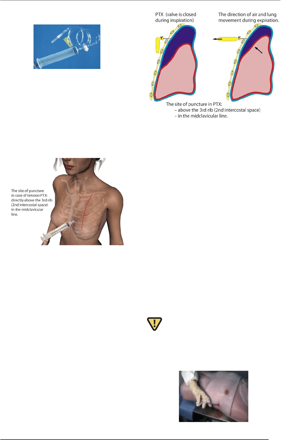
Decompression catheters are placed in the midcla-
vicular line in the 2nd rib interspace. Placement in
the middle third of the clavicle minimizes the risk of
injury to the internal mammary artery.
A puncture is made through the skin, 1–2 cm from
the sternum.
A needle (14–16 gauge) or needle catheter (Braunule;
2 inch/5 cm) is used, perpendicular to the skin, just
above the cephalad border of the 3rd rib (the intercos-
tal vessels are largest on the lower edge of the rib).
Once the needle is in the pleural space, the hissing
sound of escaping air is listened for, and the needle
is removed while the catheter is le in place.
Option I: Removal of the needle; the plastic cathe-
ter stays in place; preparation for placement of the
chest tube.
Option II: A plastic or rubber condom is placed on
the end of the needle, which acts as a valve (before
the definite management of the PTX).
Complications
Injury of intercostal vessels and nerves.
PTX (if the procedure is performed in patients without
PTX, the risk rate of lung injury and PTX is 10–20%).
Infection.
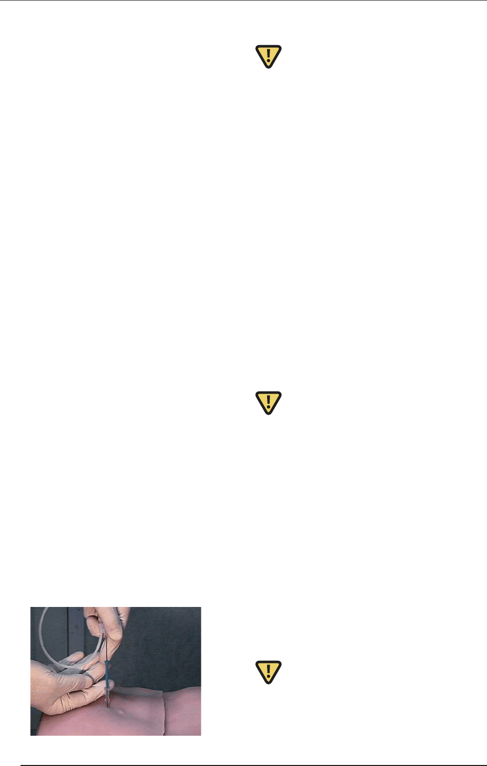
5.5. Percutaneous thoracocentesis
for the treatment of PTX
Equipments
18 G Braunule, pneumocath (9 F), Seldinger catheter
(6–16 F), chest tube (18–36 F) and water seal/Heimlich
valve (see later).
5.6. Chest drain – chest tubes
Indications
Fluid and air should be evacuated from the pleural space
and a negative intrapleural pressure reestablished to reex-
pand the lungs, with needle decompression management.
ADVANCED MEDICAL SKILLS
109
VII. BASIC THORACIC SURGICAL PRACTICALS