Boros Mihaly. Surgical Techniques
Подождите немного. Документ загружается.


Preface
e second part of “Surgical Techniques” is the subject-matter of the “Advanced Medical Skills” course. In these mod-
ules, the Institute of Surgical Research introduces surgical principles and techniques, and advanced interventions
such as surgical operations, e.g. laparotomy, appendectomy, intestinal resection, bowel anastomosis, thoracocentesis
and thoracotomy, to interested students. ese procedures are taught in simulated real-life, clinical surroundings and
circumstances.
is curricular structure is used to teach and update scientific and medical findings relevant to surgical practice,
to enhance clinical reasoning and decision-making, and to provide individual feedback and career advice. Typical fu-
ture careers of participants of this course include surgery and surgical specialties, such as gynecology, head and neck
surgery, neurosurgery, oncology, ophthalmology, orthopedics, plastic surgery, thoracic surgery, urology, vascular sur-
gery, anesthesiology, emergency medicine, critical care and cardiology.
e goals are to foster skills-based decision-making, and to broaden the correlation of physiology, anatomy and
pharmacology to acute clinical care. Emphasis is placed on procedures, critical thinking and the assessment of skills,
in order to develop the knowledge and skills to support a career choice in those specialties in which expertise in surgi-
cal anatomy is critical.
ADVANCED MEDICAL SKILLS
90

I. Laparotomy
“We took out fieen pounds of a dirty, gelatinous
looking substance. Aer which we cut through the fal-
lopian tube, and extracted the sac, which weighed sev-
en pounds and one half… In five days I visited her, and
much to my astonishment found her making up her bed.”
(McDowell E. ree cases of extirpation of diseased
ovaria. Eclectic Repertory Anal Rev. 1817; 7:242–244.)
Terms and definitions
Laparo or lapar (Greek: λ α π α ρ α, λ α π α ρ ο σ) means
the so part of the body between the ribs and the hip; it
denotes the flank or loins and the abdominal wall. is
term is sometimes used loosely (and incorrectly) in ref-
erence to the abdomen in general. Laparotomy therefore
means a surgical incision through the flank; less cor-
rectly, but more generally, it is an abdominal section at
any point to gain access to the peritoneal cavity.
1. History of abdominal surgery
1809 On Christmas morning, Dr. Ephraim McDow-
ell (1771–1830) in Danville (Kentucky, USA)
successfully removed an ovarian tumor from
Mrs. Crawford without anesthetic or antisep-
sis. e risk of fatal infection was very high –
the operation was bitterly criticized.
1879 Jules Émile Péan (1830–1898) opened the ab-
domen of a patient with cancer of the pylorus.
e diseased section was cut out; the remain-
der was sewn to the duodenum. e patient
died 5 days later.
1880 Ludwig Rydyger (1850–1920) carried out the same
procedure, but it had been planned in advance;
the patient died within 12 hr, of “exhaustion.”
1881 Christian Albert eodor Billroth (1829–1894)
performed a successful operation (the patient
died 4 months later due to the propagation of
the tumor). Two other, fatal operations followed:
Billroth was stoned on the streets of Vienna.
1885 Billroth II (pylorus cc): Successful operations
were achieved.
Today, emergency admissions account for 50% of
the general surgical work load and abdominal pain is
the leading cause of 50% of emergency admissions. It
should be noted that 70% of the diagnoses can be made
on the basis of the history alone, and 90% of the diagno-
ses can be established if the history is supplemented by
physical examination. e expensive and complicated
diagnostic tests and instrumental procedures oen (>
50%) merely confirm the results of the anamnesis and
physical examination (!).
Abdominal pain is frequently (35%) ‘aspecific’; it can
be caused by viral infections, bacterial gastroenteritis,
helminths, irritable bowel syndrome, gynecological dis-
eases, psychosomatic pain, abdominal wall pain, iatro-
genic peripheral nerve lesion, hernias or radiculopathy.
e frequency of acute appendicitis and ileus is 15–17%;
they are followed in frequency by urological diseases
(6%), cholelithiasis (5%) and colon diverticulum (4%).
e frequency of abdominal traumas, malignant dis-
eases, peptic ulcer perforation and pancreatitis is 2–3%,
while that of rupture of an aorta aneurysm, inflamma-
tory bowel disease, gastroenteritis and mesenteric isch-
emia is < 1%.
2. Technical background of
laparotomies
Abdominal incisions are based on anatomical prin-
ciples.
ey must allow adequate access to the abdomen.
ey should be capable of being extended if required.
Ideally, muscle fibers should be split rather than cut;
nerves should not be divided.
e rectus muscle has a segmental nerve supply. It
can be cut transversally without weakening a dener-
vated segment. Above the umbilicus, tendinous in-
tersections prevent retraction of the muscle.
3. Basic principles determining
the type of laparotomy
e disease process
e body habitus
e operative exposure and simplicity
Previous scars and cosmetic factors
e need for quick entry into the abdominal cavity
ADVANCED MEDICAL SKILLS
91
I. LAPAROTOMY
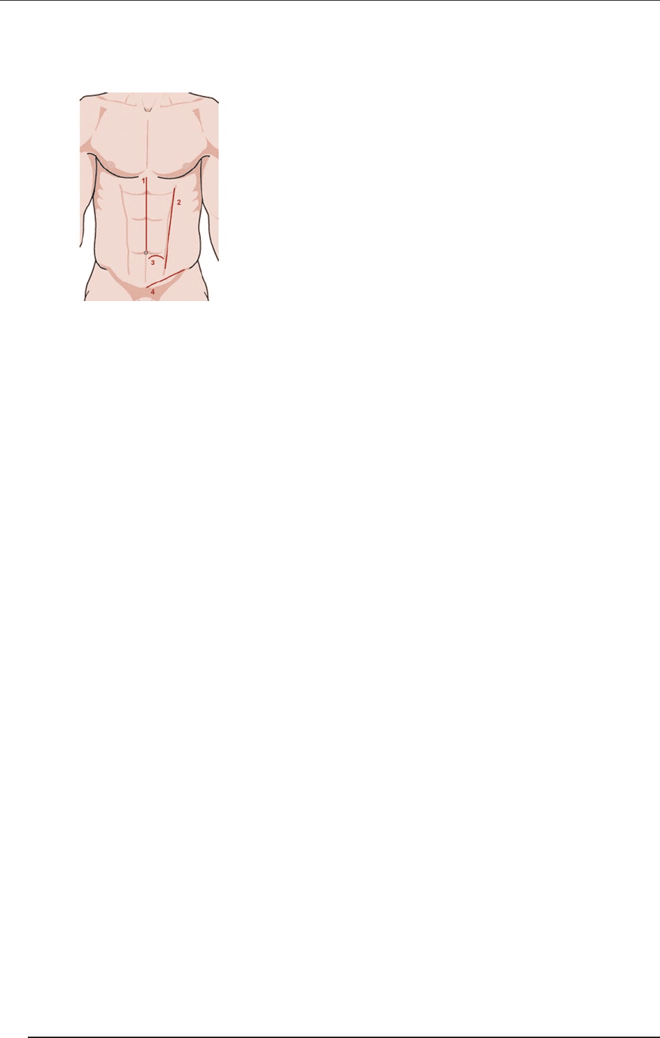
4. Recapitulation: Anatomy of
the abdominal wall
From le to right: 1. the linea alba; 2. the linea semilu-
naris; 3. the lig. arcuatum; and 4. the abdominal projec-
tion of the lig. inguinale. During laparotomy, different
anatomic structures are cut in the upper or lower ab-
dominal regions at various distances from the midline
(anterior vs lateral regions). During a midline incision,
the following tissue layers and structures are divided:
the skin,
the superficial fascia (Camper’s),
the deep fascia (Scarpa’s),
the anterior rectus sheath,
the rectus abdominis muscle
the posterior rectus sheath down to arcuate line,
the transversal fascia,
the extraperitoneal connective tissue,
the peritoneum.
Recapitulation: Important things about nerves
Transverse incision is least likely to injure nerves.
e iliohypogastric (ih) and ilioinguinal (ii) nerves
are sensory:
ih injury leads to a loss of sensation in the skin
over the mons;
ii injury leads to a loss of sensation in the labia
majora.
Both ih and ii nerves supply the lower fibers of the
internal oblique and transverses; if divided, these fi-
bers undergo denervation, which can increase the
risk of inguinal hernia.
5. Principles of healing of
laparotomy
Patient risk factors that negatively affect wound
healing:
Diabetes and obesity
Poor nutrition
Prior radiation or chemotherapy
Age
Alcohol
Ascites and malignancy
Immunosuppression
Coughing, retching
Hospital factors that affect wound healing negatively
Long operations
Along period of hospitalization preoperatively
Drains through incision
Shaving prior to surgery
Type of suture
Closure technique
6. Prevention of wound
complications
e scalpels should not be the same for the skin and
deep incisions.
A scalpel should be used to cut skin and fascia and
not diathermy; the infection rate aer diathermy is
twice as high.
Deep sc. sutures should be avoided, but absorbable
synthetic material (e.g. 4.0 Dexon) may be used sub-
cutaneously to decrease tension on the skin.
Use of catgut (for fascia or sc. suturing) should be
avoided.
Contaminated or dirty wounds:
delayed closure,
staples with saline-soaked gauze.
Opening of a bacteria-containing organ:
delayed closure,
irrigation of all layers,
monofilament, nonabsorbable suture,
systemic antibiotics 30 min before operation or as
soon as possible, and repeat in a prolonged case.
ADVANCED MEDICAL SKILLS
92
I. LAPAROTOMY
II. Incisions
e term incision originates from the Latin (in + cidere
→ incisio). An incision can be longitudinal, oblique or
transverse. e most important types are demonstrat-
ed in association with abdominal operations; the prin-
ciples are identical in the other body regions (extremi-
ties, chest, neck, etc.).
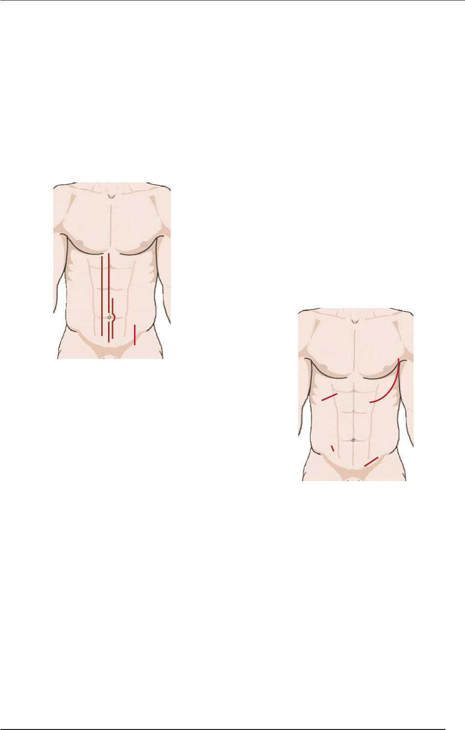
1. Longitudinal incisions
1. Midline; 2. supraumbilical (upper midline); 3. in-
fraumbilical (lower midline); 4. right paramedian; 5.
McEvedy preperitoneal approach for inguinal and fem-
oral hernia repair (McEvedy PG: Femoral hernia. Ann R
Coll Surg Engl, 1950).
1.1. Characteristics of longitudinal
incisions
Median incision
is was the commonest abdominal intervention be-
fore the era of minimally invasive surgery. e umbi-
licus and the falciform ligament above the umbilicus
should not be incised. Meticulous, careful handling
of bleeding is necessary in the superficial layers before
the peritoneum is opened. e urinary bladder can be
reached through the Retzius space (spatium retropu-
bicum Retzii); if there has previously been an opera-
tion in this field, a more caudal entry is necessary (the
chance of scar formation and adhesions is less).
Advantages: ere is excellent exposure to the abdo-
men and pelvis, which can easily be extended, and
also rapid entry into the abdominal cavity; the mid-
line is the least hemorrhagic incision, and is easy to
perform; the linea alba is the guide to the midline.
Disadvantages: e scar may be wide and not beau-
tiful, with a possible increase in hernias and dehis-
cence with the midline.
Paramedian incision
e site is parallel to and ~ 3 cm from the midline.
e following structures are divided: skin – anteri-
or rectus sheath (the m. rectus is retracted laterally)
– posterior rectus sheath (above the arcuate line) –
transversalis fascia – extraperitoneal fat – peritone-
um. Closure is performed in layers.
Indication: If excellent exposure is needed to one
side of the abdomen or pelvis.
Advantages: A lower incidence of incisional hernias.
Disadvantages: It takes longer to make and close this
incision, there is an increased risk of infection, and
intraoperative bleeding, and a risk of nerve damage;
if sited beside the midline, and it can compromise
the blood supply in the middle.
2. Oblique incisions
(1). Kocher incision for cholecystectomy (sec. eodor
Kocher (1841–1917), Nobel Prize for medicine and
physiology in 1909, mainly for thyroid surgery); (2).
McBurney incision for appendectomy (aer Charles
McBurney (1845–1913), who performed his first op-
eration for appendicitis in 1897); (3). le inguinal; (4).
thoraco-abdominal
2.1. e basic type of oblique
incisions
Indications for McBurney muscle-splitting incision
(see later): Appendicitis, pelvic abscess and extra-
peritoneal drainage.
2
4
3
1. medián
2. fels
� medián
3. alsó medián
4. j.o. paramedián
5. McEvedy
1
5
2
4
1. Kocher
2. McBurney
3. b.o. inguinalis
4. thoraco-abdominalis
1
3
ADVANCED MEDICAL SKILLS
93
II. INCISIONS
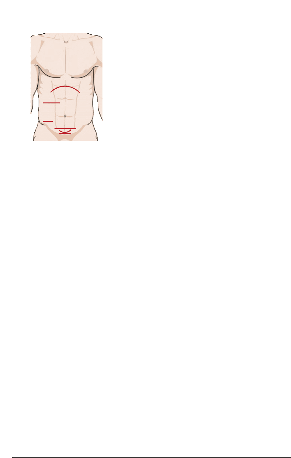
3. Transverse incisions
1. Gable incision, 2. transverse muscle splitting, 3. Lanz
incision, 4. Maylard incision, 5. Pfannenstiel incision, 6.
Cherney incision
3.1. Basic characteristics of
transverse incisions
Advantages: ese incisions give the best cosmetic
results, they give a much stronger scar than midline
incisions and less painful than longitudinal inci-
sions, and there is less interference with respiration.
ere is no difference in dehiscence rate.
Disadvantages: ey are more time-consuming, and
more hemorrhagic; nerves are sometimes divided,
spaces are opened and there is a potential for hema-
tomas; upper abdominal access is limited.
Main types:
Pfannenstiel incision: For gynecological indica-
tions. Advantages: Most wound security (in pelvic
incisions), least exposure, usually 10-15 cm long.
Disadvantages: Separates the perforating nerves
and small vessels from the ant. rectus, and this
may weaken the strength of the wound healing. If
extended past the m. rectus, it can damage the ih
and ii nerves.
Maylard incision: is gives excellent exposure to
the lower pelvis; it is used for radical pelvic sur-
gery; it is a true transverse muscle-cutting inci-
sion, 3–8 cm above the symphysis.
Cherney incision: is is like a Pfannenstiel inci-
sion, but divides the m. rectus at the tendinous in-
sertion to the symphysis. It gives excellent access
to the space of Retzius. During closure, re-attach-
ment of muscle tendons to the rectus sheath, and
not the symphysis, should be performed in order
to avoid osteomyelitis.
Rockey Davis (Elliot) incision: is alternative to
the McBurney incision, extends to the lateral bor-
der of the rectus (it was described first by JW El-
liot in 1896, then by AE Rockey in 1905, and fi-
nally by GG Davis in 1906).
Lanz incision: is is a special incision at the
right fossa iliaca. As compared with the McBur-
ney incision, it is transverse, more medial toward
the rectus, and closer to the iliac crest (spina ili-
aca anterior superior), and gives better cosmet-
ic results. Due to its transverse direction, the ih
and ii nerves can be damaged, and the incidence
of hernia is higher. e main indication is expo-
sure of the appendix and cecum; the mirror im-
age (le iliac fossa) can be used for the le colon
(not for the rectum).
4. Special extraperitoneal
incisions for staging
J-shaped incision: 3 cm medial to the iliac crest; this
allows the extraperitoneal removal of para-aortic
nodes; it can also be le-sided; but the right is easier.
“Sunrise” incision: 6 cm above the umbilicus, per-
mitting the extraperitoneal removal of para-aortic
nodes, and allowing immediate irradiation.
2
1. Gable
2. haránt rácsmetszés
3. Lanz
4. Maylard
5. Pfannenstiel
6. Cherney
1
3
4
5
6
ADVANCED MEDICAL SKILLS
94
II. INCISIONS

III. Laparotomy in
surgical training
Median laparotomy is indicated when the whole ab-
dominal part of the gastrointestinal tract should be ex-
plored. is will be a task in the surgical techniques
practicals (the following operative description is related
to animal (e.g. pig) interventions, in which the steps are
identical to those of human operations).
1. General rules
Anesthesia
Method: General anesthesia.
Equipment: Typical monitors, a respirator and a
warming blanket. Insertion of a Foley catheter, and
application of an electrodispersive pad. e anesthesi-
ologist will insert a nasogastric tube aer intubation.
Positioning
Supine, with arms on armboards.
Special considerations: High-risk areas (for geriatric
patients, particular attention should be paid to the
skin and joints).
Skin preparation
Method of hair removal: Clippers or wet, with a razor.
Anatomic perimeters: Traditionally from the nipple
line across the chest from the table side to the table
side to mid-thigh.
Solution options: Betadine (povidone-iodine) or an
alternative (e.g. Hibiclens in USA).
Draping/incision
In explorations, usually 4 towels (USA: a laparoto-
my T-sheet) are used in the midline (but the isola-
tion depends on the location of the lesion; it could
be paramedian or oblique, etc; see above).
Supplies
General: Blades (3) #10 and (1) #15, scissors, forceps, elec-
tric unit pencil, suction tubing, hemostats (Péan, all sizes),
staples (optional), retractors (Gosset) and sutures (ample
supply of free ties; sizes 2-0 and 3-0 are most common).
Specific: Catheters, drains, etc.
2. Middle median laparotomy
is can be applied when the diagnosis is uncertain.
Its advantages are that a large area can be examined
through a small incision and, aer the abdomen
has been opened, the incision can be lengthened
both cranially and caudally if necessary and can be
quickly closed. e disadvantages are that the com-
mon aponeurosis of six different strong flat abdom-
inal muscles is cut, the statics of the abdominal wall
is greatly impaired, and this predisposes to wound
disruption and scar hernia oen occurs.
e first step is the scrub preparation of the opera-
tive field from the xyphoid process to the symphy-
sis; draping should be performed as described ear-
lier. e midline is shown by the umbilicus; two
laparatomy sponges are placed, one on each side of
the planned incision. Generally a short, 10–15-cm-
long incision is made, partly above and partly below
the umbilicus, going round the umbilicus at a dis-
tance of 1–2 cm from the le (not to injure the falci-
form ligament and the ligamentum teres hepatis).
In the first phase of the operation, the skin and the
sc. fat, and then the aponeurosis of the linea alba are
cut. e linea alba is a line-like sheet some tenths
of a mm thick below the umbilicus, while it is wid-
er, strong and tendinous above it. e incision cuts
the posterior rectus sheet, transversal fascia, preperi-
toneal fat and parietal peritoneum. Below the umbi-
licus, the arcuate linea (linea semilunaris Douglasi)
borders the area below which there is no rectus sheet.
Aer the skin incision, Doyen clamps are placed on
the wound edges and the wound towels. e sc. fat
is usually cut with a diathermy pencil; bleeding can
be stopped by compression and, if necessary, by lig-
atures and stitches, or the preventive handling of
bleeding is used. Recapitulation:
a. A ligature is applied to cuts, or bleeding vessels if the
bleeding cannot be stopped by compression. Cut ves-
sels are grasped with hemostatic clamps (Péan or mos-
quito). e position is checked by wiping off the blood.
ADVANCED MEDICAL SKILLS
95
III. LAPAROTOMY IN SURGICAL TRAINING
If the grasping is not successful, a second hemostat
is placed deeper. e vessel is then ligated below the
clamp. Aer the first half-hitch has been tied, the he-
mostat is removed and the second half-hitch is tied.
b. Preventive hemostasis: e vessel to be cut is closed
with two hemostats in advance. e vessel is sepa-
rated between them, and the two vessel ends are
then ligated separately.
c. Suture for hemostasis: A double, 8-form stitch is
placed below the bleeding vessel, and the thread is
knotted. is suture is applied if a hemostat cannot be
used, e.g. in the cases of vessels that are thin-walled or
lie in a fascia layer, or retract deep into the tissues.
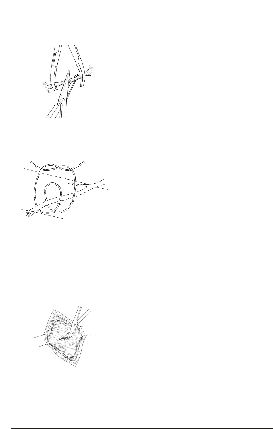
When the sc. connective tissues are divided, the
wound edges are lied up with two tissue forceps or
clamps, and the tissues are cut transversally, layer by
layer with Mayo scissors.
During the blunt dissection of tissues, the closed tips
of Mayo scissors (or Péan, dissector) are pushed into
the tissues. e tissues are dissected by the opening
of the instrument with its blunt outer edges. ese
steps are repeated as necessary.
e incision is deepened until the linea alba is reached,
the linea is then picked up with two tissue forceps
above the umbilicus and a small incision is made be-
tween them (this can be done with Mayo scissors). e
opening is then lengthened cranially and caudally with
Mayo scissors while the abdominal wall is lied up.
If the incision is made exactly in the midline, the
rectus sheet will not be opened, and the muscles will
not be severed. Above the umbilicus, care should be
taken not to injury the ligamentum falciforme hepa-
tis. e thick, fatty ligament can be clamped with
two Péan hemostats and cut between them, a better
exploration being achieved in this way.
eperitoneal cavity is isolatedfrom thesc.layer by mak-
ing a second draping. Two laparotomy sponges are placed
on each side of the incision and fastened to the edges of
the peritoneum with Mikulicz clamps on both sides.
e abdominal wall is elevated with the surgeon’s in-
dex and middle fingers or with the help of the assis-
tant, and the incision of the linea alba is lengthened
with Mayo scissors (or a diathermy knife) both cra-
nially and caudally to the corners of the skin wound.
During this, the peritoneum edges are fixed to the
sponges with Mikulicz clamps.
A Gosset self-retaining retractor is placed into the
abdominal wound. e greater omentum or intes-
tines should not be allowed to come between the
jaws of the retractor and the abdominal wall. e
abdominal organs can be moved only with warm sa-
line-moistened laparotomy sponges.
Aer median laparotomy, the following organs can
be examined: 1. the greater omentum; 2. the spleen;
3. the liver, gall bladder and bile ducts; 4. the stomach;
5. the small intestine and mesenteric lymph nodes; 6.
the appendix (cecum); 7. the large intestines; 8. the
pancreas; 9. the adrenal glands; and 10. the kidneys.
e abdominal wall is closed in layers. Sutures of ap-
propriate size should be selected to close the differ-
ent layers, and the wound edges should be exactly
approximated. It should be checked that no foreign
body has been le in the peritoneal cavity. All wound
towels, sponges and instruments should be count-
ed. During abdominal operations, sponges clamped
with an instrument (a sponge-holding clamp) can be
used only for wiping, and instruments are placed on
the ends of laparotomy sponges.
e Gosset self-retaining retractor is removed, and the
laparotomy sponges isolating the peritoneal cavity are
released from the Mikulicz clamps and removed, but
the edges of the peritoneum are clamped again.
e wound of the peritoneum is closed with a half-cir-
cle muscle needle, with a continuous running suture (in
pigs with #40 linen thread). Tissue forceps can be used
for the first stitch, but in most cases the wound of the
peritoneum can be well explored with Mikulicz clamps.
Suturing is usually done towards the umbilicus; the first
ADVANCED MEDICAL SKILLS
96
III. LAPAROTOMY IN SURGICAL TRAINING
stitch is inserted at the cranial wound corner, but it can
also be performed in the opposite direction, i.e. toward
the xyphoid process. If the abdominal wall is closed in
multiple layers, the first row of stitches closes the poste-
rior rectus sheet together with the peritoneum.
e assistant ties a knot on the short free end of
the thread. He/she keeps the suture under continu-
ous tension with his/her right hand and helps with
the closing of the wound edges. When the peritone-
um has been closed, only one-third of the thread is
pulled through the wound and a doubled thread is
le on the other side. e single and double ends of
the thread are knotted and cut short.
e anterior rectus sheet and sc. wound are closed
with interrupted sutures. e skin is closed with Do-
nati stitches, using a skin (1/4 or 3/8) needle and #40
linen thread. e wound is disinfected with Beta-
dine and covered with a bandage.
3. Some important details
e principles of closing the fascia
e fascia should be closed with the minimum num-
ber of stitches, at least 1 cm from the edges, since ne-
crosis may occur (each stitch 1 cm from another and
from the edges).
Each stitch should be closed with the same strength;
the wound edges should only be approximated (!);
sewing in fat or connective tissues should be avoided
(except in cases of en masse closure).
e Smead Jones technique involves a “far to far,
near to near” suture (en masse far stitches on both
sides, then near stitches involving the fascia only).
e healing tendency is theoretically good, and this
technique decreases tension, but it is time-consum-
ing and rarely used in clinical practice.
Drainage
is may be passive or active (see earlier; the passive
drain is never brought out in the line of the incision
(danger of infection!).
e most frequent indications are infection, oozing,
and the need to eliminate a cavity.
For clean wounds, the prophylactic use of drainage
can be controversial; closed suction can be useful in
the case of clean/contaminated wounds (especially if
no antibiotics are given).
Wound irrigation
Irrigation with physiological saline to prevent infec-
tion (motto: “e solution to pollution is dilution”).
Irrigation with antiseptic solution (e.g. 1% povi-
done-iodine) is effective, but can be cytotoxic (e.g.
fibroblasts can be damaged).
Closing the skin
None of the methods (wound clips, suturing, etc.) is
substantially better than the others.
To cover an abdominal skin wound, Opsite, Telfa, etc. can
be applied; the bandage can stay in place for 2–3 days.
In the event of irradiation, abdominal clips should
stay in the wound longer.
Special case: the obese patient
According to international standards, a subject whose
body weight exceeds the ideal by 25–30% is overweight;
an excess of 30–60% means that the subject is obese; in
extreme obesity the body weight exceeds the ideal by
100%. e obesity is “morbid” if the weight excess is
greater than 130%.
e Pickwick syndrome received its name aer Joe, the
somnolent, red-faced, fat boy character of Charles Dickens.
It was given by Sir William Osler (1918): “A remarkable phe-
nomenon associated with excessive fat in young persons is an
uncontrollable tendency to sleep like the fat boy in Pickwick.”
Modified routine in operations on obese patients
Extensive cleansing of the umbilicus and preopera-
tive bath(s)
5000–8000 U/12 h heparin 2 h before surgery
Elastic bandage and stockings
Removal of abdominal hair with an electric razor only
A very extensive scrub preparation (under skin
wrinkles also), pulling the pannus caudally
Transverse incisions should always be made far from
the wet, warm fatty skin wrinkles (duplicates)
En masse closure with a continuous running suture
Drain and suction bottle over the fascia, removal 72
h later, or if the volume is < 50 mℓ/day
Removal of wound clips aer 14 days.
ADVANCED MEDICAL SKILLS
97
III. LAPAROTOMY IN SURGICAL TRAINING
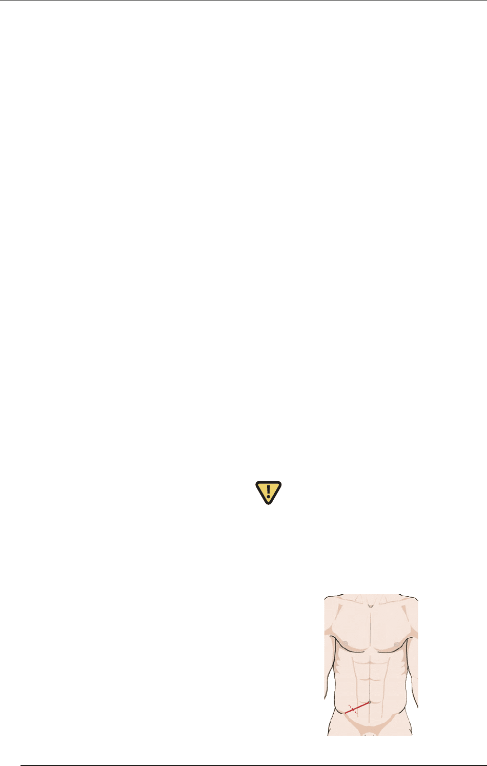
McBurney pont
(a köldök és a spina
iliaca ant. sup. közötti
lateralis harmadoló pont)
X
X
IV. Basic surgical
procedures on
the intestines.
Appendectomy
Motto: “If in doubt, take it out.”
Open appendectomy was earlier one of the first opera-
tions of the young surgeon, but recently it is increasing-
ly performed with minimal invasive methods (see lat-
er). e intervention is relatively simple in the majority
of the cases; the consecutive steps are built on each other
and illustrate the classical, well-planned and safe surgical
technique. At present the urgent operation is still the only
safe method of treatment of appendicitis (it must be per-
formed even if there is only a reasonable suspicion).
1. e history of appendectomy
1521 Jacopo Berengario da Capri (1460–1530)
described the appendix as an anatomical
structure
1500s Vidus Vidius’s (Guido Guidi, 1500–1569)
book of anatomy: the term appendix was
in general use
1710 Philippe Verheyen (1648–1710) coined
the term appendix vermiformis
1800s “Lower abdominal pain” as a medical di-
agnosis
1812 A connection was found between peritonitis
and necrotic appendix (John Parkinson).
1824 A connection between periappendicu-
lar inflammation and a necrotic appen-
dix (Jean Baptiste de Louyer-Villermay)
1827 A connection between a periappendicular
abscess and the appendix (François Melier)
1848 Surgical drainage of a periappendicular
abscess (Henry Hancock)
1867 Several successful drainages of a periap-
pendicular abscess (Willard Parker)
1882 Death of Leon Gambetta, Prime Minis-
ter of France. Autopsy proved a periap-
pendicular abscess
1886 Reginald H. Fitz (pathologist) suggested
that “lower abdominal pain” is “appen-
dicitis”, and proposed urgent surgery in
the event of signs and symptoms
1887 April 27 George omas Morton performed the
first successful human appendectomy:
removal of a perforated appendix
1889 John B. Murphy performed a series of
100 successful appendectomies
1902 A successful operation on the British
Crown Prince Edward (VII) before his
coronation ceremony.
1.1. Recapitulation: relevant
anatomy
e appendix does not elongate as rapidly as the rest
of the colon, thereby forming a wormlike structure.
e average length is 10 cm (2–20) with inner circu-
lar and outer longitudinal (continuation of the tae-
niae coli) muscle layers. Submucosal lymphoid folli-
cles enlarge (peak in 12–20 years) and then decrease
in size, correlating with the incidence of appendici-
tis. e blood supply is from the appendicular artery
(branch of the ileocolic artery).
e location of the base is constant, whereas the po-
sition of the tip of the appendix varies: 65% retroce-
cal position; 30% at the brim or in the true pelvis;
and 5% extraperitoneal, behind the cecum, ascend-
ing colon, or distal ileum. e location of the tip of
the appendix determines early signs and symptoms.
Even in a case of surgically verified appendicitis, the
Meckel diverticulum should be looked for at the an-
timesenteric edge of the ileum, orally 40–100 cm
from the appendix. Both can be considered to be
developmental rudiments; their inflammation of-
ten develops simultaneously. Meckel diverticulum
should be suspected if there is a long-lasting umbili-
cal discharge in the anamnesis.
1.2. Open appendectomy
An “RLQ” (right-lower quadrant) incision over the Mc-
Burney point (2/3 of the distance between the umbili-
cus and the anterior superior iliac spine). e incision
was described by Lewis L. McArthur in June 1894, but
named aer Charles McBurney, who presented a case
in the July 1894 issue of Annals of Surgery.
ADVANCED MEDICAL SKILLS
98
IV. BASIC SURGICAL PROCEDURES ON THE INTESTINES. APPENDECTOMY
e sc. tissue and Scarpa fascia are dissected until the
external oblique aponeurosis is identified. is aponeu-
rosis is divided sharply along the direction of its fibers.
A muscle-splitting technique is then used to gain ac-
cess to the peritoneum.
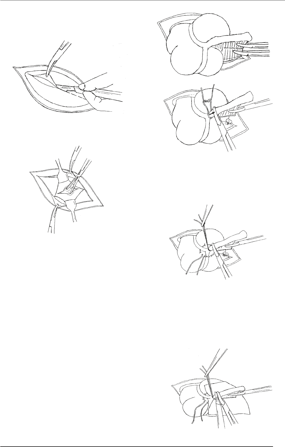
e peritoneum is lied up with two forceps or he-
mostats in order to avoid damage to the underlying
viscera. A small incision is made in the peritoneum
and, aer entry to the peritoneal cavity (if a purulent
fluid sample should be taken for bacteriological di-
agnosis), the appendix is sought out.
Retractors are placed into the peritoneum, and the
cecum is identified and partially exteriorized, us-
ing a moist gauze pad. e taenia coli is followed to
the point where it converges with the other taenia,
leading to the base of the appendix. e appendix is
brought into the field of vision. Gentle manipulation
may be required for the blunt dissection of any in-
flammatory adhesions.
“Skeletization” includes cutting of the mesoappen-
dix and ligation of the appendicular artery (this is
the branch of the ileocolic artery originating from
the superior mesenteric artery; if it is ligated central-
ly, the terminal ileum can be necrotized). e meso-
appendix is cut between Péan hemostats in several
steps (clamping–cutting–ligating), care being taken
that a tissue collar should be le on the remaining
proximal stump). Generally 3–0 absorbable thread
is used. Finally, the appendix is completely mobi-
lized step by step.
e base of the appendix is crushed with a straight
Kocher clamp (aer this step, the operation can-
not be regarded as “sterile”), and then ligated with
a thin absorbable thread (in animals, #40 linen
thread is used).
A seromuscular, purse-string suture is placed around
the stump of the appendix, using 3 or 4–0 thread,
with a round-bodied serosa needle. Care should be
taken as to the depth of the stitches: if they are too
deep, the infected bowel content can pass into the
abdominal cavity; if they are too superficial, they
can be torn out.
ADVANCED MEDICAL SKILLS
99
IV. BASIC SURGICAL PROCEDURES ON THE INTESTINES. APPENDECTOMY