BD Diagnostic Systems (publ.). Difco Manual (Manual of Microbiological Culture)
Подождите немного. Документ загружается.

166 The Difco Manual
plating method, and found the latter method resulted in better
recovery of fecal streptococci. They developed m E Agar as a primary
isolation medium for enterococci, and Esculin Iron Agar as an
in situ substrate test medium for identifying organisms capable of
hydrolyzing esculin.
6
Two research projects by the Environmental Protection Agency (EPA)
evaluated the relationships between swimming-associated illness
and the ambient densities of indicator bacteria.
7,8
The studies
demonstrated that enterococci have a better correlation with
swimming-associated illness for both marine and fresh waters than
fecal coliforms. Escherichia coli has a correlation in fresh water equal
to enterococci but does not correlate as well in marine waters.
7,8
This
suggests that enterococci may be better indicator organisms for some
recreational waters.
7,8
m E Agar and Esculin Iron Agar are prepared according to the
formulas specified in Standard Methods.1 These media are used in
the membrane filter technique for the isolation of fecal streptococcus
and enterococcus groups.
1
This procedure can be used to test marine
and fresh water sources.
m E Agar with the addition of 0.075% indoxyl B-D glucoside
(m EI Agar) is recommended by the U.S. EPA as a one step procedure
for the isolation and identification of enterococci in recreational
water.
9
This method is used in the EPA Beaches Environmental
Assessment Closure and Health (BEACH) Program. m EI Agar
eliminates the necessity of transferring the incubated membrane to
Esculin Iron Agar.
Principles of the Procedure
m E Agar is a highly selective and differential primary isolation
medium that supports good growth of enterococci. Bacto Peptone
and Yeast Extract provides carbon, nitrogen, minerals, vitamins
and other growth factors for organism growth. Sodium Chloride
maintains the osmotic balance of the medium. Nalidixic Acid and
Sodium Azide act as selective agents to inhibit gram negative
bacteria. Actidione
®
inhibits fungi. At the concentration in the
formula, 2,3,5 triphenyl tetrazolium chloride (TTC) dyes enterococci
colonies. TTC slightly inhibits growth of other microorganisms.
In addition, the elevated incubation temperature of 41°C inhibits
some indigenous microbial flora. Esculin is hydrolyzed by enterococci
to form esculetin and dextrose. The esculetin reacts with the iron
salt (ferric ammonium citrate) contained in the medium to produce
a black to reddish brown complex that appears in the medium
surrounding the colonies. The production of black to reddish brown
complex verifies the colonies as enteroccci and facilitates their
enumeration. Bacto Agar is the solidifying agent in the medium.
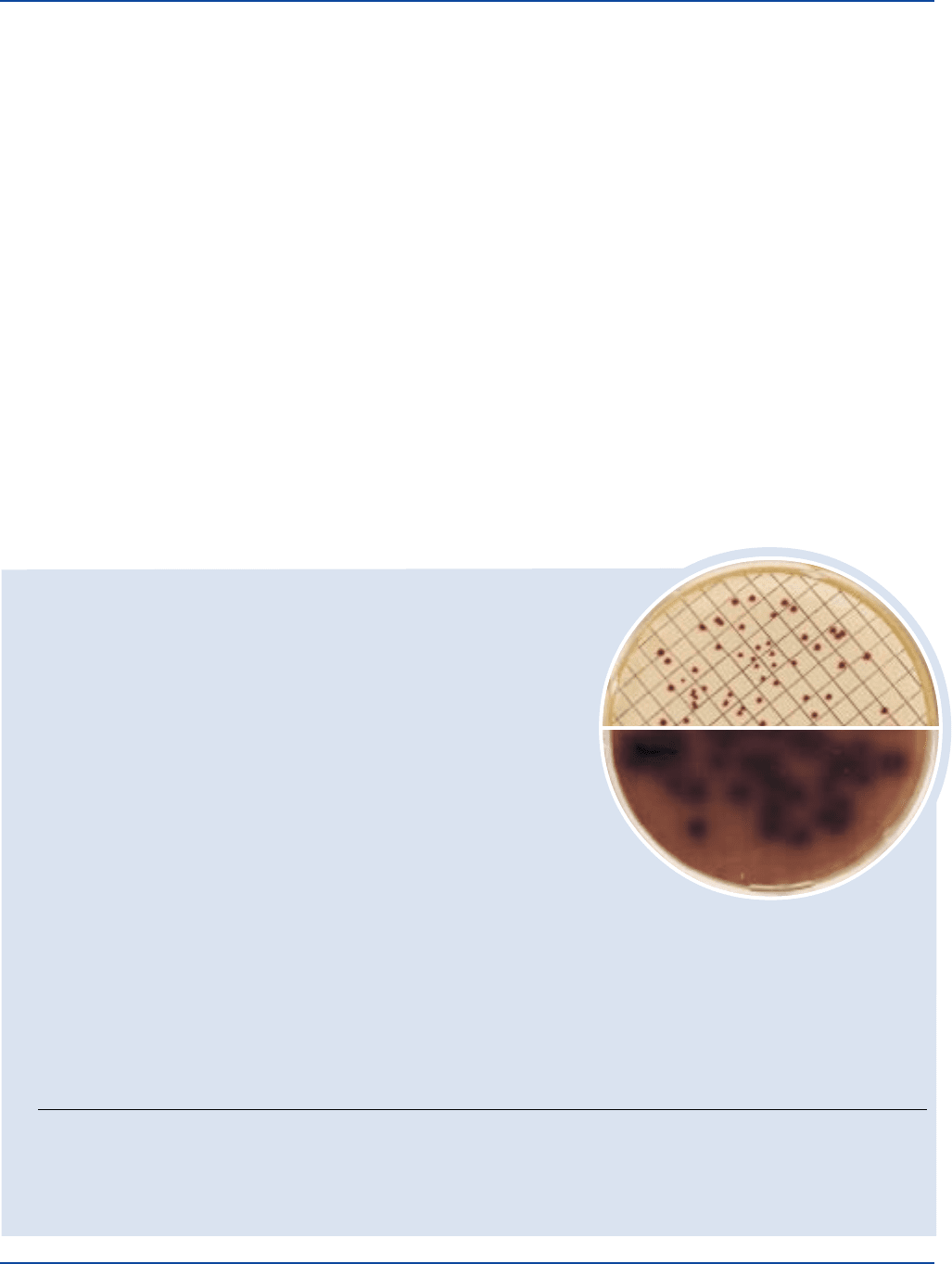
Esculin Iron Agar
Enterococcus faecalis
ATCC
®
29212
Cultural Response
Prepare m E Agar per label directions and pour into 9 x 50 mm plates. Dilute the test organisms and filter through membrane filters.
Place the filters on m E Agar plates and incubate the plates in an upright position for 48 hours at 41 ± 0.5°C. Remove the filters and
place over prepared Esculin Iron Agar plates. After 20 minutes incubation at 41 ± 0.5°C, count colonies giving positive esculin
reaction (formation of black or reddish brown precipitate).
ORGANISM ATCC
®
INOCULUM CFU/10 ml GROWTH ON m E AGAR REACTION ON ESCULIN IRON AGAR
Enterococcus faecalis 29212* 20-60 good/pink to red colonies black or red/brown ppt
Enterococcus faecalis 33186 20-60 good/pink to red colonies black or red/brown ppt
Escherichia coli 25922* 20-60 marked to complete inhibition inhibited
The cultures listed are the minimum that should be used for performance testing.
*These cultures are available as Bactrol
TM
Disks and should be used as directed in Bactrol Disks Technical Information.
User Quality Control
Identity Specifications
m E Agar
Dehydrated Appearance: Light beige, free-flowing, homogeneous.
Solution: 7.12% solution, soluble in distilled or deionized water
upon boiling. Light to medium amber with bluish cast,
very slightly opalescent.
Prepared Medium: Light to medium amber with blue cast, slightly opalescent.
Reaction of 7.12%
Solution at 25°C: pH 7.1 ± 0.2
Esculin Iron Agar
Dehydrated Appearance: Tan to dark tan, free-flowing, homogeneous.
Solution: 1.65%, soluble in distilled or deionized water upon boiling. Medium
amber with blue cast, very slightly opalescent without significant precipitate.
Prepared Medium: Medium amber with blue cast, slightly opalescent without precipitate.
Reaction of 1.65%
Solution at 25°C: pH 7.1 ± 0.2
m E Agar & Esculin Iron Agar Section II
m E Agar
Enterococcus faecalis
ATCC
®
29212

The Difco Manual 167
Formula
m E Agar
Formula Per Liter
Bacto Yeast Extract . . . . . . . . . . . . . . . . . . . . . . . . . . . . . . . 30 g
Bacto Peptone . . . . . . . . . . . . . . . . . . . . . . . . . . . . . . . . . . . 10 g
Sodium Chloride . . . . . . . . . . . . . . . . . . . . . . . . . . . . . . . . . 15 g
Esculin. . . . . . . . . . . . . . . . . . . . . . . . . . . . . . . . . . . . . . . . . . 1 g
Actidione
®
. . . . . . . . . . . . . . . . . . . . . . . . . . . . . . . . . . . . . 0.05 g
Sodium Azide . . . . . . . . . . . . . . . . . . . . . . . . . . . . . . . . . . 0.15 g
Bacto Agar . . . . . . . . . . . . . . . . . . . . . . . . . . . . . . . . . . . . . 15 g
Final pH 7.1 ± 0.2 at 25°C
Esculin Iron Agar
Formula Per Liter
Esculin. . . . . . . . . . . . . . . . . . . . . . . . . . . . . . . . . . . . . . . . . . 1 g
Ferric Ammonium Citrate . . . . . . . . . . . . . . . . . . . . . . . . . 0.5 g
Bacto Agar . . . . . . . . . . . . . . . . . . . . . . . . . . . . . . . . . . . . . 15 g
Final pH 7.1 ± 0.2 at 25°C
Precautions
1. For Laboratory Use.
2. m E Agar
HARMFUL BY INHALATION AND IF SWALLOWED.
(US) IRRITATING TO EYES, RESPIRATORY SYSTEM AND
SKIN. (US) Avoid contact with skin and eyes. Do not breathe dust.
Wear suitable protective clothing. Keep container tightly closed.
TARGET ORGAN(S): Cardiovascular, Lungs, Nerves
FIRST AID: In case of contact with eyes, rinse immediately
with plenty of water and seek medical advice. After contact with
skin, wash immediately with plenty of water. If inhaled,
remove to fresh air. If not breathing, give artificial respiration.
If breathing is difficult, give oxygen. Seek medical advice. If
swallowed seek medical advice immediately and show this
container or label.
3. Follow proper established laboratory procedures in handling and
disposing of infectious materials.
Storage
Store the dehydrated medium below 30°C. The dehydrated medium is
very hygroscopic. Keep container tightly closed.
Expiration Date
The expiration date applies to the product in its intact container
when stored as directed. Do not use a product if it fails to meet
specifications for identity and performance.
Procedure
Materials Provided
m E Agar
Esculin Iron Agar
Materials Required But Not Provided
Bacto TTC Solution 1%
Nalidixic acid
Indoxyl β-D glucoside (optional)
Sterile Petri dishes, 50 x 9 mm
Membrane filter equipment
Sterile pipettes
Sterile 47 mm, 0.45 µm, gridded membrane filters
Autoclave
Glassware
Dilution blanks
41°C incubator or waterbath
Fluorescent lamp
Magnifying lens
Method of Preparation
m E Agar
1. Suspend 7.12 grams in 100 ml distilled or deionized water.
2. Boil to dissolve completely.
3. Autoclave at 121°C for 15 minutes. Cool to 45°C.
4. Add 0.024 grams of nalidixic acid and 1.5 ml TTC Solution 1%
(0.015 grams triphenyl tetrazolium chloride).
5. Adjust to pH 7.1 if necessary.
6. Dispense 4-5 ml into 9 x 50 mm Petri dishes.
Note: Nalidixic acid is soluble in water with an alkaline pH.
Esculin Iron Agar
1. Suspend 1.65 grams in 100 ml distilled or deionized water.
2. Boil to dissolve completely.
3. Autoclave at 121°C for 15 minutes.
4. Dispense 4-5 ml into 9 x 50 mm Petri dishes.
Specimen Collection and Preparation
Collect water samples as described in Standard Methods for the
Examination of Water and Wastewater.
1
Test Procedure
1. Follow the membrane filter procedure described in Standard
Methods for the Examination of Water and Wastewater.
1
2. Choose a sample size so that 20-60 colonies will result.
3. Place the filter on an m E Agar plate and incubate for 48 hours
at 41 ± 0.5°C.
4. After incubation, remove the filter from m E Agar and place
it on Esculin Iron Agar plate. Retain at room temperature for
approximately 20-30 minutes.
5. Incubate Esculin Iron Agar at 41 ± 0.5°C for 20 minutes.
Results
Pink to red enterococci develop a black or reddish-brown
precipitate on the underside of the filter.
1
Count colonies using a
fluorescent lamp and a magnifying lens.
1
Report results as estimated
number or organisms per 100 ml of water.
Limitations of the Procedure
1. Since the nutritional requirements of organisms vary, some
strains may be encountered that fail to grow or grow poorly on
this medium.
2. m E Agar and Esculin Iron Agar should be used in sequence.
Section II m E Agar & Esculin Iron Agar
168 The Difco Manual
Escherichia coli
ATCC
®
25922
Uninoculated
tube
3. Incubation at 41°C is recommended.
4. Approximately 10% false-positive esculin reactions may be expected.
When used as m EI Agar, U.S. EPA reports a 6.0% false positive
and 6.5% false negative rate with mE Agar.
References
1. Eaton, A. D., L. S. Clesceri, and A. E. Greenberg (ed.). 1995.
Standard methods for the examination of water and wastewater,
19th ed. American Public Health Association, Washington, D.C.
2. Slanetz, L. W., and C. H. Bartley. 1957. Numbers of enterococci
in water, sewage, and feces determined by the membrane filter
technique with an improved medium. J. Bacteriol. 74:591-595.
3. ASTM. 1996. Annual book of ASTM standards. Section 11,
Water and environmental technology. PCN: 01-110296-16.
ASTM, West Conshohocken, PA.
4. Kenner, R. A., H. F. Clark, and P. W. Kabler. 1960. Fecal
streptococci I. Cultivation and enumeration of streptococci in
surface waters. Appl. Microbiol. 9:15-20.
5. Isenberg, H. D., D. Goldberg, and J. Sampson. 1970.
Laboratory studies with a selective enterococcus medium. Appl.
Microbiol. 20:433-436.
6. Levin, M. A., J. R. Fischer, and V. J. Cabelli. 1975. Membrane
filter technique for enumeration of enterococci in marine waters.
Appl. Microbiol. 30:66-70.
7. Cabelli, V. J. 1981. Health effects criteria for marine recreational
waters. U.S. Environmental Protection Agency. EPA-600/1-80-031.
Cincinnati, OH.
8. Dufour, A. P. 1983. Health effects criteria for fresh recreational
waters. U.S. Environmental Protection Agency. Cincinnati, OH.
9. U.S. Environmental Protection Agency. 1997. EPA method 1600:
Membrane filter test method for enterococci in water. U.S. Environ-
mental Protection Agency. EPA-821-R-97-004. Washington, D.C.
Packaging
m E Agar 100 g 0333-15
500 g 0333-17
Esculin Iron Agar 100 g 0488-15
EC Medium Section II
Bacto
®
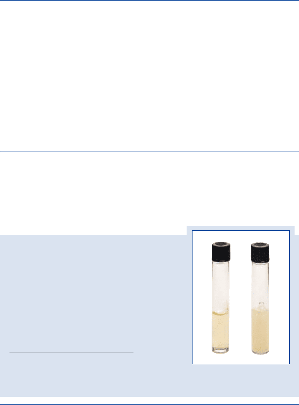
EC Medium
User Quality Control
Identity Specifications
Dehydrated Appearance Light beige, free-flowing,
homogeneous.
Solution: 3.7% solution, soluble in distilled or
deionized water. Light amber, clear.
Prepared Medium: Light amber, clear.
Reaction of 3.7%
Solution at 25°C: pH 6.9 ± 0.2
Cultural Response
Prepare EC Medium per label directions. Inoculate tubes with
the test organisms, and incubate at 44.5 ± 0.2°C for 24 ± 2
hours. Read tubes for growth and gas production.
INOCULUM GAS
ORGANISM ATCC
®
CFU GROWTH PRODUCTION
Enterococcus faecalis 19433 1,000 inhibited –
Escherichia coli 25922* 1,000 good +
Escherichia coli 8739 1,000 good +
The cultures listed are the minimum that should be used for performance testing.
*This culture is available as Bactrol
™
Disk and should be used as directed in Bactrol Disk Technical Information.
Intended Use
Bacto EC Medium is used for differentiating and enumerating
coliforms in water, wastewater, shellfish and foods.
Also Known As
EC Medium is also referred to as EC Broth. EC is an abbreviation for
Escherichia coli.
Summary and Explanation
EC Medium was developed by Hajna and Perry1 in an effort to
improve the methods for the detection of the coliform group and
E. coli. This medium consists of a buffered lactose broth with the
addition of 0.15% Bile Salts No. 3. Growth of spore forming
bacteria and fecal streptococci is inhibited by the bile salts, while
growth of E.coli is enhanced by its presence. The medium can be

The Difco Manual 169
Section II EC Medium
used at 37°C for the detection of coliform organisms or at 45.5°C
for the isolation of E. coli.
In a further evaluation of EC Medium and Lauryl Tryptose Broth,
Perry and Hajna
2
reported the results obtained from eleven
different laboratories examining a variety of waters, milk and
shellfish. The results indicate that the media are highly specific
for coliform bacteria. Fishbein and Surkiewicz
3
used the EC
confirmation test for recovery of E. coli from frozen foods and nut
meats. This study
3
showed that the test is optimal when conducted
at 45.5°C, with incubation limited to 24 hours.
EC Medium is employed in elevated-temperature tests for
distinguishing organisms of the total coliform group that also
belong to the fecal coliform group.
4
The fecal coliform test, using
EC Medium, is applicable to investigations of drinking water,
stream pollution, raw water sources, wastewater treatment systems,
bathing waters, seawaters and general water-quality monitoring.
Prior enrichment in presumptive media is required for optimum
recovery of fecal coliforms when using EC Medium.
EC Medium is used in standard methods for food and water testing.
4,5,6
Principles of the Procedure
Tryptose provides the nitrogen, vitamins and amino acids in
EC Medium. Lactose is the carbon source. Bile Salts No. 3 is the
selective agent against gram positive bacteria, particularly
bacilli and fecal streptococci. Dipotassium Phosphate and
Monopotassium Phosphate are the buffering agents. Sodium Chloride
maintains the osmotic balance of the medium.
Formula
EC Medium
Formula Per Liter
Bacto Tryptose . . . . . . . . . . . . . . . . . . . . . . . . . . . . . . . . . . 20 g
Bacto Lactose . . . . . . . . . . . . . . . . . . . . . . . . . . . . . . . . . . . . 5 g
Bacto Bile Salts No. 3 . . . . . . . . . . . . . . . . . . . . . . . . . . . . 1.5 g
Dipotassium Phosphate . . . . . . . . . . . . . . . . . . . . . . . . . . . . 4 g
Monopotassium Phosphate. . . . . . . . . . . . . . . . . . . . . . . . . 1.5 g
Sodium Chloride . . . . . . . . . . . . . . . . . . . . . . . . . . . . . . . . . . 5 g
Final pH 6.9 ± 0.2 at 25°C
Precautions
1. For Laboratory Use.
2. Follow proper established laboratory procedures in handling and
disposing of infectious materials.
Storage
Store the dehydrated medium below 30°C. The dehydrated medium is
very hygroscopic. Keep container tightly closed.
Expiration Date
The expiration date applies to the product in its intact container
when stored as directed. Do not use a product if it fails to meet the
specifications for identity and performance.
Procedure
Materials Provided
EC Medium
Materials Required But Not Provided
Glassware
Fermentation vials
Autoclave
Incubator or waterbath
Method of Preparation
1. Suspend 37 grams in 1 liter distilled or deionized water.
2. Warm slightly to dissolve completely.
3. Dispense into tubes containing inverted fermentation vials.
4. Autoclave at 121°C for 15 minutes.
Specimen Collection and Preparation
Obtain and process specimens according to the procedures established
by laboratory policy or standard methods.
4,5,6
Test Procedure
Follow the methods and procedures as stated in standard methods.
4,5,6
Results
Gas production with growth in EC Medium within 24 hours or less is
considered a positive fecal coliform reaction. Failure to produce gas
with little or no growth, is a negative reaction.
4
Limitations of the Procedure
1. Since the nutritional requirements of organisms vary, some strains
may be encountered that fail to grow or grow poorly on this medium.
2. False-negative reactions in recovering coliforms from water supplies
can occur due to low pH, refrigeration and use of bactericidal or
bacteriostatic agents.
7
References
1. Hajna and Perry. 1943. Am. J. Public Health 33:550.
2. Hajna and Perry. 1944. Am. J. Public Health 34:735.
3. Fishbein and Surkiewicz. 1964. Appl. Microbiol. 12:127.
4. Eaton, A. D., L. S. Clesceri, and A. E. Greenberg (ed.). 1995.
Standard methods for the examination of water and wastewater,
19th ed. American Public Health Association, Washington, D.C.
5. Association of Official Analytical Chemists. 1995. Bacteriological
analytical manual, 8th ed. AOAC International, Gaithersburg, MD.
6. Vanderzant, C., and D. F. Splittstoesser (ed.). 1992.
Compendium of methods for the microbiological examination of
food, 3rd ed. American Public Health Association, Washington, D.C.
7. Ray, B. 1986. Impact of bacterial injury and repair in food
microbiology: Its past, present and future. J. Food Prot. 49:651.
Packaging
EC Medium 100 g 0314-15
500 g 0314-17
10 kg 0314-08
170 The Difco Manual
Escherichia coli
ATCC
®
25922
Bacto
®
EC Medium with MUG
User Quality Control
Identity Specifications
Dehydrated Appearance: Light beige, free-flowing, homogeneous.
Solution: 3.71% solution, soluble in distilled or deionized water; light amber, clear.
Prepared Medium: Light amber, clear.
Reaction of 3.71%
Solution at 25°C: pH 6.9 ± 0.2
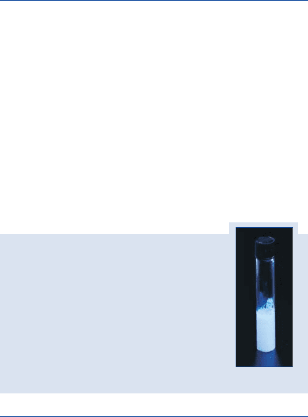
Cultural Response
Prepare EC Medium with MUG per label directions. Inoculate tubes in duplicate. Incubate the
first set at 35 ± 2°C for 24 hours and the second set at 44.5 ± 0.2°C. Read fluorescence under
a long-wave UV light.
INOCULUM GROWTH AT GROWTH AT
ORGANISM ATCC
®
CFU 35°C/GAS 44.5°C/GAS FLUORESCENCE
Enterobacter aerogenes 13048* 100-1,000 good/+ inhibited/– –
Escherichia coli 25922* 100-1,000 good/+ good/+ +
Enterococcus faecalis 19433* 100-1,000 inhibited/– inhibited/– –
The cultures listed are the minimum that should be used for performance testing.
*These cultures are available as Bactrol
™
Disks and should be used as directed in Bactrol Disks
Technical Information.
Intended Use
Bacto EC Medium with MUG is used for detecting Escherichia coli
in water, food and milk.
Also Known As
EC is an abbreviation for Escherichia coli.
Summary and Explanation
EC Medium was developed by Hajna and Perry
1
to improve the
methods for the detection of coliforms and E. coli. This medium
consists of a buffered lactose broth with the addition of 0.15% Bile
Salts No. 3. Growth of spores formers and fecal streptococci were
inhibited by the bile salts, while growth of E. coli is enhanced. EC
Medium with MUG is the same formula as EC Medium with the
addition of 4-methylumbelliferyl-ß-D-glucuronide.
Feng and Hartman
2
developed a rapid assay for E. coli by incorporating
4-methylumbelliferyl-ß-D-glucuronide (MUG) into Lauryl Tryptose
Broth at a final concentration of 100 µg/ml. Robison
3
compared the
fluorogenic assay with present methodology and found that total
agreement between the two methods was 94.8%. Moburg
4
determined
the amount of MUG could be reduced to a final concentration of
50 µg/ml without adversely affecting results. Koburger and Miller
5
recommended the incorporation of MUG into EC Broth for use in
testing shellfish.
EC Medium with MUG is prepared according to the formula specified
by US EPA
6
and standard methods for water and food testing.
7,8
Principles of the Procedure
Tryptose provides the nitrogen, vitamins and amino acids in EC
Medium with MUG. Lactose is the carbon source in this medium. Bile
Salts No. 3 is the selective agent against gram-positive bacteria,
particularly bacilli and fecal streptococci. Dipotassium Phosphate and
Monopotassium Phosphate are buffering agents. Sodium Chloride
maintains the osmotic balance of the medium.
E. coli produces the enzyme glucuronidase that hydrolyzes MUG to
yield a fluorogenic product that is detectable under long-wave (366 nm)
UV light. The addition of MUG to EC Medium provides another
criterion, in addition to growth response and gas production, to
determine the presence of E. coli in food and environmental samples.
Formula
EC Medium with MUG
Formula Per Liter
Bacto Tryptose . . . . . . . . . . . . . . . . . . . . . . . . . . . . . . . . . . 20 g
Bacto Lactose . . . . . . . . . . . . . . . . . . . . . . . . . . . . . . . . . . . . 5 g
Bacto Bile Salts No. 3 . . . . . . . . . . . . . . . . . . . . . . . . . . . . 1.5 g
Dipotassium Phosphate . . . . . . . . . . . . . . . . . . . . . . . . . . . . 4 g
Monopotassium Phosphate. . . . . . . . . . . . . . . . . . . . . . . . . 1.5 g
Sodium Chloride . . . . . . . . . . . . . . . . . . . . . . . . . . . . . . . . . . 5 g
MUG (4-methylumbelliferyl-ß-D-glucuronide) . . . . . . . 0.05 g
Final pH 6.9 ± 0.2 at 25°C
Precautions
1. For Laboratory Use.
2. Follow proper established laboratory procedures in handling and
disposing of infectious materials.
EC Medium with MUG Section II

The Difco Manual 171
Storage
Store the dehydrated medium below 30°C. The dehydrated medium is
very hygroscopic. Keep container tightly closed.
Expiration Date
The expiration date applies to the product in its intact container when
stored as directed. Do not use a product if it fails to meet specifications
for identity and performance.
Procedure
Materials Provided
EC Medium with MUG
Materials Required But Not Provided
Test tubes
Fermentation vials
Sterile pipettes
Incubator 35°C, 44.5°C
Long-wave UV lamp
Autoclave
Method of Preparation
1. Suspend 37.1 grams in 1 liter distilled or deionized water.
2. Warm slightly to dissolve completely.
3. Dispense into test tubes containing inverted fermentation vials.
4. Autoclave at 121°C for 15 minutes.
5. Before opening the autoclave, allow the temperature to drop below
75°C to avoid entrapping air bubbles in the fermentation vials.
Specimen Collection and Preparation
Collect food, water or other environmental samples in accordance with
recommended procedures.
6,7,8
Test Procedure
Follow the methods and procedures as stated in appropriate references.
6,7,8
Results
Following incubation, observe tubes for growth, production of gas and
fluorescence. Positive gas production is demonstrated by displacement
of the medium from the fermentation vial. Positive MUG reactions
exhibit a bluish fluorescence under long-wave (approximately 366 nm)
UV light. Typical strains of E. coli are positive for both gas production
and fluorescence. Non-E. coli coliforms that grow may exhibit
fluorescence but will not produce gas.
Limitations of the Procedure
1. Since the nutritional requirements of organisms vary, some strains may
be encountered that fail to grow or grow poorly on this medium.
2. Strains of E. coli that fail to grow in EC Medium with MUG, fail to
produce gas, or fail to produce glucuronidase may infrequently be
encountered.
3. Strains of Salmonella, Shigella and Yersinia that produce glucu-
ronidase may be encountered. These strains must be distinguished
from E. coli on the basis of other parameters, i.e., gas production,
growth at 44.5°C.
4. The presence of endogenous glucuronidase in shellfish samples
may cause false positive fluorescent reactions at the presumptive
stage. To prevent this problem, the use of EC Medium with MUG
in the confirmatory stage has been recommended.
5
References
1. Hajna and Perry. 1943. Am. J. Public Health 33:550.
2. Feng, P. C. S., and P. A. Hartman. 1982. Fluorogenic assays for
immediate confirmation of Escherichia coli. Appl. Environ.
Microbiol. 43:1320-1329.
3. Robison, B. J. 1984. Evaluation of a fluorogenic assay for detection
of Escherichia coli in foods. App. Environ. Microbiol. 48:285-288.
4. Moberg, L. J. 1985. Fluorogenic assay for rapid detection of
Escherichia coli in food. Appl. Environ. Microbiol. 50:1383-1387.
5. Koburger, J. A., and M. L. Miller. 1985. Evaluation of a
fluorogenic MPN procedure for determining Escherichia coli in
oysters. J. Food Prot. 48:244-245.
6. Federal Register. 1991. National primary drinking water regulation;
analytical techniques; coliform bacteria. Fed. Regist. 56:636-643.
7. Eaton, A. D., L. S. Clesceri, and A. E. Greenberg (ed.). 1995.
Standard methods for the examination of water and wastewater,
19th ed. American Public Health Association, Washington, D.C.
8. Vanderzant, C., and D. F. Splittstoesser (ed.). 1992. Compen-
dium of methods for the microbiological examination of food,
3rd ed. American Public Health Association, Washington, D.C.
Packaging
EC Medium with MUG 100 g 0022-15
500 g 0022-17
Section II EE Broth Mossel
Bacto
®
EE Broth Mossel
Intended Use
Bacto EE Broth Mossel is used for selectively enriching and detecting
Enterobacteriaceae, particularly from foods.
Summary and Explanation
EE Broth Mossel is prepared according to the formula of Mossel,
Visser and Cornelissen.
1
The formula contains dextrose to facilitate
growth of most Enterobacteriaceae, thus insuring the detection of
Salmonella and other lactose- negative organisms. EE Broth Mossel
should be used as an enrichment broth, followed by a selective
medium, e.g., Violet Red Bile Agar.
The enumeration of Enterobacteriaceae is of great concern in monitoring
the sanitary condition of food. Enterobacteriaceae can be injured in
food-processing procedures, which include exposure to low temperatures,
sub-marginal heat, drying, radiation, preservatives or sanitizers.
2
Recovery relies on proper resuscitation of damaged cells.
Principles of the Procedure
Tryptose provides nitrogen, vitamins and amino acids. Dextrose
is a carbon source. Disodium Phosphate and Monopotassium
172 The Difco Manual
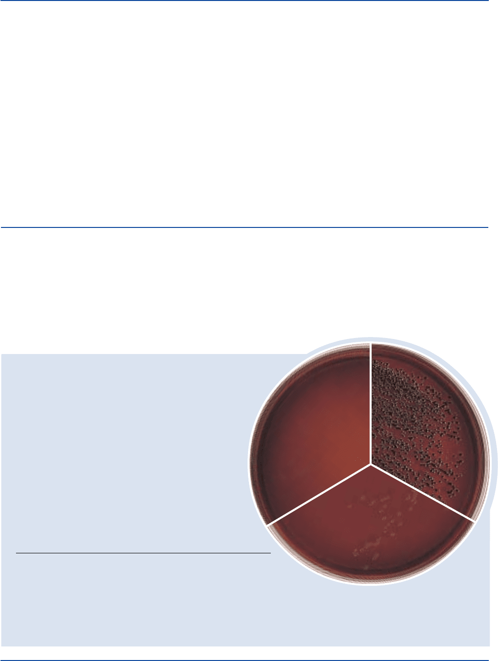
Escherichia coli
ATCC
®
25922
Uninoculated
tube
Phosphate are buffering agents. Brilliant Green and Oxgall are
selective agents.
Formula
EE Broth Mossel
Formula Per Liter
Bacto Tryptose . . . . . . . . . . . . . . . . . . . . . . . . . . . . . . . . . . 10 g
Bacto Dextrose . . . . . . . . . . . . . . . . . . . . . . . . . . . . . . . . . . . 5 g
Disodium Phosphate . . . . . . . . . . . . . . . . . . . . . . . . . . . . . . . 8 g
Monopotassium Phosphate. . . . . . . . . . . . . . . . . . . . . . . . . . 2 g
Brilliant Green . . . . . . . . . . . . . . . . . . . . . . . . . . . . . . . 0.0135 g
Bacto Oxgall . . . . . . . . . . . . . . . . . . . . . . . . . . . . . . . . . . . . 20 g
Final pH 7.2± 0.2 at 25°C
Precautions
1. For Laboratory Use.
2. IRRITANT. IRRITATING TO EYES, RESPIRATORY SYSTEM
AND SKIN. Avoid contact with skin and eyes. Do not breathe dust.
Wear suitable protective clothing. Keep container tightly closed.
FIRST AID: In case of contact with eyes, rinse immediately with
plenty of water and seek medical advice. After contact with skin,
wash immediately with plenty of water. If inhaled, remove to fresh
air. If not breathing, give artificial respiration. If breathing is diffi-
cult, give oxygen. Seek medical advice. If swallowed seek medical
advice immediately and show this container or label.
3. Follow proper established laboratory procedures in handling and
disposing of infectious materials.
Storage
Store the dehydrated medium below 30°C. The dehydrated medium is
very hygroscopic. Keep container tightly closed.
User Quality Control
Identity Specifications
Dehydrated Appearance: Light green, homogeneous, free-flowing.
Solution: 4.5% solution, soluble in distilled or deionized water;
emerald green and clear.
Prepared Medium: Emerald green, clear.
Reaction of 4.5%
Solution at 25°C pH 7.2 ± 0.2
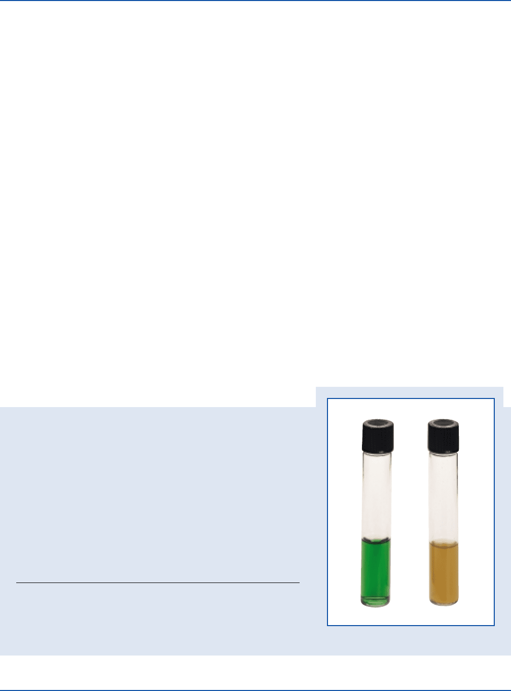
Cultural Response
Prepare EE Broth Mossel per label directions. Inoculate the medium and incubate
at 35 ± 2°C for 18-24 hours and up to 48 hours if necessary.
INOCULUM ACID
ORGANISM ATCC
®
CFU GROWTH PRODUCTION
Escherichia coli 25922* 100-1,000 good + (yellow)
Staphylococcus aureus 25923* 100-1,000 inhibited –
The cultures listed are the minimum that should be used for performance testing.
*These cultures are available as Bactrol
™
Disks and should be used
as directed in Bactrol Disks Technical Information.
Expiration Date
The expiration date applies to the product in its intact container when
stored as directed. Do not use a product if it fails to meet specifications
for identity and performance.
Procedure
Materials Provided
EE Broth Mossel
Materials Required But Not Provided
Glassware
Incubator (35°C)
Waterbath
Method of Preparation
1. Dissolve 45 grams in 1 liter distilled or deionized water.
2. Dispense 120 ml amounts into 250 ml flasks.
3. Heat at 100°C (waterbath or flowing steam) for 30 minutes. Do
Not Autoclave.
Specimen Collection and Preparation
Obtain and process specimens according to the techniques and
procedures established by laboratory policy.
Test Procedure
1. Inoculate flasks of EE Broth Mossel with approximately 10 grams
of homogenized food or other material to be tested.
2. Shake the inoculated medium thoroughly for a few seconds to mix well.
3. Incubate for a total of 20-24 hours at 35-37°C. Shake the flasks
after the first 3 hours of incubation.
EE Broth Mossel Section II
The Difco Manual 173
4. Prepare plates such as Violet Red Bile Agar for streaking. To ensure
recovery of dextrose fermenters, add 1% dextrose before boiling.
5. Streak a loopful of the enrichment culture onto the prepared plates.
6. Incubate the plates for 18-24 hours at 35-37°C. Examine for the
presence of coliforms which appear pink to purplish-red on Violet
Red Bile Agar. The color of coliform colonies may vary if a different
medium is used.
For a complete discussion on Enterobacteriaceae in food testing, refer
to procedures in Standard Methods.
3,4
Results
Acid production causes the color of EE Broth Mossel to become
yellow. A negative reaction results in no color change and the medium
remains green.
Limitations of the Procedure
1. Since the nutritional requirements of organisms vary, some strains
may be encountered that fail to grow or grow poorly on this medium.
References
1. Mossel, D. A. A., M. Vissar, and A. M. R. Cornellisen. 1963. The
examination of foods for Enterobacteriaceae using a test of the
type generally adopted for the detection of salmonellae. J. Appl.
Bacteriol. 26:444-452.
2. Hartman, P. A., and S. A. Minnich. 1981. Automation for rapid
identification of Salmonella in foods. J. Food Prot. 44:385-386.
3. Association of Official Analytical Chemists. 1995. Bacteriological
analytical manual, 8th ed. AOAC International, Gaithersburg, MD.
4. Vanderzant, C., and D. F. Splittstoesser (ed.). 1992.
Compendium of methods for the microbiological examination
of food, 3rd ed. American Public Health Association,
Washington, D.C.
Packaging
EE Broth Mossel 500 g 0566-17
10 kg 0566-08
Section II EMB Agar
Bacto
®
EMB Agar
User Quality Control
Identity Specifications
Dehydrated Appearance: Pinkish purple, free flowing, homogeneous.
Solution: 3.6% solution, soluble in distilled or
deionized water on boiling. Solution
is green with orange cast, opalescent
with a uniform flocculent precipitate.
Prepared Plates: Purple with a greenish-orange cast,
opalescent, may have a fine
precipitate.
Reaction of 3.6%
Solution at 25°C: pH 7.2 ± 0.2
Cultural Response
Prepare EMB Agar per label directions. Inoculate and incubate
at 35 ± 2°C for 18-24 hours.
INOCULUM
ORGANISM ATCC
®
CFU GROWTH APPEARANCE
Enterococcus 29212* 1,000 partial colorless
faecalis inhibition
Escherichia 25922* 100-1,000 good blue-black w/dark centers
coli and green metallic sheen
Salmonella 14028* 100-1,000 good colorless to amber
typhimurium
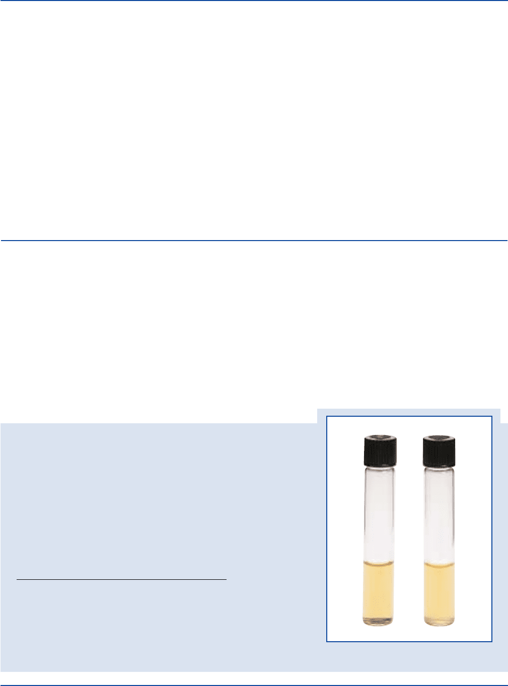
Escherichia coli
ATCC
®
25922
Uninoculated
plate
Salmonella typhimurium
ATCC
®
14028
The cultures listed are the minimum that should be used for performance testing.
*These cultures are available as Bactrol
™
Disks and should be used as directed in Bactrol Disks Technical Information.
Intended Use
Bacto EMB Agar is used for isolating and differentiating gram-
negative enteric bacilli.
Also Known As
EMB Agar is also known as Eosin Methylene Blue Agar
Summary and Explanation
The original Eosin Methylene Blue Agar was the formulation of Holt-
Harris and Teague.
1
The use of eosin and methylene blue as indicators
gave sharp and distinct differentiation between colonies of lactose
fermenting and nonfermenting organisms. Sucrose was included in the
medium to detect members of the coliform group that fermented
sucrose more readily than lactose. Lactose-positive colonies were black

174 The Difco Manual
or possessed dark centers with transparent, colorless peripheries.
Lactose- or sucrose-negative colonies were colorless. The Eosin
Methylene Blue Agar of Holt-Harris and Teague had definite
advantages over the Fuchsin Sulfite Agar of Endo. The EMB Agar
formulation was more sensitive, more accurate, more stable, and gave
an earlier differentiation between the lactose fermenters and lactose
and sucrose nonfermenters.
Two years after Holt-Harris and Teague had introduced their new
medium, Levine
2
described an Eosin Methylene Blue Agar for
differentiating fecal and nonfecal coliforms. Levine’s medium
differentiated salmonellae and other lactose nonfermenters from the
coliform organisms.
EMB Agar is a combination of the Levine and the Holt-Harris and
Teague formulae. EMB Agar is selective due to the presence of inhibitors
and differential based on the ability of some organisms to ferment
carbohydrates with the absorption of eosin and methylene blue.
EMB Agar is recommended for use in examining clinical specimens
for enteric pathogens.
3,4,5
The medium enables the isolation and differ-
entiation of gram-negative enteric bacilli.
Principles of the Procedure
Peptone is a source of nitrogen and other nutrients in the formulation.
Eosin and methylene blue are dyes which combine to form a precipitate
at an acid pH. The dyes act both as pH indicators and inhibitors.
Gram-positive bacteria are partially inhibited on the medium. Lactose
and Sucrose are fermentable carbohydrates. Phosphate acts as a buffer.
Bacto Agar is a solidifying agent.
Formula
EMB Agar
Formula Per Liter
Bacto Peptone . . . . . . . . . . . . . . . . . . . . . . . . . . . . . . . . . . . 10 g
Bacto Lactose . . . . . . . . . . . . . . . . . . . . . . . . . . . . . . . . . . . . 5 g
Bacto Sucrose . . . . . . . . . . . . . . . . . . . . . . . . . . . . . . . . . . . . 5 g
Dipotassium Phosphate . . . . . . . . . . . . . . . . . . . . . . . . . . . . 2 g
Bacto Agar . . . . . . . . . . . . . . . . . . . . . . . . . . . . . . . . . . . . 13.5 g
Eosin Y . . . . . . . . . . . . . . . . . . . . . . . . . . . . . . . . . . . . . . . . 0.4 g
Methylene Blue . . . . . . . . . . . . . . . . . . . . . . . . . . . . . . . . 0.065 g
Final pH 7.2 ± 0.2 at 25°C
Precautions
1. For Laboratory Use.
2. Follow proper established laboratory procedure in handling and
disposing of infectious materials.
Storage
Store the dehydrated medium below 30°C. The dehydrated medium is very
hygroscopic. Keep container tightly closed. Store prepared plates at 2-8°C.
Expiration Date
Expiration date applies to the product in its intact container when stored
as directed. Do not use a product if it fails to meet specifications for
identity and performance.
Procedure
Materials Provided
EMB Agar
Materials Required But Not Provided
Flasks with closures
Distilled or deionized water
Bunsen burner or magnetic hot plate
Autoclave
Waterbath (45-50)°C
Petri dishes
Incubator (35°C)
Method of Preparation
1. Suspend 36 grams in 1 liter distilled or deionized water.
2. Heat to boiling to dissolve completely.
3. Autoclave at 121°C for 15 minutes. Avoid overheating.
4. Cool to 45-50°C in a waterbath.
5. Dispense into sterile Petri dishes. Evenly disperse the precipitate
when dispensing.
Specimen Collection and Preparation
1. Collect specimens in sterile containers or with sterile swabs and
transport immediately to the laboratory following recommended
guidelines.
3,4,5
2. For specific information about specimen preparation and inoculation
of clinical specimens, consult appropriate references.
3,4,5
Test Procedure
For isolation of enteric pathogens from clinical specimens, inoculate
fecal specimens and rectal swabs onto a small area of one quadrant
of the EMB Agar plate and streak for isolation. This will permit
development of discrete colonies. Incubate plates at 35°C. Examine
plates at 24 hours and again at 48 hours for colonies with characteristic
morphologies associated with potential pathogens.
Results
Salmonella and Shigella colonies are translucent and amber colored or
colorless. Coliforms that use lactose and/or sucrose produce blue-black
colonies with dark centers and greenish metallic sheen. Other coliforms
such as Enterobacter form mucoid, pink colonies. Strains of
Enterococcus faecalis are partially inhibited on this medium and
appear as colorless colonies.
Limitations of the Procedure
1. EMB Agar is only moderately inhibitory. Some staphylococci,
streptococci and yeast may grow. They will appear as small,
pinpoint colonies. Gram-negative nonfermenting bacilli may grow
and appear as non-lactose fermenters. Biochemical tests are necessary
for further identification to genus or species.
6
2. Some strains of Salmonella and Shigella may not grow on EMB
Agar.
6
It is recommended that a nonselective, differential medium
(MacConkey Agar or Hektoen Enteric Agar) and a selective medium
(Bismuth Sulfite Agar, SS Agar or Desoxycholate Citrate Agar) be
run in parallel with EMB Agar.
3. Sterilization reduces the methylene blue, leaving the medium
orange in color. The normal purple color of the medium may be
restored by gentle mixing. If the reduced medium is not shaken to
oxidize the methylene blue, a dark zone beginning at the top and
extending downward through the medium will gradually appear.
The sterilized medium normally contains a flocculent precipitate
which should not be removed. By cooling to 50°C and gently
EMB Agar Section II
The Difco Manual 175
mixing the medium before pouring it into plates, the flocculation
will be finely dispersed.
4. Greenish metallic sheen is not always present. The presence of the
greenish metallic sheen is not diagnostic for E. coli.
6
5. Store and incubate EMB Agar plates in the dark. Visible light can
alter the ability of the medium to support microbial growth,
especially of Proteus spp.
7
References
1. Holt-Harris, J. E., and O. Teague. 1916. A new culture medium
for the isolation of Bacillus typhosa from stools. J. Infect. Dis.
18:596-600.
2. Levine, M. M. 1918. Differentiation of E. coli and B. aerogenes
on a simplified Eosin-Methylene Blue Agar. J. Infect. Dis. 23:43.
3. Gray, L. D. 1995. Escherichia, Salmonella, Shigella and Yersinia,
p. 450-456. In P. R. Murray, E. J. Baron, M. A. Pfaller, F. C.
Tenover, and R. H. Yolken (ed.), Manual of clinical microbiology,
6th ed. American Society for Microbiology, Washington, D.C.
4. Pezzlo, M. (ed.). 1992. Aerobic bacteriology, p. 1.0.1-1.20.47. In
H. D. Isenberg, (ed.), Clinical microbiology procedures handbook,
vol. 1. American Society for Microbiology, Washington, D.C.
5. Baron, E. J., L. R. Peterson, and S. M. Finegold. 1994. Bailey &
Scott’s diagnostic microbiology, 9th ed. Mosby-Year Book, Inc.,
St. Louis, MO.
6. MacFaddin, J. F. 1985. Media for isolation-cultivation-
identification-maintenance of medical bacteria, vol. 1. Williams &
Wilkins, Baltimore, MD.
7. Girolami, R. L., and J. M. Stamm. 1976. Inhibitory effect of light
on growth-supporting properties of Eosin Methylene Blue Agar.
Appl. Environ. Microbiol. 31:141.
Packaging
EMB Agar 100 g 0076-15
500 g 0076-17
2 kg 0076-07
10 kg 0076-08
Section II EVA Broth
Bacto
®
EVA Broth
Enterococcus faecalis
ATCC
®
29212
Uninoculated
tube
User Quality Control
Identity Specifications
Dehydrated Appearance: Light beige, free-flowing, homogeneous.
Solution: 3.58% solution, soluble in distilled or deionized water.
Solution is light amber, clear to very slightly opalescent.
Reaction of 3.58%
Solution at 25°C: pH 7.0 ± 0.2
Cultural Response
Prepare EVA Broth per label directions. Inoculate and incubate the tubes at
35 ± 2°C for 18-48 hours.
INOCULUM
ORGANISM ATCC
®
CFU GROWTH
Enterococcus faecalis 19433* 100-1,000 good
Enterococcus faecalis 29212* 100-1,000 good
Escherichia coli 25922* 1,000-2,000 inhibited
The cultures listed are the minimum that should be used for performance testing.
*These cultures are available as Bactrol
™
Disks and should be used as directed in
Bactrol Disks Technical Information.
Intended Use
Bacto EVA Broth is used for detecting and confirming enterococci in
water and other specimens as an indication of fecal contamination.
Also Known As
EVA Broth is also known as Ethyl Violet Azide Broth.
Summary and Explanation
The presence of enterococci in water and other specimens indicates
fecal contamination. Mallmann and Seligmann
1
compared various
enrichment media for detecting fecal streptococci and found that Azide
Dextrose Broth presumptively identified the streptococci. However,
because gram-positive bacteria other than enterococci grow in the
medium, confirmation is necessary. Litsky et al.
2
studied various dyes
and selective agents and formulated a medium using ethyl violet and
sodium azide as selective agents. The medium known as Ethyl
Violet Azide (EVA) Broth is specific for enterococci. In conjunction
with Azide Dextrose Broth, EVA Broth is used to confirm the presence
of enterococci.
Principles of the Procedure
EVA Broth contains Tryptose as a source of carbon, nitrogen, vitamins
and minerals. Dextrose is the carbohydrate. Sodium Azide and
