BD Diagnostic Systems (publ.). Difco Manual (Manual of Microbiological Culture)
Подождите немного. Документ загружается.


156 The Difco Manual
Chloride maintains the osmotic balance of the medium. Sodium
Desoxycholate and Sodium Citrate inhibit growth of gram-positive
bacteria. Neutral Red is a pH indicator. Bacto Agar is a solidifying
agent.
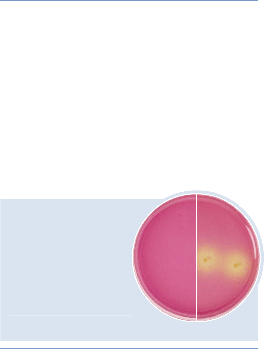
Differentiation of enteric bacilli is based on fermentation of lactose.
Bacteria that ferment lactose produce acid and, in the presence of
neutral red, form red colonies. Bacteria that do not ferment lactose
form colorless colonies. The majority of normal intestinal bacteria
ferment lactose (red colonies) while Salmonella and Shigella species
do not ferment lactose (colorless colonies).
Formula
Bacto Desoxycholate Lactose Agar
Formula Per Liter
Bacto Proteose Peptone . . . . . . . . . . . . . . . . . . . . . . . . . . . 10 g
Bacto Lactose . . . . . . . . . . . . . . . . . . . . . . . . . . . . . . . . . . . 10 g
Sodium Desoxycholate . . . . . . . . . . . . . . . . . . . . . . . . . . . . 0.5 g
Sodium Chloride . . . . . . . . . . . . . . . . . . . . . . . . . . . . . . . . . . 5 g
Sodium Citrate . . . . . . . . . . . . . . . . . . . . . . . . . . . . . . . . . . . 2 g
Bacto Agar . . . . . . . . . . . . . . . . . . . . . . . . . . . . . . . . . . . . . 15 g
Neutral Red . . . . . . . . . . . . . . . . . . . . . . . . . . . . . . . . . . . . 0.03 g
Final pH 7.1 ± 0.2 at 25°C
Precautions
1. For Laboratory Use.
2. Follow proper established laboratory procedures in handling and
disposing of infectious materials.
Storage
Store the dehydrated medium below 30°C. The dehydrated medium is
very hygroscopic. Keep container tightly closed.
Expiration Date
The expiration date applies to the product in its intact container when
stored as directed. Do not use a product if it fails to meet specifications
for identity and performance.
Procedure
Materials Provided
Bacto Desoxycholate Lactose Agar
Materials Required but not Provided
Glassware
Distilled or deionized water
Bunsen burner or heating plate
Incubator(35°C)
Petri dishes
Method of Preparation
1. Suspend 42.5 grams in 1 liter distilled or deionized water.
2. Boil 1 minute with frequent, careful agitation to dissolve
completely. Avoid overheating.
3. DO NOT AUTOCLAVE.
Specimen Collection and Preparation
Refer to appropriate references for specimen collection and preparation.
Test Procedure
See appropriate references for specific procedures.
Results
Refer to appropriate references and procedures for results.
User Quality Control
Identity Specifications
Dehydrated Appearance: Pinkish beige, free-flowing, homogeneous.
Solution: 4.25% solution, soluble in distilled or
deionized water on boiling. Solution
is pinkish-red, very slightly to slightly
opalescent.
Prepared Medium: Pinkish-red, very slightly to slightly
opalescent.
Reaction of 4.25%
Solution at 25°C: pH 7.1 ± 0.2
Cultural Response
Prepare Desoxycholate Lactose Agar per label directions. Inoculate
the medium and incubate at 35 ± 2°C for 18-24 hours.
INOCULUM
ORGANISM ATCC
®
CFU GROWTH APPEARANCE
Enterococcus faecalis 29212* 1000-2,000 markedly inhibited –
Escherichia coli 25922* 30-300 good pink w/bile precipitate
Salmonella typhimurium 14028* 30-300 good colorless
The cultures listed are the minimum that should be used for performance testing.
*These cultures are available as Bactrol
™
Disks and should be used as directed in Bactrol Disks Technical Information.
Escherichia coli
ATCC
®
25922
Uninoculated
plate
Desoxycholate Lactose Agar Section II

The Difco Manual 157
References
1. MacFaddin, J. F. 1985. Media for isolation-cultivation-
identification-maintenance of medical bacteria, p. 269-275, vol 1.
Williams & Wilkins, Baltimore, MD.
2. Leifson, E. 1935. New culture media based on sodium
desoxycholate for the isolation of intestinal pathogens and for the
enumeration of colon bacilli in milk and water. J. Pathol. Bacteriol.
40:581-599.
3. American Public Health Association. 1960. Standard methods
for the examination of dairy products microbiological and
chemical, 11th ed. American Public Health Association,
Washington, D.C.
4. American Public Health Association. 1960. Standard methods
for the examination of water and wastewater, 11th ed. American
Public Health Association, Washington, D.C.
Packaging
Desoxycholate Lactose Agar 500 g 0420-17
Bacto
®
Dextrose Agar
Bacto Dextrose Broth
Intended Use
Bacto Dextrose Agar is used for cultivating a wide variety of
microorganisms with or without added blood.
Bacto Dextrose Broth is used for cultivating fastidious microorganisms
and for detecting gas from enteric bacilli.
Summary and Explanation
In 1932, Norton
1
recommended a basal medium containing 0.5-1%
dextrose with approximately 5% defibrinated blood for the isolation of
many fastidious bacteria, including Haemophilus and Neisseria.
Dextrose is an energy source used by many organisms. The high
concentration of this ingredient makes Dextrose Agar a suitable
medium for the production of early, abundant organism growth and
User Quality Control
Identity Specifications
Dextrose Agar
Dehydrated Appearance: Medium beige, homogeneous,
free-flowing.
Solution: 4.3% solution, soluble in distilled or
deionized water on boiling; medium
amber, very slightly to slightly
opalescent.
Prepared Medium: Plain - Medium amber, slightly
opalescent without significant
precipitate.
With blood - Cherry-red, opaque.
Reaction of 4.3%
Solution at 25°C pH 7.3 ± 0.2
Dextrose Broth
Dehydrated Appearance: Light tan, homogeneous, free-flowing.
Solution: 2.3% solution, soluble in distilled or
deionized water; light to medium amber,
clear without significant precipitate.
Prepared Medium: Light to medium amber.
Reaction of 2.3%
Solution at 25°C: pH 7.2 ± 0.2
Cultural Response
Dextrose Agar
Prepare Dextrose Agar per label directions with and without sterile
defibrinated sheep blood. Inoculate and incubate at 35 ± 2°C
under proper atmospheric conditions for 18-48 hours.
INOCULUM GROWTH
ORGANISM ATCC
®
CFU w/o BLOOD w/5% SHEEP BLOOD
Neisseria meningitidis 13090* 100-1,000 poor good
Staphylococcus aureus 25923* 100-1,000 good good
Streptococcus pyogenes 19615* 100-1,000 good good
Dextrose Broth
Prepare Dextrose Broth per label directions with and without
0.1% Bacto Agar; dispense into tubes containing fermentation
vials. Inoculate and incubate at 35 ± 2°C under proper
atmospheric conditions. Read growth and gas production at
15-24 and 40-48 hours.
GAS GROWTH
ORGANISM ATCC
®
GROWTH CFU PRODUCTION w/1% AGAR
Escherichia coli 25922* good 100-1,000 + good
Neisseria meningitidis 13090* good 100-1,000 – good
Streptococcus pyogenes 19615* good 100-1,000 – good
Staphylococcus aureus 25923* good 100-1,000 – good
The cultures listed are the minimum that should be used for
performance testing.
*This culture is available as Bactrol
™
Disks and should be used as
directed in Bactrol Disks Technical Information.
shortening the lag periods of older cultures. Because of the increased
dextrose content, Dextrose Agar is not suitable for observation of
hemolysis when supplemented with 5% sheep, rabbit or horse blood.
Dextrose Broth is a highly nutritious broth suitable for the isolation of
fastidious organisms and specimens containing a low inoculum. The
addition of 0.1-0.2% agar to Dextrose Broth facilitates anaerobic
growth and aids in dispersion of reducing substances and CO
2
formed
in the environment.
2
The low agar concentration provides suitable
conditions for both aerobic growth in the clear upper zone and for
microaerophilic and anaerobic growth in the lower, flocculent agar zones.
Dextrose Agar and Dextrose Broth are specified in the Compendium
of Methods for the Microbiological Examination of Foods.
3
Principles of the Procedure
Beef Extract and Tryptose provide nitrogen, amino acids and vitamins.
Dextrose is a carbon source, and the increased concentration is a dis-
tinguishing characteristic of this medium from other formulations used
Section II Dextrose Agar & Dextrose Broth

158 The Difco Manual
as blood agar bases. Bacto Agar is a solidifying agent.
Supplementation with 5% blood provides additional growth factors for
fastidious microorganisms.
Formula
Dextrose Agar
Formula Per Liter
Bacto Beef Extract . . . . . . . . . . . . . . . . . . . . . . . . . . . . . . . . 3 g
Bacto Tryptose . . . . . . . . . . . . . . . . . . . . . . . . . . . . . . . . . . 10 g
Bacto Dextrose . . . . . . . . . . . . . . . . . . . . . . . . . . . . . . . . . . 10 g
Sodium Chloride . . . . . . . . . . . . . . . . . . . . . . . . . . . . . . . . . . 5 g
Bacto Agar . . . . . . . . . . . . . . . . . . . . . . . . . . . . . . . . . . . . . 15 g
Final pH 7.3 ± 0.2 at 25°C
Dextrose Broth
Formula Per Liter
Bacto Beef Extract . . . . . . . . . . . . . . . . . . . . . . . . . . . . . . . . 3 g
Bacto Tryptose . . . . . . . . . . . . . . . . . . . . . . . . . . . . . . . . . . 10 g
Bacto Dextrose . . . . . . . . . . . . . . . . . . . . . . . . . . . . . . . . . . . 5 g
Sodium Chloride . . . . . . . . . . . . . . . . . . . . . . . . . . . . . . . . . . 5 g
Final pH 7.2 ± 0.2 at 25°C
Precautions
1. For Laboratory Use.
2. Follow proper established laboratory procedures in handling and
disposing of infectious materials.
Storage
Store the dehydrated medium below 30°C. The dehydrated medium is
very hygroscopic. Keep container tightly closed.
Expiration Date
The expiration date applies to the product in its intact container when
stored as directed. Do not use a product if it fails to meet specifications
for identity and performance.
Procedure
Materials Provided
Dextrose Agar
Dextrose Broth
Materials Required But Not Provided
Glassware
Autoclave
Incubator (35°C)
Waterbath (45-50°C) (optional)
Sterile defibrinated blood (optional)
Sterile Petri dishes
Method of Preparation
Dextrose Agar
1. Suspend 43 grams in 1 liter distilled or deionized water.
2. Heat to boiling to dissolve completely.
3. Autoclave at 121°C for 15 minutes. Cool to 45-50°C.
4. OPTIONAL: To prepare blood agar, aseptically add 5% sterile
defibrinated blood to the medium at 45-50°C. Mix well.
5. Dispense into sterile Petri dishes or as desired.
Dextrose Broth
1. Suspend 23 grams in 1 liter distilled or deionized water.
2. Dissolve in distilled or deionized water.
3. Autoclave at 121°C for 15 minutes. Cool to 45-50°C.
4. Dispense into tubes.
Specimen Collection and Preparation
Specimens are obtained and processed according to the techniques and
procedures established by institutional policy.
Test Procedure
For a complete discussion on microorganism isolation and identifica-
tion, refer to appropriate references.
Results
Refer to appropriate references and procedures for results.
Limitations of the Procedure
1. Because the nutritional requirements of organisms vary, some strains
may be encountered that fail to grow or grow poorly on this medium.
References
1. Norton. 1932. Bacteriology of pus. J. Lab. Clin. Med. 17:558-565.
2. MacFaddin, J. D. 1985. Media for isolation-cultivation-
identification- maintenance of medical bacteria, vol. 1, p. 802-804.
Williams & Wilkins, Baltimore, MD.
3. Vanderzant, C. and D. F. Splittstoesser (ed.). 1992. Compendium
of methods for the microbiological examination of food, 3rd ed.
American Public Health Association, Washington, D.C.
Packaging
Dextrose Agar 500 g 0067-17
Dextrose Broth 500 g 0063-17
Bacto
®
Dextrose Starch Agar
Intended Use
Bacto Dextrose Starch Agar is used for cultivating pure cultures of
Neisseria gonorrhoeae and other fastidious microorganisms.
Summary and Explanation
Dextrose Starch Agar is recommended as a complete solid medium for
the propagation of pure cultures of Neisseria gonorrhoeae. This
highly nutritious medium without additives will also support
excellent growth of N. meningitidis, Streptococcus pneumoniae and
S. pyogenes. Dextrose Starch Agar, in half concentration, is
recommended as a Stock Culture Agar for the maintenance of
N. gonorrhoeae, N. meningitidis and other organisms not capable
Dextrose Starch Agar Section II

The Difco Manual 159
User Quality Control
Identity Specifications
Dehydrated Appearance: Beige, free-flowing, homogeneous.
Solution: 6.5 % solution, soluble in distilled
or deionized water on boiling; light
amber, opalescent with a precipitate.
Prepared Medium: Light amber, opalescent with a
precipitate.
Reaction of 6.5%
Solution at 25°C pH 7.3 ± 0.2
Cultural Response
Prepared Dextrose Starch Agar per label directions. Incubate
inoculated medium at 35 ± 2°C for 18-48 hours under
5-10% CO
2
.
INOCULUM
ORGANISM ATCC
®
CFU GROWTH
Neisseria gonorrhoeae CDC 98* 100-1,000 good
Neisseria meningitidis 13090 98* 100-1,000 good
Streptococcus pyogenes 19615 98* 100-1,000 good
The cultures listed are the minimum that should be used for
performance testing.
*These cultures are available as Bactrol
™
Disks and should be used
as directed in Bactrol Disks Technical Information.
of hydrolyzing starch. This medium cannot be used to maintain
stock cultures of organisms capable of splitting starch; acid production
from starch will create an unsatisfactory environment.
Dextrose Starch Agar was used by Wilkins, Lewis and Barbiers
1
in an
agar dilution procedure to test the activity of antibiotics against
Neisseria species.
Principles of the Procedure
Proteose Peptone No. 3 and Gelatin provide the nitrogen, vitamins and
amino acids in Dextrose Starch Agar. Soluble Starch improves growth
response. Dextrose is a carbon source. Sodium chloride maintains the
osmotic balance of the medium, and disodium phosphate is a buffering
agent. Bacto Agar is the solidifying agent.
Formula
Dextrose Starch Agar
Formula Per Liter
Bacto Proteose Peptone No. 3 . . . . . . . . . . . . . . . . . . . . . . 15 g
Bacto Dextrose . . . . . . . . . . . . . . . . . . . . . . . . . . . . . . . . . . . 2 g
Bacto Soluble Starch . . . . . . . . . . . . . . . . . . . . . . . . . . . . . 10 g
Sodium Chloride . . . . . . . . . . . . . . . . . . . . . . . . . . . . . . . . . . 5 g
Disodium Phosphate . . . . . . . . . . . . . . . . . . . . . . . . . . . . . . . 3 g
Bacto Gelatin . . . . . . . . . . . . . . . . . . . . . . . . . . . . . . . . . . . 20 g
Bacto Agar . . . . . . . . . . . . . . . . . . . . . . . . . . . . . . . . . . . . . 10 g
Final pH 7.3 ± 0.2 at 25°C
Precautions
1. For Laboratory Use.
2. Follow proper established laboratory procedures in handling and
disposing of infectious materials.
Storage
Store the dehydrated medium below 30°C. The dehydrated medium is
very hygroscopic. Keep container tightly closed.
Expiration Date
The expiration date applies to the product in its intact container when
stored as directed. Do not use a product if it fails to meet specifications
for identity and performance.
Procedure
Material Provided
Dextrose Starch Agar
Materials Required But Not Provided
Glassware
Autoclave
Incubator (35°C)
Waterbath (45-50°C) (optional)
Sterile Petri dishes
Method of Preparation
1. Suspend 65 grams in 1 liter distilled or deionized water.
2. Heat to boiling to dissolve completely.
3. Autoclave at 121°C for 15 minutes. Cool to 45-50°C.
4. Dispense into sterile Petri dishes.
Specimen Collection and Preparation
Obtain and process specimens according to the techniques and
procedures established by institutional policy.
Test Procedure
For a complete discussion of the isolation and identification of
N. gonorrhoeae and other fastidious pathogens, refer to the procedures
described in Clinical Microbiology Procedures Handbook
2
and Manual
of Clinical Microbiology.
3
Results
Refer to appropriate references and procedures for results.
Limitations of the Procedure
1. Since the nutritional requirements of organisms vary, some strains may
be encountered that fail to grow or grow poorly on this medium.
2. This medium is not recommended for isolation of gonococci from
mixed cultures.
References
1. Wilkins, Lewis, and Barbiers. 1956. Antibiot. Chemother. 6:149.
2. Isenberg, H. D. (ed.) 1992. Clinical microbiology procedures hand-
book, vol.1. American Society for Microbiology, Washington, D.C.
3. Murray, P. R., E. J. Baron, M. A. Pfaller, F. C. Tenover and
R. H. Yolken (ed.). 1995. Manual of clinical microbiology, 6th ed.
American Society for Microbiology, Washington, D.C.
Packaging
Dextrose Starch Agar 500 g 0066-17
10 kg 0066-08
Section II Dextrose Starch Agar
160 The Difco Manual
Bacto
®
Dextrose Tryptone Agar
User Quality Control
Identity Specifications
Dehydrated Appearance: Light, greenish-beige, free-flowing,
homogeneous.
Solution: 3.0% solution, soluble in distilled or
deionized water on boiling; purple, very
slightly to slightly opalescent without
significant precipitate.
Prepared Medium: Purple, slightly opalescent without
significant precipitate.
Reaction of 3.0%
Solution at 25°C: pH 6.7 ± 0.2
Cultural Response
Prepare Dextrose Tryptone Agar per label directions. Inoculate
plates and incubate at 55°C for 36-48 hours. Examine cultures for
growth. A change in the color of the medium from purple to yellow
indicates dextrose fermentation.
INOCULUM
ORGANISM ATCC
®
CFU GROWTH APPEARANCE
Bacillus coagulans 7050 100-1,000 good yellow
Bacillus stearothermophilus 7953 100-1,000 good yellow
The cultures listed are the minimum that should be used for performance testing.
Bacillus coagulans
ATCC
®
7050
Uninoculated
plate
Intended Use
Bacto Dextrose Tryptone Agar is used for cultivating thermophilic
“flat-sour” microorganisms associated with food spoilage.
Summary and Explanation
In the 1930’s, the National Canners Association specified the use of
Dextrose Tryptone Agar for isolating “flat sour” organisms from food
products.
1
“Flat sour” spoilage of canned foods is caused by Bacillus
coagulans (Bacillus thermoacidurans). Bacterial growth results in a
0.3-0.5 drop in pH, while the ends of the can remain flat. B. coagulans
is a soil microorganism that can be found in canned tomato products
and dairy products. Conditions favorable for multiplication of the
bacterium can result in spoilage of the food product.
2
Dextrose Tryptone Agar can also be used to isolate other food spoilage
bacteria: mesophilic aerobic spore formers in the genera Bacillus and
Sporolactobacillus and thermophilic flat sour spore formers such as
B. stearothermophilus.
2
Principles of the Procedure
Dextrose Tryptone Agar contains Tryptone to provide carbon and nitrogen
sources for general growth requirements. Dextrose is the carbohydrate
source. Brom Cresol Purple is the pH indicator. Bacto Agar is the
solidifying agent.
Formula
Bacto Dextrose Tryptone Agar
Formula Per Liter
Bacto Tryptone . . . . . . . . . . . . . . . . . . . . . . . . . . . . . . . . . . 10 g
Bacto Dextrose . . . . . . . . . . . . . . . . . . . . . . . . . . . . . . . . . . . 5 g
Bacto Agar . . . . . . . . . . . . . . . . . . . . . . . . . . . . . . . . . . . . . 15 g
Bacto Brom Cresol Purple . . . . . . . . . . . . . . . . . . . . . . . . 0.04 g
Final pH 6.7 ± 0.2 at 25°C
Precautions
1. For Laboratory Use.
2. Follow proper established laboratory procedure in handling and
disposing of infectious materials.
Storage
Store the dehydrated medium below 30°C. The dehydrated medium is
very hygroscopic. Keep container tightly closed.
Expiration Date
The expiration date applies to the product in its intact container when
stored as directed. Do not use a product if it fails to meet specifications
for identity and performance.
Procedure
Materials Provided
Bacto Dextrose Tryptone Agar
Materials Required but not Provided
Glassware
Distilled or deionized water
Autoclave
Petri dishes
Incubator
Dextrose Tryptone Agar Section II

The Difco Manual 161
Method of Preparation
1. Suspend 30 grams in 1 liter distilled or deionized water.
2. Heat to boiling to dissolve completely.
3. Autoclave at 121°C for 15 minutes.
4. Cool to room temperature.
Specimen Collection and Preparation
Refer to appropriate references for specimen collection and preparation.
Test Procedure
See appropriate references for specific procedures.
Section II Differential Reinforced Clostridial Agar
Results
Refer to appropriate references and procedures for results.
References
1. National Canners Association. 1933. Bacterial standards
for sugar.
2. Vanderzant, C., and D. F. Splittstoesser (ed.). 1992. Compen-
dium of methods for the microbiological examination of foods,
3rd ed. American Public Health Association, Washington, D.C.
Packaging
Dextrose Tryptone Agar 500 g 0080-17
Bacto
®
Differential Reinforced Clostridial Agar
User Quality Control
Identity Specifications
Dehydrated Appearance: Light tan, free-flowing, homogeneous.
Solution: 4.25% solution, soluble in distilled or
deionized water on boiling. Solution
is light to medium amber, clear to
slightly opalescent while hot; upon
cooling, solution becomes light red.
Prepared Medium: Light pink, very slightly to slightly
opalescent.
Reaction of 4.25%
Solution at 25°C: pH 7.1 ± 0.2
Cultural Response
Prepare Differential Reinforced Clostridial Agar per label
directions. Inoculate and incubate at 35°C in an anaerobic
environment for 72 hours.
APPROXIMATE
ORGANISM ATCC
®
INOCULUM CFU RECOVERY BLACK COLONIES
Clostridium bifermentans 638 100 Good +
Clostridium perfringens 12924 100 Good +
Clostridium septicum 12464 100 Good +
Clostridium septicum
ATCC
®
12464
Uninoculated
plate
The cultures listed are the minimum that should be used for performance testing.
*These cultures are available as Bactrol
™
Disks and should be used as directed in Bactrol Disks Technical Information.
Intended Use
Bacto Differential Reinforced Clostridial Agar is used for enumerating
and cultivating sulfite-reducing clostridia.
Also Known As
Differential Reinforced Clostridial Agar is also known as DRCA.
Summary and Explanation
Differential Reinforced Clostridial Medium was developed by Gibbs
and Freame in 1965
1
. The medium could be used to enumerate
clostridia in foods using the Most Probable Number (MPN) method.
Differential Reinforced Clostridial Agar (DRCA) is based on
Differential Reinforced Clostridial Medium, but with the addition
of agar.
The assay is performed using unheated and heat shocked tubes of
DRCA containing replicate dilutions of the test sample. Blackening of
the medium is presumptive evidence for the presence of sulfite-reducing
clostridia. In this method, heat shocked tubes showing blackening are
confirmatory for clostridia. Non-heat shocked tubes showing blackening

162 The Difco Manual
must be heat shocked to kill off vegetative cells and subcultured into
DRCA to confirm the presence of sulfite-reducing clostridia.
Principles of the Procedure
Tryptone, Bacto Peptone, Beef Extract, Yeast Extract, Starch, and
L-Cysteine provide nutrients and co-factors required for good growth
of clostridia. Dextrose is included in the medium as an energy source.
Partial selectivity of the medium is achieved through the addition of
Sodium Acetate. Bacto Agar has been incorporated into this medium
as a solidifying agent. Anaerobiosis in the medium is detected by the
redox indicator Resazurin. The addition of Ferric Ammonium Citrate
to the medium is used to detect sulfite reduction. Blackening of the
medium is due to the formation of iron sulfide.
Formula
Differential Reinforced Clostridial Agar
Formula Per Liter
Bacto Tryptone . . . . . . . . . . . . . . . . . . . . . . . . . . . . . . . . . . . 5 g
Bacto Peptone . . . . . . . . . . . . . . . . . . . . . . . . . . . . . . . . . . . . 5 g
Bacto Beef Extract, Desiccated . . . . . . . . . . . . . . . . . . . . . . 8 g
Bacto Yeast Extract . . . . . . . . . . . . . . . . . . . . . . . . . . . . . . . . 1 g
L-Cysteine HCl . . . . . . . . . . . . . . . . . . . . . . . . . . . . . . . . . . 0.5 g
Starch . . . . . . . . . . . . . . . . . . . . . . . . . . . . . . . . . . . . . . . . . . 1 g
Dextrose . . . . . . . . . . . . . . . . . . . . . . . . . . . . . . . . . . . . . . . . 1 g
Sodium Acetate . . . . . . . . . . . . . . . . . . . . . . . . . . . . . . . . . . . 5 g
Sodium Bisulfite . . . . . . . . . . . . . . . . . . . . . . . . . . . . . . . . . 0.5 g
Ferric Ammonium Citrate . . . . . . . . . . . . . . . . . . . . . . . . . 0.5 g
Resazurin. . . . . . . . . . . . . . . . . . . . . . . . . . . . . . . . . . . . . 0.002 g
Bacto Agar . . . . . . . . . . . . . . . . . . . . . . . . . . . . . . . . . . . . . 15 g
Final pH 7.1 ± 0.2 at 25°C
Precautions
1. For Laboratory Use.
2. Follow proper, established laboratory procedures in handling and
disposing of infectious materials.
Storage
Store the dehydrated medium below 30°C. The powder is very
hygroscopic. Keep container tightly closed. Store prepared medium
at 2-8°C.
Expiration Date
The expiration date applies to the product in its intact container when
stored as directed. Do not use a product if it fails to meet specifications
for identity and performance.
Procedure
Materials Provided
Differential Reinforced Clostridial Agar
Material Required But Not Provided
Anaerobic Jar Complete
Flasks with closures
Distilled or deionized water
Autoclave
Incubator (35°C)
Ringer’s solution or 0.1% peptone water
Method of Preparation
1. Suspend 42.5 grams in 1 liter of distilled or deionized water.
2. Heat to boiling to dissolve completely.
3. Dispense 10 ml portions into tubes.
4. Autoclave at 121°C for 15 minutes.
Specimen Collection and Preparation
1. Collect samples in sterile containers and transport immediately to
the laboratory following recommended guidelines.
4,5
2. Process each sample using procedures appropriate for that
sample.
4,5
Test Procedure
1. Prepare serial 10-fold dilutions of the sample in 1/4 strength
Ringer’s solution or 0.1% peptone water.
2. Depending on the amount of the initial sample, transfer 1 ml or 0.1
ml of the appropriate dilution, prepared in step 1, to the bottom of
a molten (45-50°C) DRCA tube. Prepare a duplicate tube using the
same procedure.
3. Tighten the caps on the tubes.
4. Heat one of the duplicate DRCA tubes prepared in step 2 to 80 ± 1°C
for 10 minutes to kill vegetative cells.
5. Incubate both tubes, heat-shocked and non-heat-shocked, at 35 ± 1°C
for 5 days; examine for sulfite reduction.
Results
The presence of clostridia is presumptively indicated by blackening in
the medium. Heat-shocked tubes showing blackening should be
considered confirmatory for the presence of sulfite-reducing clostridia.
Limitations of the Procedure
1. Non-heat-shocked cultures showing blackening must be heat
shocked and subcultured to DRCA for confirmation.
References
1. Gibbs, B. M., and B. Freame. 1965. Methods for the recovery of
clostridia from foods. J. Appl. Microbiol. 28:95-143.
2. Miller, N. J., O. W. Gerrett, and T. S. Prickett. 1939. Anaerobic
technique, a modified deep agar shake. Food Research 4:447-51.
3. Mikrobiologische Untersuchungsverfahren gemäß Anlage 3 (zu §
4 Abs. 3) der Mineral-und Tafelwasserverordnung vom 1.8. 1984,
Untersuchung auf sulfitreduzierende, sporenbildende Anaerobier.
4. Murray, P. R., E. J. Baron, M. A. Pfaller, F. C. Tenover, and
R. H. Yolken (ed.). 1995. Manual of clinical microbiology, 6th ed.
American Society for Microbiology, Washington, D.C.
5. MacFaddin, J. F. 1985. Media for isolation-cultivation-identification-
maintenance of medical bacteria, vol. 1. Williams & Wilkins,
Baltimore, MD.
Packaging
Differential Reinforced Clostridial Agar 500 g 0641-17
Differential Reinforced Clostridial Agar Section II

The Difco Manual 163
Section II Dubose Albumin Broth, Dubos Broth Base, Dubos Medium Albumin, Dubos Oleic Agar Base & Dubos Oleic Albumin Complex
Bacto
®
Dubos Albumin Broth
.
Bacto Dubos Broth Base
Bacto Dubos Medium Albumin
.
Bacto Dubos Oleic Agar Base
Bacto Dubos Oleic Albumin Complex
User Quality Control
Identity Specifications
Dubos Albumin Broth
Appearance: Almost colorless, clear to very
slightly opalescent.
Reaction of
Solution at 25°C: pH 7.0 ± 0.2
Dubos Broth Base
Dehydrated Appearance: Light beige, free-flowing, homogeneous.
Solution: 0.65% solution, soluble in distilled or
deionized water. Solution is very light
to light amber, clear, may have a
slight precipitate.
Reaction of 0.65%
Solution at 25°C: pH 6.6 ± 0.2
Dubos Medium Albumin
Appearance: Very light amber, clear liquid.
Reaction of
Solution at 25°C: pH 6.6 ± 0.2
Dubos Oleic Agar Base
Dehydrated Appearance: Beige, free-flowing, homogeneous.
Solution: 2% solution, soluble in distilled or
deionized water upon boiling. Solution
is light amber, slightly opalescent to
opalescent with fine precipitate.
Reaction of 2%
Solution at 25°C: pH 6.6 ± 0.2
Dubos Oleic Albumin Complex
Appearance: Light amber, clear liquid without
precipitate.
Reaction of
Solution at 25°C: pH 6.8 ± 0.2
Intended Use
Bacto Dubos Albumin Broth is used for rapidly cultivating
Mycobacterium tuberculosis.
Bacto Dubos Broth Base is used with Bacto Dubos Medium Albumin
for rapidly cultivating pure cultures of Mycobacterium tuberculosis.
Bacto Dubos Oleic Agar Base is used with Bacto Dubos Oleic
Albumin Complex and penicillin for isolating and determining the
susceptibility of Mycobacterium tuberculosis.
Summary and Explanation
Mycobacterial infections, particularly tuberculosis, are a worldwide
health problem. Almost three million people worldwide die of
tuberculosis each year.
1
During the mid 1980s, the number of tuber-
culosis (TB) cases in the U.S. began increasing. Prior to this time, the
number of cases in the U.S. had been decreasing, reaching a low in
1984.
2
Non-tuberculous mycobacteria infections have also increased
since the mid 1980s.
3
Dubos Broth is prepared according to the Dubos, Fenner and Pierce
4
modification of the medium originally described by Dubos and Davis
5
and Dubos and Middlebrook.
6
Dubos and Middlebrook
6
described Dubos Oleic Medium Albumin as
suitable for primary isolation and cultivation of the tubercle bacillus
and for studying colony morphology. In comparative studies, Dubos
Oleic Albumin Agar Medium was superior to other media studied for
primary isolation.
7,8
There are two types of solid culture media for the primary isolation of
mycobacteria, those that have coagulated egg as a base and those that
have agar. Lowenstein formulations are examples of media that
contain egg; Middlebrook and Dubos formulations contain agar.
Agar based media are not liquified by contaminating proteolytic
organisms but overgrowth may occur. These media are recommended
for specimens from nonsterile sites.
9
The medium is clear so colonies
of mycobacteria can be viewed through a stereo microscope even if
contaminating organisms are present. Colonies can be observed in 10
to 12 days.
Drugs may be added to Dubos media in exact concentrations because
the medium is solidified with agar rather than by inspissation. Also,
there is less drug inactivation when egg ingredients are not present.
Mycobacteria grow more rapidly in broth media. Primary culture of all
specimens in broth media is recommended.
10
Tween 80 in the medium
acts as a surfactant, dispersing the bacilli, which increases growth.
Principles of the Procedure
Casitone and Asparagine are sources of nitrogen. Disodium Phosphate
and Monopotassium Phosphate are sources of phosphates and,
along with Calcium Chloride, help maintain the pH of the medium.
Magnesium Sulfate, Ferric Ammonium Sulfate, Zinc Sulfate and
Copper Sulfate are sources of trace metals and sulfates. Bacto Agar is
the solidifying agent.
Formula
Dubos Albumin Broth
Formula Per Liter
Bacto Dubos Broth Base . . . . . . . . . . . . . . . . . . . . . . . . . . 6.5 g
Distilled or Deionized Water . . . . . . . . . . . . . . . . . . . . . . 900 ml
Bacto Dubos Medium Albumin . . . . . . . . . . . . . . . . . . . . 100 ml
continued on following page

164 The Difco Manual
Dubos Broth Base
Formula Per Liter
Bacto Casitone . . . . . . . . . . . . . . . . . . . . . . . . . . . . . . . . . . 0.5 g
Bacto Asparagine . . . . . . . . . . . . . . . . . . . . . . . . . . . . . . . . . 2 g
Tween
®
80 . . . . . . . . . . . . . . . . . . . . . . . . . . . . . . . . . . . . . . 0.2 g
Monopotassium Phosphate. . . . . . . . . . . . . . . . . . . . . . . . . . 1 g
Disodium Phosphate (Anhyd.) . . . . . . . . . . . . . . . . . . . . . . 2.5 g
Ferric Ammonium Citrate . . . . . . . . . . . . . . . . . . . . . . . . . 50 mg
Magnesium Sulfate . . . . . . . . . . . . . . . . . . . . . . . . . . . . . . . 10 mg
Calcium Chloride . . . . . . . . . . . . . . . . . . . . . . . . . . . . . . . . 0.5 mg
Zinc Sulfate . . . . . . . . . . . . . . . . . . . . . . . . . . . . . . . . . . . . . 0.1 mg
Copper Sulfate . . . . . . . . . . . . . . . . . . . . . . . . . . . . . . . . . . 0.1 mg
Dubos Medium Albumin
A 5% solution of albumin fraction V from bovine plasma and
7.5% dextrose in normal saline.
Dubos Oleic Agar Base
Formula Per Liter
Bacto Casitone . . . . . . . . . . . . . . . . . . . . . . . . . . . . . . . . . . 0.5 g
Bacto Asparagine . . . . . . . . . . . . . . . . . . . . . . . . . . . . . . . . . 1 g
Monopotassium Phosphate. . . . . . . . . . . . . . . . . . . . . . . . . . 1 g
Disodium Phosphate (Anhyd.) . . . . . . . . . . . . . . . . . . . . . . 2.5 g
Ferric Ammonium Citrate . . . . . . . . . . . . . . . . . . . . . . . . . 50 mg
Magnesium Sulfate . . . . . . . . . . . . . . . . . . . . . . . . . . . . . . . 10 mg
Calcium Chloride . . . . . . . . . . . . . . . . . . . . . . . . . . . . . . . . 0.5 mg
Zinc Sulfate . . . . . . . . . . . . . . . . . . . . . . . . . . . . . . . . . . . . . 0.1 mg
Copper Sulfate . . . . . . . . . . . . . . . . . . . . . . . . . . . . . . . . . . 0.1 mg
Bacto Agar . . . . . . . . . . . . . . . . . . . . . . . . . . . . . . . . . . . . . 15 g
Dubos Oleic Albumin Complex
A 0.05% solution of alkalinized oleic acid in a 5% solution of albumin
fraction V in normal saline (0.85%).
Precautions
1. For Laboratory Use.
2. Follow proper, established laboratory procedures in handling and
disposing of infectious materials.
3. Dubos Broth Base
IRRITATING TO EYES, RESPIRATORY SYSTEM AND
SKIN. Avoid contact with skin and eyes. Do not breathe dust. Wear
suitable protective clothing. Keep container tightly closed.
Dubos Oleic Agar Base
MAY BE IRRITATING TO EYES, RESPIRATORY SYSTEM
AND SKIN. Avoid contact with skin and eyes. Do not breathe dust.
Wear suitable protective clothing. Keep container tightly closed.
FIRST AID: In case of contact with eyes, rinse immediately with
plenty of water and seek medical advice. After contact with skin,
wash immediately with plenty of water. If inhaled, remove to fresh
air. If not breathing, give artificial respiration. If breathing is diffi-
cult, give oxygen. Seek medical advice. If swallowed seek medical
advice immediately and show this container or label.
Storage
Store Dubos Broth Base and Dubos Oleic Agar Base dehydrated
below 30°C. The dehydrated medium is very hygroscopic. Keep
container tightly closed.
Store Dubos Albumin Broth, Dubos Medium Albumin and Dubos
Oleic Albumin Complex at 2-8°C.
Store prepared media at 2-8°C.
Expiration Date
The expiration date applies to the product in its intact container when
stored as directed. Do not use a product if it fails to meet specifications
for identity and performance.
Procedure
Materials Provided
Dubos Albumin Broth
Dubos Broth Base
Dubos Medium Albumin
Dubos Oleic Agar Base
Dubos Oleic Albumin Complex
Materials Required but not Provided
Glycerol
Penicillin (for preparing Dubos Oleic Agar Base)
Glassware
Distilled or deionized water
Autoclave
Incubator (CO
2
, 35°C)
Method of Preparation
Dubos Broth
1. Dissolve 1.3 grams Dubos Broth Base in 180 ml distilled or
deionized water (or 170 ml water and 10 ml Glycerol).
User Quality Control
Cultural Response
Dubos Albumin Broth
Prepare medium from Dubos Broth Base and Dubos Medium
Albumin per label directions or use prepared Dubos Albumin
Broth. Inoculate and incubate at 35 ± 2°C under 5-10% CO
2
for up to 21 days.
INOCULUM
ORGANISM ATCC
®
CFU GROWTH
Mycobacterium fortuitum 6841 300-1,000 good
Mycobacterium intracellulare 13950 300-1,000 good
Mycobacterium kansasii 12478 300-1,000 good
Mycobacterium scrofulaceum 19981 300-1,000 good
Mycobacterium tuberculosis H37 Ra 25177 300-1,000 good
Dubos Oleic Agar
Prepare medium from Dubos Oleic Agar Base and Dubos Oleic
Albumin Complex per label directions. Inoculate and incubate
at 35 ± 2°C under 5-10% CO
2
for up to 21 days.
INOCULUM
ORGANISM ATCC
®
CFU GROWTH
Escherichia coli 25922* 1,000-2,000 partial
inhibition
Mycobacterium fortuitum 6841 300-1,000 good
Mycobacterium intracellulare 13950 300-1,000 good
Mycobacterium kansasii 12478 300-1,000 good
Mycobacterium scrofulaceum 19981 300-1,000 good
Mycobacterium tuberculosis H37 Ra 25177 300-1,000 good
The cultures listed are the minimum that should be used for
performance testing.
*These cultures are available as Bactrol
™
Disks and should be used
as directed in Bactrol Disks Technical Information.
Dubose Albumin Broth, Dubos Broth Base, Dubos Medium Albumin, Dubos Oleic Agar Base & Dubos Oleic Albumin Complex Section II

The Difco Manual 165
2. Autoclave at 121°C for 15 minutes.
3. Cool below 50°C.
4. Aseptically add 20 ml Dubos Medium Albumin and mix thoroughly.
5. Dispense into tubes.
Dubos Oleic Agar
1. Suspend 4 grams Dubos Oleic Agar Base in 180 ml distilled or
deionized water.
2. Heat to boiling to dissolve completely.
3. Autoclave at 121°C for 15 minutes.
4. Cool to 50-55°C.
5. Aseptically add 20 ml Dubos Oleic Albumin Complex and 5,000
-10,000 units penicillin (25-50 units per ml medium).
6. Mix thoroughly.
7. Dispense into sterile tubes or plates.
Specimen Collection and Preparation
7
1. Collect specimens in sterile containers and transport immediately
to the laboratory following recommended guidelines.
2. Process each specimen as appropriate for that specimen.
Test Procedure
1. Inoculate the specimen onto/into the medium and incubate tubes
for up to eight weeks.
2. Examine tubes for growth.
Results
Mycobacteria grow on the medium or in the broth.
Limitations of the Procedure
1. Negative culture results do not rule out active infection by mycobac-
teria. Some factors that are responsible for unsuccessful cultures are:
• The specimen was not representative of the infectious material,
i.e., saliva instead of sputum.
• The mycobacteria were destroyed during digestion and
decontamination of the specimen.
• Gross contamination interfered with the growth of the
mycobacteria.
• Proper aerobic conditions and increased CO
2
tension were not
provided during incubation.
2. Mycobacteria are strict aerobes and growth is stimulated by
increased levels of CO
2
. Screw caps on tubes or bottles should
remain loose for a free exchange of CO
2
.
References
1. Musser, J. M. 1995. Antimicrobial agent resistance in Mycobacteria:
molecular genetic insights. Clin. Microbiol. Rev. 8:496-514.
2. Klietmann, W. 1995. Resistance and susceptibility testing for
Mycobacterium tuberculosis. Clin. Microbiol. Newsletter 17:65-69.
3. Nolte, F. S., and B. Methcock. 1995. Mycobacterium, p. 400-437.
In P. R. Murray, E. J. Baron, M. A. Pfaller, F. C. Tenover, and R. H.
Yolken (ed.), Manual of clinical microbiology, 6th ed. American
Society for Microbiology, Washington, D.C.
4. Am. Rev. Tuberculosis, 1950, 61:66.
5. J. Exp. Med., 1946, 83:409.
6. Am. Rev. Tuberc., 1947, 56:334.
7. A. Rev. Tuberculosis, 1950, 61:563.
8. Am. J. Clin. Path., 1950, 20:678.
9. Isenberg, H. D. (ed.). 1994. Clinical microbiology procedures hand-
book, suppl. 1. American Society for Microbiology, Washington, D.C.
10. Tenover, F. C., J. T. Crawford, R. E. Huebner, L. J. Geiter,
C. R. Horsburgh, Jr., and R. C. Good. 1993. The resurgence of
tuberculosis: is your laboratory ready? J. Clin. Microbiol. 31:767-770.
Packaging
Dubos Albumin Broth 20 tubes 1022-39
Dubos Broth Base 500 g 0385-17
Dubos Medium Albumin 12 x 20 ml 0309-64
Dubos Oleic Agar Base 500 g 0373-17
Dubos Oleic Albumin Complex 12 x 20 ml 0375-64
Section II m E Agar & Esculin Iron Agar
Bacto
®
m E Agar
Bacto Esculin Iron Agar
Intended Use
Bacto m E Agar is used with nalidixic acid and triphenyl tetrazolium
chloride in isolating and differentiating enterococci from water by
membrane filtration and in an in situ esculin test on Bacto Esculin Iron
Agar.
Bacto Esculin Iron Agar is used for enumerating enterococci from water
by membrane filtration based on esculin hydrolysis.
Also Known As
Esculin Iron Agar is abbreviated as EIA.
Summary and Explanation
Enterococcus species are a subgroup of fecal streptococci that
includes E. faecalis, E. faecium, E. gallinarum, and E. avium.
1
Enterococci are differentiated from other streptococci by their
ability to grow in 6.5% sodium chloride, at pH 9.6, and at 10°C and
45°C.
1
The enterococci portion of the fecal streptococcus group
is a valuable bacterial indicator for determining the extent of fecal
contamination of recreational surface waters.
1
Slanetz and Bartley
2
first reported quantitating enterococci by the
membrane filter method in 1957. A wide range of levels of enterococci
in water can be enumerated and detected because small or large
volumes of water can be analyzed by the membrane filter technique.
3
In 1961, Kenner et al.
4
described the KF method for detecting
and quantitating fecal streptococci. In 1966, Isenberg et al.
5
reported
a plating procedure with differentiation based on esculin hydrolysis.
Levin, Fischer and Cabelli
6
compared the KF method with Isenberg’s
