Baeckvall J.-E. (ed.) Modern Oxidation Methods
Подождите немного. Документ загружается.

E, L, and I), which is surrounded by the helices J and K and two sets of b-sheets. These
regions comprise (a) the heme-binding loop, containing the most characteristic P450
consensus sequence (Phe-Gly/Ser-X-Gly-X-His/Arg-X-Cys-X-Gly-X-Ile/Leu/Phe-X)
with the absolutely conserved cysteine that serves as a fifth ligand to the heme iron,
(b) the conserved Glu-X-X-Arg motif, probably needed to stabilize the core structure
through a salt bridge, and (c) the consensus sequence, considered as P450 signature
(Ala/Gly-Gly-X-Asp/Glu-Thr), which is thought to play a role in oxygen activation
through proton transfer [23]. Next to these organized elements, two further regions
can be found: an unstructured region called meander and the cysteine pocket [24].
Besides the highly conserved structural regions, there also exist some extremely
variable ones. These constitute the substrate-binding site that causes the acceptance
of a wide range of chemically different molecules. Other flexible regions are the B-C
and F-G loops, which are located along the substrate access channel and therefore
situated distal of the protoporphyrin system. Substrate recognition and binding is
mainly arranged through six substrate recognition sites (SRS): the B
0
helix (SRS1),
parts of helixes F (SRS2), G (SRS3), and I (SRS4), as well as the b4-hairpin (SRS5) and
the b2-loop (SRS6) [22]. Mutations in these regions have a high impact on substrate
specificity. Crystal structures obtained from X-ray analysis of P450s with bound
substrate indicate that the substrate-binding region is very flexible and often
susceptible to structural reorganization upon substrate binding, encouraging an
induced-fit model [25] accounting for the broad substrate spectra of many P450
monooxygenases, especially the microsomal ones.
The first structure of a P450 to be uncovered was that of P450
cam
from Pseudomonas
putida (CYP101) in 1985 [26]. For a long time only the structures of soluble, microbial
P450s were resolved, for example, those from P450
BM3
[27], P450
terp
[28],
P450
eryF
[29], or P450
nor
[30]. Eukaryotic P450s are membrane-bound and therefore
more difficult to crystallize. Nevertheless, in 2000 the first structure of a mammalian
P450, CYP2C5 from Oryctolagus cuniculus, was uncovered [31], followed by the first
structure of a human P450, CYP2C9 in 2003 [32]. This led to great developments in
the crystallization and structure determination of eukaryotic P450s, for example that
of CYP3A4 in 2004 [33], CYP2D6 in 2006 [34], CYP46A1 in 2008 [35], or recently
CYP19A1 in 2009 [36]. At least thirty crystal structures of eight mammalian
cytochrome P450s (CYP 2C5, 2C8, 2C9, 3A4, 2D6, 2B4, 2A6 and 1A2) have been
published [37].
12.2.2
Enzymology
The vast majority of cytochrome P450 monooxygenases catalyze the reductive
scission of dioxygen, which requires the consecutive delivery of two electrons to
the heme iron. P450s utilize reducing equivalents (electrons in the form of hydride
ions) ultimately derived from the pyridine cofactors NADH or NADPH and trans-
ferred to the heme via special redox proteins [38, 39].
The P450 catalytic cycle was recently revised by Sligar and colleagues [40] and is
shown in Scheme 12.1. Substrate binding in the active site induces the dissociation of
12.2 Properties of Cytochrome P450 Monooxygenases
j
423
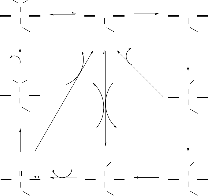
the water molecule that is bound as the sixth coordinating ligand to the heme iron (1).
This, in turn, induces a shift of the heme iron spin state from low-spin to high-spin
accompanied by a positive shift of the reduction potential in the order of
130–140 mV [41]. The increased potential allows the delivery of the first electron,
which reduces the heme iron from the ferric Fe
III
(2) to the ferrous Fe
II
form (3). After
the first electron transfer, the Fe
II
(3) binds dioxygen, resulting in a ferrous dioxygen
complex (4). The consecutive delivery of the second electron converts this species into
a ferric peroxy anion (5a). Subsequent steps in the P450 cycle are considered to be
relatively fast with respect to the electron transfer. The ferric peroxy species is
protonated to a ferric hydroperoxy complex (5b; also referred to as compound 0). The
following protonation of this complex results in a high-valent ferryl-oxo complex
(6; also referred to as compound I) accompanied by the release of a water molecule
31
4
5a
7
5b6
2
RH
RH
e
-
O
2
e
-
(2-)
H
+
H
2
O
H
+
ROH
H
2
O
2
H
+
pero
xid
e
shunt
H
2
O
2
H
+
H
2
O
2 e
-
2 H
+
autoxidation
shunt
O
2
-
(-)
Fe
III
S
O
HH
Fe
III
S
RH
Fe
II
S
RH
Fe
III
S
O
O
RH
Fe
III
S
O
O
(-)
RH
Fe
III
S
O
OH
RH
Fe
IV
S
O
oxidase
shunt
Fe
III
S
O
HR
Scheme 12.1 The catalytic cycle of cytochrome P450 monooxygenases (reproduced from Ref. [44],
with permission).
424
j
12 Biooxidation with Cytochrome P450 Monooxygenases
through heterolytic scission of the dioxygen bond in the preceding intermediate (7).
Compound I (6) is considered to be the intermediate catalyzing the majority of
P450 reactions; however, compound 0 (5b) may also be important for some P450-
dependent catalytic reactions [42], for example, the epoxidation of C¼C double
bonds [43].
Under certain conditions P450s can enter one of three so-called uncoupling
pathways (Scheme 12.1). Autoxidation shunt occurs if the second electron is not
delivered to reduce the ferrous dioxygen complex (4), which can decay forming
superoxide. The inappropriate positioning of the substrate in the active site is often the
molecular reason for the two other uncoupling cycles. The ferric hydroperoxy complex
(5b) can collapse and release hydrogen peroxide (peroxide shunt), while decay of
compound I (6) is accompanied by the release of water (oxidase shunt). For industrial
applications, it is particularly important to note that the uncoupling pathways in all
cases consume reducing equivalents from NAD(P)H without product formation.
Catalysis with cytochrome P450 monooxygenases requires two electrons to be
transferred to the heme via redox proteins. Depending on the topology of the protein,
components involved in the electron transfer to the heme, P450s can be grouped, for
example by a classification system with ten different classes as has recently been
suggested by Bernhardt and colleagues [39]. For industrial applications the fusion
enzymes of class VIII are of particular interest, since they come along with their
natural redox proteins. This group covers enzymes consisting of a P450 monooxy-
genase fused to a CPR-like reductase. The best studied representatives are P450
BM3
(CYP102A1) from Bacillus megaterium [45, 46] and its two homologs CYP102A2 [47]
and CYP102A3 [48] from Bacillus subtilis, as well as their eukaryotic counterpart,
CYP505A1 (P450
foxy
) from the ascomycete fungus Fusarium oxysporum [49].
It is notable that there are other P450s not requiring electron transfer proteins,
for example natural P450 peroxygenases, which employ the peroxide shunt for
catalysis [50]. Three enzymes with a potential for biocatalytic applications are the
H
2
O
2
-utilizing fatty acid hydroxylases of the CYP152 family, namely CYP152B1 (SP
a
)
from Sphingomonas paucimobilis [51], CYP152A1 (P450
Bsb
) from Bacillus subtilis [52],
and CYP152A2 (P450
CLA
) from Clostridium acetobutylicum [53].
12.2.3
Reactions Catalyzed by P450s
The ability of P450s to catalyze a large variety of oxidative and also some reductive
reactions – collectively involving thousands of substrates – has been discussed in
a number of reviews [7, 11, 54–57], so here we will reflect only the most important
oxidative reactions catalyzed by P450s. These reactions include hydroxylation of
nonactivated sp
3
-hybridized carbon atoms, epoxidation, aromatic hydroxylation,
CC bond cleavage, heteroatom oxygenation, heteroatom release (dealkylation),
oxidative ester cleavage, oxidative phenol- and ring-coupling, isomerization via
(abortive) oxidation, oxidative dehalogenation, and other complex reactions like
dimer formation via Diels-Alder reactions of products or Baeyer-Villiger-type
oxidations.
12.2 Properties of Cytochrome P450 Monooxygenases
j
425
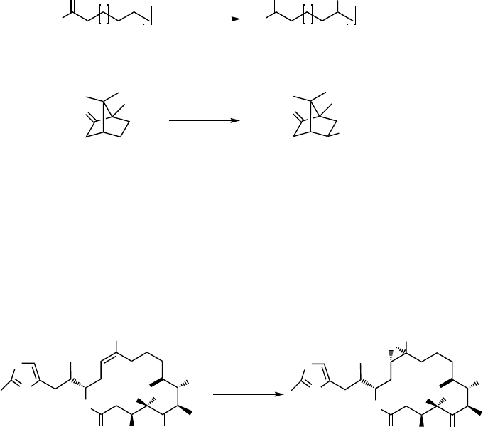
Hydroxylation of nonactivated sp
3
-hybridized carbon atoms belongs to the clas-
sical oxidative reactions catalyzed by P450s. Examples of this reaction include the
hydroxylation of saturated fatty acids (8) to hydroxy fatty acids (9) catalyzed, for
example, by eukaryotic CYP4 and bacterial CYP102 enzymes [58], as well as the
stereospecific hydroxylation of
D-( þ )-camphor (10)to5-exo-hydroxycamphor (11)
through P450
cam
[59] (Scheme 12.2).
Epoxidation of C¼C double bonds is another major reaction type catalyzed by
P450s. Particularly attractive in this regard is P450
EpoK
from Sorangium cellulosum,
which catalyzes the epoxidation of the thiazole-containing macrolactone epothilone
D(12) to epothilone B (13) [60] (Scheme 12.3). Epothilones are important anti-tumor
polyketides with high microtubule stabilizing activity.
Aromatic hydroxylation also belongs to the common P450 reactions. P450
NikF
from Streptomyces tendae T
€
u901 has been claimed to catalyze hydroxylation of
pyridylhomothreonine (14) to form hydroxypyridylhomothreonine (15) in the bio-
synthesis of nikkomycin, an inhibitor of chitin synthase [61] (Scheme 12.4). Many
P450s have been engineered toward aromatic hydroxylation, since this ability makes
them attractive candidates for the production of fine chemicals (see Sections 12.4
and 12.5.3).
10
11
P450
cam
O
HO
n
m
O
HO
n
m
P450
BM3
8 9
O
O
OH
OH
Scheme 12.2 Fatty acid hydroxylation catalyzed by P450
BM3
and camphor hydroxylation catalyzed
by P450
cam
.
O
OH
O
O
S
N
OH
CH
3
O
OH
O
O
S
N
OH
CH
3
O
P450
EpoK
12
13
Scheme 12.3 Epoxidation of Epothilon D (12) to Epothilon B (13) by P450
EpoK
.
426
j
12 Biooxidation with Cytochrome P450 Monooxygenases
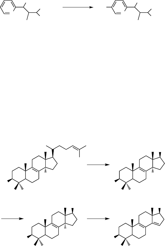
Some P450s are able to catalyze the cleavage of C–C bonds via multiple substrate
oxidations. For example, the demethylation of lanosterol (16) to a precursor of
cholesterol, 4,4-dimethyl-5a-cholesta-8,14,24-diene-3b-ol (19), by a lanosterol 14a-
demethylase (CYP51) [62] includes three steps and proceeds via initial hydroxylation
of the C14 methyl group followed by further oxidation of the alcohol (17) to the
aldehyde (18). Finally, acyl cleavage occurs leading to formation of a double bond in
the steroid (Scheme 12.5). A similar cascade of reactions is assumed to be catalyzed
by CYP107H1 (P450
BioI
) from Bacillus subtilis during conversion of long-chain fatty
acyl CoA esters to pimeloyl CoA in the biotin biosynthesis pathway [63].
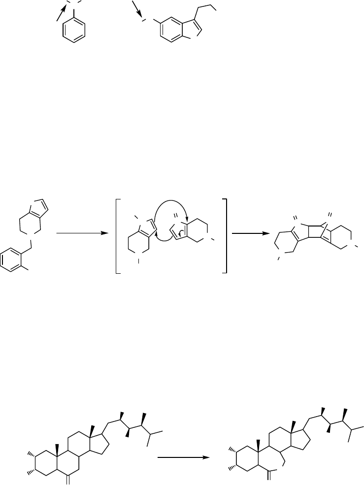
P450s catalyze oxidations, not only at carbon-atoms, but also at N-, S-, and O-atoms,
and they also catalyze dealkylations, which are believed to be the next step in the
hydroxylation of an a-carbon atom. Examples include the N-oxygenation of N,N-
dimethylaniline (20) and N,N-dialkylarylamines by mammalian CYP2B1 and
CYP2B4, respectively [64, 65], and O-demethylation of 5-methoxytryptamine (21)
by CYP2D6 [66] (Scheme 12.6).
N
OH
COOH
NH
2
N
OH
COOH
NH
2
HO
P450
NikF
15
14
Scheme 12.4 Aromatic hydroxylation of pyridylhomothreonine (14) to hydroxypyridylhomothreo-
nine (15) by P450
NikF
during nikkomycin biosynthesis.
HO
CH
3
16
17
+ HCOOH
1918
CYP51
HO
R
CH
2
OH
HO
R
CHO
HO
R
CYP51 CYP51
Scheme 12.5 Demethylation of lanosterol (16) to 4,4-dimethyl-5a-cholesta-8,14,24-diene-3b-ol
(19) catalyzed by lanosterol 14a-demethylase (CYP51).
12.2 Properties of Cytochrome P450 Monooxygenases
j
427
Some P450s are able to catalyze oxidative phenol coupling, a reaction usually
carried out by peroxidases. Three independent P450 monooxygenases with such
activity have been shown to be involved in the synthesis of vancomycin-type
antibiotics in Amycolatopsis balhimycina [67].
Dimerization of thiophene S-oxide (23 ) via a Diels-Alder reaction was observed
when ticlodipine (22) was oxidized by CYP2C19 or CYP2D6 [68] (Scheme 12.7).
Baeyer-Villiger-type oxidations can also be catalyzed by some P450s, for example
CYP85A2 from Arabidopsis thaliana, which catalyzes the conversion of castasterone
(24) to brassinolide (25) [69] (Scheme 12.8).
N
H
3
C CH
3
20
N
H
O
H
3
C
NH
2
21
Scheme 12.6 N-oxygenation of N,N-dimethylaniline (20) and O-demethylation of
5-methoxytryptamine (21); reaction sides are indicated by arrows.
N
S
Cl
R
N
S
+
R
N
S
O
-
O
2322
R
N
S
R
N
O
S
O
or CYP2D6
CYP2C19
Scheme 12.7 Dimerization of thiophene S-oxide (23) via a Diels-Alder reaction through oxidation
of ticlodipine (22).
HO
OH
HO
HO
O
HO
OH
HO
HO
O
O
5242
CYP85A2
Scheme 12.8 Conversion of castasterone (24) to brassinolide (25) via Baeyer-Villiger-type
oxidation.
428
j
12 Biooxidation with Cytochrome P450 Monooxygenases
Many other unusual types of oxidative and also some reductive reactions catalyzed
by P450s have been described in the literature, including oxidative deamination,
desulfurylation, oxidative dehalogenation, isomerization, dehydrogenation, dehy-
dration, reductive dehalogenation, epoxide reduction, and others [54, 57, 70].
12.2.4
P450s as Industrial Biocatalysts
12.2.4.1 Advantages
P450 biocatalysts operate – like any enzyme applied for industrial biocatalysis – under
ambient conditions, thereby often exhibiting exquisite substrate specificity as well as
regio- and/or stereoselectivity. Compared with other biocatalysts, however, P450s
potentially have additional advantages for industrial applications:
.
Since their discovery, P450s have been studied in enormous detail because of their
involvement in a plethora of crucial cellular roles – from carbon source assim-
ilation, through biosynthesis of hormones, to carcinogenesis, drug activation, and
degradation of xenobiotics.
.
As mentioned above, P450s are able to catalyze more than 20 different reaction
types and can oxidize a wide range of molecules [3]. Many of the compounds occur
naturally and can be important industrial precursors.
.
P450s can be produced industrially by fermentation. Considerable progress
has been made during the last decade for recombinant expression of P450s in
the well-studied hosts Escherichia coli, Pseudomonas putida and yeasts Saccharo-
myces cerevisiae and Pichia pastoris, which facilitates their use as industrial
catalysts [71–77].
.
The number of identified P450s is huge and constantly increasing as a result of
microbial screenings and increasing information on sequenced genomes. The
collection of P450s in (recombinant) libraries allows high-throughput screenings
as well as functional characterization of new members of the P450 family and
offers a route to diverse building blocks.
12.2.4.2 Challenges in the Development of Technical P450 Applications
Besides the que stions concerning process stability a nd activity of P450s, which
apply to all industrial biocatalysts, t he development of technical appl ications for
P450s faces specific problems. Probably the most import ant drawback restricting
industrial applications is the fact that nearly all P450s require costly cofactors
NADPH or N ADH, which makes their application impossible if the cofactor h as to
be added in a stoichiometric amount. Closely linked to this cofactor dependency is
the challenge (a) to find suitable redox proteins that can adequately deliver the
electrons to the heme and (b) to constru ct auxil iary redox modules. However, m any
efforts have b een made to overcome these hurdles by either minimizing or
removing the need for NAD(P)H or by d esigning new s trategies for simplified
transmission of reducing power [11, 78, 79], s ome of these are discussed in the
following sections.
12.2 Properties of Cytochrome P450 Monooxygenases
j
429

12.2.4.3 General Aspects of Industrial Application and Engineering of P450s
Because of the cofactor dependence of P450s, their industrial applications have so far
been restricted to whole-cell systems, which take advantage of the hosts endogenous
cofactor regeneration systems. In such instances, however, physiological effects like
limited substrate uptake, toxicity of substrate or product, product degradation, and
elaborate downstream processing must be taken into account [80]. Moreover, when
concentrations of recombinant P450 biocatalysts within the cell reach a certain level,
the cofactor concentration may again become a bottleneck for the overall process.
Another factor important for efficient biocatalysis with P450s is the yield of product
based on NAD(P)H consumed, or the coupling efficiency. Besides reducing the
efficiency of cofactor usage, uncoupling between NADPH oxidation and product
formation results in reactive oxygen species (such as superoxide anions and hydro-
gen peroxide) that cause oxidative destruction of the heme and oxidative damage of
the protein.
Thus, optimization strategies for cytochrome P450 monooxygenases target di-
verse areas. These include identification of key residues involved in substrate
binding, the extension of substrate spectra, the sub stitution or regeneration of the
cofactor NAD(P)H, and the enhancement of enzyme stability, activity, selectivity, and
coupling efficiency (Figure 12.2). To achieve these objectives, va rious techniques of
protein engineering have been applied, for example site-directed and random
mutagenesis, DNA recombination, or combinations of these methods. The applied
strategies have their advantages and drawbacks, which are discussed in numerous
excellent reviews and books [81– 85].
12.3
Application and Engineering of P450s for the Pharmaceutical Industry
P450s have a central role in drug metabolism, where they catalyze a large proportion
of the most complex and chemically challenging steps in the biosynthesis of many
naturalproductsused inmedicinetoday.Thus,potentialapplicationsof P450sconcern
their involvement in drug biosynthesis, as well as strategies for developing new
derivatives of drugs based on P450 engineering [86]. Given the diversity of reactions
catalyzed by P450s, however, few of them have been exploited in industry so far.
Optimization strategies
Optimization strategies
for P450s
for P450s
Altered substrate specificity
Altered substrate specificity
Enhanced activity
Enhanced activity
Enhanced solvent tolerance
Enhanced solvent tolerance
Enhanced enzyme stability
Enhanced enzyme stability
Improved regio- and stereoselectivity
Improved regio- and stereoselectivity
Cofactor replacement
Cofactor replacement
Enhanced coupling efficiency
Enhanced coupling efficiency
Figure 12.2 P450 optimization strategies for potential biotechnological application.
430
j
12 Biooxidation with Cytochrome P450 Monooxygenases
12.3.1
Microbial Oxidations with P450s for Synthesis of Pharmaceuticals
Drug development is based on the detailed characterization of metabolic pathways
and their relevance for drug safety. This type of analysis often requires milligram
quantities of metabolites, which are difficult to synthesize by chemical routes,
especially when the metabolites result from stereoselective oxidations. Microbial
oxidations using fungi, yeast, archea, and bacteria can be performed either by using
native P450-producing strains – which can be altered by metabolic engineering – or
with the aid of recombinant whole-cells harboring microbial P450s. Microbial
equivalents of human P450-catalyzed oxidations are an additional alternative, espe-
cially of interest when several hundred milligrams to many grams of metabolites are
requested, both for identification purposes and for production of non-human
metabolites with new biological properties.
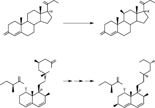
Microbial oxidations of steroids represent very well-established large-scale com-
mercial applications of P450 monooxygenases. The 11b-hydroxylation of 11-deoxy-
cortisol (26) to hydrocortisone (27) using a P450 from Curvularia sp. [87]
(Scheme 12.9) is applied by Schering AG (in 2006 acquired by Merck, Germany)
at an industrial scale of approximately 100 tonnes per year [80]. Another example is
the regioselective hydroxylation of progesterone to 11a-hydroxyprogesterone by
Rhizopus sp. developed in the 1950s by Pharmacia & Upjohn (later acquired
by Pfizer Inc., USA) [88, 89]. Both processes are one-step biotransformations, which
cannot be achieved by chemical routes.
CH
3
O
O
H
O
O
H
HO
H
CH
3
O
O
H
HOOC
HO
OH
H
HO
H
28
29
Mucor
hiemalis
O
OH
O
OH
O
OH
O
OH
HO
Curvularia
sp.
26
27
P450
P450
Scheme 12.9 Two examples of microbial oxidations of steroids: 11b-hydroxylation of
11-deoxycortisol (26) to hydrocortisone (27), and production of pravastatin (29) by oxidation
of compactin (28).
12.3 Application and Engineering of P450s for the Pharmaceutical Industry
j
431
Production of the cholesterol-reducing pravastatin (29) by oxidation of compactin
(28) catalyzed by a P450 monooxygenase from Mucor hiemalis (Daiichi Sankyo Inc.,
USA, and Bristol-Myers Squibb, USA) is another example of a commercial appli-
cation of microbial oxidations [90, 91] (Scheme 12.9). The same reaction can be
catalyzed by Streptomyces sp. Y-110. In a batch culture with continuous feeding of
compactin into the culture medium a conversion rate of 15 mg L
1
h
1
pravastatin
and a final concentration of 1 g L
1
pravastatin were achieved [92].
Diverse activities of microbial cytochrome P450 monooxygenases have potential
applications in the synthesis of new antibiotics, especially in view of the widespread
resistance to bacterial antibiotics. Streptomyces strains and other bacterial actinomy-
cete species produce many important natural products including the majority of
known antibiotics, and P450s catalyze important biosynthetic steps [93]. Particularly
intriguing is the fact that Streptomyces has a large P450 complement reflecting the
ecological niche that the organism finds itself in. The first complete Streptomyces
genome (Streptomyces coelicolor A3(2)) was published in 2002, revealing the presence
of 18 P450 genes [94]. Subsequently, genomes of Streptomyces avermitilis (33 P450
genes [95]) and Streptomyces peucetius (15 P450 genes [96]) have been reported. Many
efforts have been undertaken to identify gene clusters involved in synthesis of
pharmaceutically important compounds and to increase the product yield of these
compounds by the use of engineered Streptomyces strains.
Recent examples include the elucidation of the biosynthetic gene cluster organi-
zation for pladienolide – a polyketide antitumor macrolide – in Streptomyces platensis
Mer-11107, where a P450 of the CYP107 family acts as a 6-hydroxylase [97].
Pladienolide B (30) and its 16-hydroxylated derivative pladienolide D (31) show
strong antitumor activity (Scheme 12.10). The original strain of S. platensis produces
mainly pladienolide B, while pladienolide D is produced to a lesser extent. Conse-
quently, to facilitate the production of pladienolide D, an engineered strain was
constructed by over-expression of a pladienolide B 16-hydroxylase PsmA (a P450
from the CYP105 family) from Streptomyces bungoensis A-1544 in S. platensis. The
recombinant strain produced pladienolide D at a production level comparable to that
of pladienolide B [98].
OH
O
R
O
O
OH
OH
O
O
6
1
16
30: R = H; 31: R = OH
Scheme 12.10 Structures of pladienolide B (30) and pladienolide D (31).
432
j
12 Biooxidation with Cytochrome P450 Monooxygenases
