ASM Metals HandBook Vol. 17 - Nondestructive Evaluation and Quality Control
Подождите немного. Документ загружается.


Internal cracks F
(c)
F F
(c)
F
(c)
F
F
(c)
Voids G G G G G
G
Thickness F G F F G
F
Metallurgical variations
F F F F F
F
Sheet and plate
Thickness G
(d)
G G
(d)
G
(d)
G
G
(d)
Laminations U U U U U
U
Voids G G G G G
G
Bars and tubes
Seams P P P P P
P
Pipe G G G G G
F
Cupping G G G G G
F
Inclusions F F F F F
F
Castings
Cold shuts G G G G G
G
Surface cracks F
(c)
F F
(c)
F
(c)
F
F
(c)
Internal shrinkage G G G G G
G
Voids, pores G G G G G
G
Core shift G G G G G
G
Forgings
Laps P
(c)
P
(c)
P
(c)
P P
U

Inclusions F F F F F
U
Internal bursts G G G F F
G
Internal flakes P
(c)
P U P
(c)
P
U
Cracks and tears F
(c)
F F
(c)
F
(c)
F
F
(c)
Welds
Shrinkage cracks G
(c)
G G
(c)
G
(c)
G
G
(c)
Slag inclusions G G G G G
G
Incomplete fusion G G G G G
G
Pores G G G G G
G
Incomplete penetration G G G G G
G
Processing
Heat-treat cracks U U U P U
U
Grinding cracks U U U U U
U
Service
Fatigue and heat cracks
F
(c)
F P
(c)
P P
P
Stress corrosion F F P F F
P
Blistering P P P P P
P
Thinning F F F F F
F
Corrosion pits F F P G G P
(a)
G, good; F, fair; P, poor; U, unsatisfactory.

(b)
Includes only visible cracks. Minute surface cracks are undetectable by radiographic inspection methods.
(c)
Radiation beam must be parallel to the cracks, laps, or flakes. In real-time radiography, the testpiece can be manipulated for proper orientation.
(d)
When calibrated using special thickness gages
Radiography can be used to inspect most types of solid material, with the possible exception of materials of very high or
very low density. (Neutron radiography, however, can often be used in such cases, as discussed in the article "Neutron
Radiography" in this Volume.) Both ferrous and nonferrous alloys can be radiographed, as can nonmetallic materials and
composites.
There is wide latitude both in the material thickness that can be inspected and in the techniques that can be used.
Numerous special techniques and special devices have been developed for the application of radiography to specific
inspection problems, including even the inspection of radioactive materials. Most of these specialized applications are not
discussed in this article, but several can be found in articles in this Volume that deal with the use of radiography in the
inspection of specific product forms.
In some cases, radiography cannot be used even though it appears suitable from Table 1, because the part is accessible
from one side only. Radiography typically involves the transmission of radiation through the testpiece, in which case both
sides of the part must be accessible. However, radiographic and radiometric inspection can also be performed with
Compton scattering, in which the scattered photons are used for imaging. With Compton scattering, inspection can be
performed when only one side is accessible. Another method of inspecting a region having one inaccessible side is to use
probes with a microfocus x-ray tube (see the section "Microfocus X-Ray Tubes" in this article).
Limitations. Compared to other nondestructive methods of inspection, radiography is expensive. Relatively large
capital costs and space allocations are required for a radiographic laboratory, although costs can be reduced when portable
x-ray or -ray sources are used in film radiography. Capital costs can be relatively low with portable units, and space is
required only for film processing and interpretation. Operating costs can be high; sometimes as much as 60% of the total
inspection time is spent in setting up for radiography. With real-time radiography, operating costs are usually much lower,
because setup times are shorter and there are no extra costs for processing or interpretation of film.
The field inspection of thick sections can be a time-consuming process because the effective radiation output of portable
sources may require long exposure times of the radiographic film. Radioactive ( -ray) sources are limited in their output
primarily because high-activity sources require heavy shielding for the protection of personnel. This limits field usage to
sources of lower activity that can be transported. The output of portable x-ray sources may also limit the field inspection
of thick sections, particularly if a portable x-ray tube is used. Portable x-ray tubes emit relatively low-energy (300 keV)
radiation and are limited in the radiation output. Both of these characteristics of portable x-ray tubes combine to limit
their application to the inspection of sections having the absorption equivalent of 75 mm (3 in.) of steel. Instead of
portable x-ray tubes, portable linear accelerators and betatrons provide high-energy (>1 MeV) x-rays for the radiographic
field inspection of thicker sections.
Certain types of flaws are difficult to detect by radiography. Cracks cannot be detected unless they are essentially parallel
to the radiation beam. Tight cracks in thick sections may not be detected at all, even when properly oriented. Minute
discontinuities such as inclusions in wrought material, flakes, microporosity, and microfissures may not be detected
unless they are sufficiently segregated to yield a detectable gross effect. Laminations are nearly impossible to detect with
radiography; because of their unfavorable orientation, delaminations do not yield differences in absorption that enable
laminated areas to be distinguished from delaminated areas.
It is well known that large doses of x-rays or -rays can kill human cells, and in massive doses can cause severe
disability or death. Protection of personnel--not only those engaged in radiographic work but also those in the vicinity of
radiographic inspection--is of major importance. Safety requirements impose both economic and operational constraints
on the use of radiography for inspection.
Radiographic Inspection
Revised by the ASM Committee on Radiographic Inspection
*
Principles of Radiography
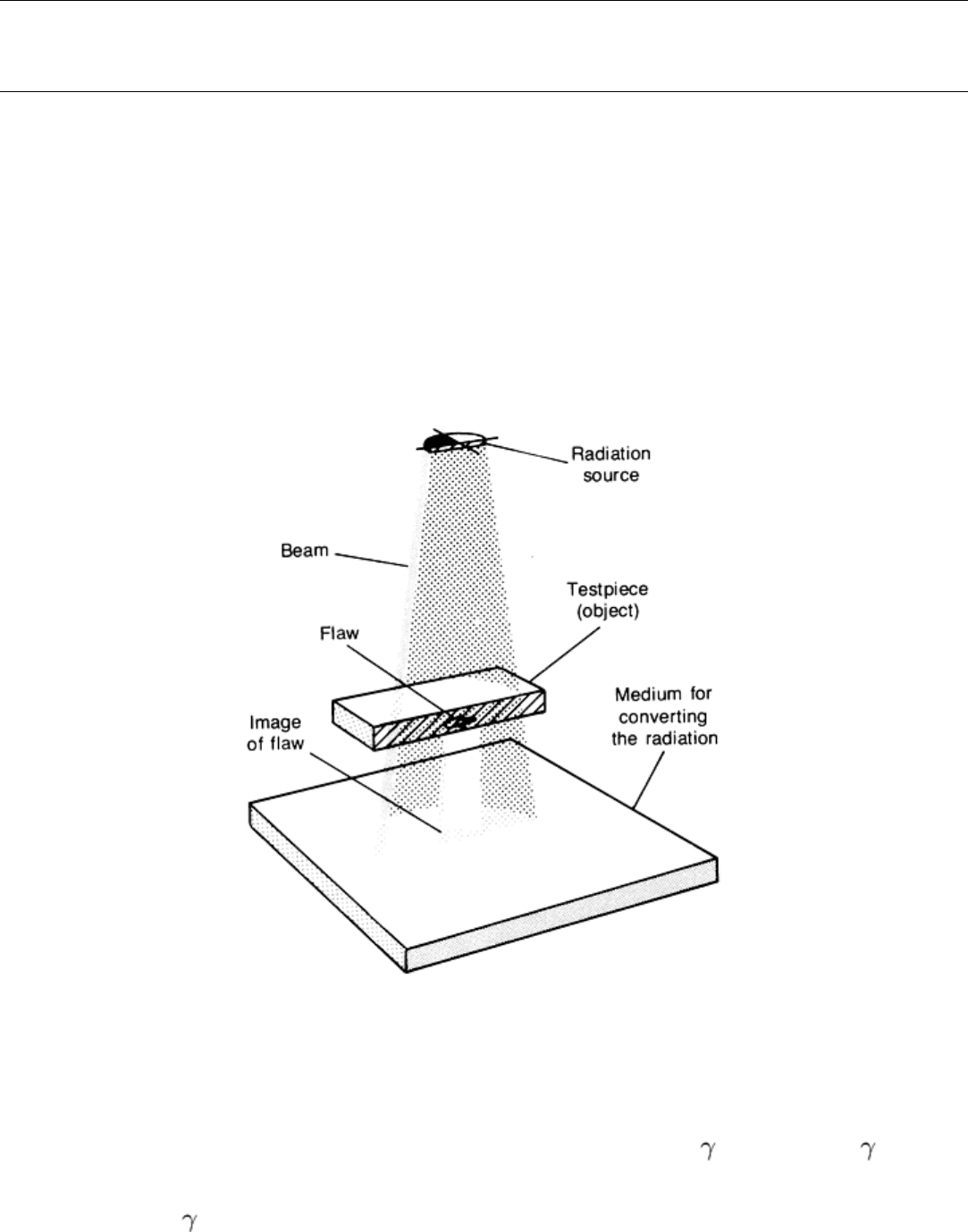
Three basic elements of radiography include a radiation source, the testpiece or object being evaluated, and a sensing
material. These elements are shown schematically in Fig. 1. The testpiece in Fig. 1 is a plate of uniform thickness
containing an internal flaw that has absorption characteristics different from those of the surrounding material. Radiation
from the source is absorbed by the testpiece as the radiation passes through it; the flaw and surrounding material absorb
different amounts of radiation. Thus, the intensity of radiation that impinges on the sensing material in the area beneath
the flaw is different from the amount that impinges on adjacent areas. This produces an image, or shadow, of the flaw on
the sensing material. This section briefly reviews the general characteristics and safety principles associated with
radiography.
Fig. 1 Schematic of the basic elements of a radiographic system showing the method of sensing the image of
an internal flaw in a plate of uniform thickness
Radiation Sources
Two types of electromagnetic radiation are used in radiographic inspection: x-rays and -rays. X-rays and -rays differ
from other types of electromagnetic radiation (such as visible light, microwaves, and radio waves) only in their
wavelengths, although there is not always a distinct transition from one type of electromagnetic radiation to another (Fig.
2). Only x-rays and -rays, because of their relatively short wavelengths (high energies), have the capability of
penetrating opaque materials to reveal internal flaws.
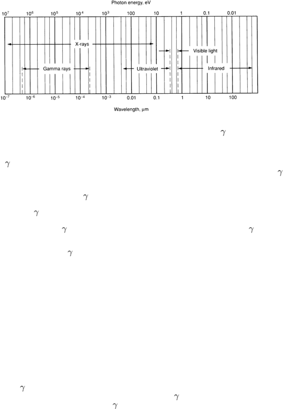
Fig. 2 Schematic of the portion of the electromagnetic spectrum that includes x-rays, -
rays, ultraviolet and
visible light, and infrared radiation showing their relationship with wavelength and photon energy
X-rays and -rays are physically indistinguishable; they differ only in the manner in which they are produced. X-rays
result from the interaction between a rapidly moving stream of electrons and atoms in a solid target material, while -
rays are emitted during the radioactive decay of unstable atomic nuclei.
The amount of exposure from x-rays or -rays is measured in roentgens (R), where 1 R is the amount of radiation
exposure that produces one electrostatic unit (3.33564 × 10
-10
C) of charge from 1.293 mg (45.61 × 10
-6
oz) of air. The
intensity of an x-ray or -ray radiation is measured in roentgens per unit time.
Although the intensity of x-ray or -ray radiation is measured in the same units, the strengths of x-ray and -ray sources
are usually given in different units. The strength of an x-ray source is typically given in roentgens per minute at one meter
(RMM) from the source or in some other suitable combination of time or distance units (such as roentgens per hour at one
meter, or RHM). The strength of a -ray source is usually given in terms of the radioactive decay rate, which has the
traditional unit of a curie (1 Ci = 37 × 10
9
disintegrations per second). The corresponding unit in the Système
International d'Unités (SI) system is a gigabecquerel (1 GBq = 1 × 10
9
disintegrations per second).
The spectrum of radiation is often expressed in terms of photon energy rather than as a wavelength. Photon energy is
measured in electron volts, with 1 eV being the energy imparted to an electron by an accelerating potential of 1 V. Figure
2 shows the radiation spectrum in terms of both wavelength and photon energy.
Production of X-Rays. When x-rays are produced from the collision of fast-moving electrons with a target material,
two types of x-rays are generated. The first type of x-ray is generated when the electrons are rapidly decelerated during
collisions with atoms in the target material. These x-rays have a broad spectrum of many wavelengths (or energies) and
are referred to as continuous x-rays or by the German word bremsstrahlung, which means braking radiation. The second
type of x-ray occurs when the collision of an electron with an atom of the target material causes a transition of an orbital
electron in the atom, thus leaving the atom in an excited state. When the orbital electrons in the excited atom rearrange
themselves, x-rays are emitted that have specific wavelengths (or energies) characteristic of the particular electron
rearrangements taking place. These characteristic x-rays usually have much higher intensities than the background of
bremsstrahlung having the same wavelengths.
Production of -Rays. Gamma rays are generated during the radioactive decay of both naturally occurring and
artificially produced unstable isotopes. In all respects other than their origin, -rays and x-rays are identical. Unlike the
broad-spectrum radiation produced by x-ray sources, -ray sources emit one or more discrete wavelengths of radiation,
each having its own characteristic photon energy (or wavelength).

Many of the elements in the periodic table have either naturally occurring radioactive isotopes or isotopes that can be
made radioactive by irradiation with a stream of neutrons in the core of the nuclear reactor. However, only certain
isotopes are extensively used for radiography (see the section "Gamma-Ray Sources" in this article).
Image Conversion
The most important process in radiography is the conversion of radiation into a form suitable for observation or further
signal processing. This conversion is accomplished with either a recording medium (usually film) or a real-time imaging
medium (such as fluorescent screens or scintillation crystals). The imaging process can also be assisted with the use of
intensifying or filtration screens, which intensify the conversion process or filter out scattered radiation.
Recording media provide a permanent image that is related to the variations in the intensity of the unabsorbed
radiation and the time of exposure. With a recording medium such as film, for example, an invisible latent image is
formed in the areas exposed to radiation. These exposed areas become dark when the film is processed (that is, developed,
rinsed, fixed, washed, and dried), with the degree of darkening (the photographic density) depending on the amount of
exposure to radiation. The film is then placed on an illuminated screen so that the image formed by the variations in
photographic density can be examined and interpreted.
Real-time imaging media provide an immediate indication of the intensity of radiation passing through a testpiece.
With fluorescent screens, for example, visible light is emitted with a brightness that is proportional to the intensity of the
incident x-ray or -ray radiation. This emitted light can be observed directly, amplified, and/or converted into a video
signal for presentation on a television monitor and subsequent recording. The various types of imaging systems are
described in the section "Real-Time Radiography" in this article.
Intensifying and filtration screens are used to improve image contrast, particularly when the radiation intensity is
low or when the radiation energy is high. The screens are useful at higher energies because the sensitivity of films and
fluorescent screens decreases as the energy of the penetrating radiation increases. The various types of screens are
discussed in the section "Image Conversion Media" in this article.
Image Quality and Radiographic Sensitivity
The quality of radiographs is affected by many variables, and image quality is measured with image-quality indicators
known as penetrameters. These devices are thin specimens made of the same material as the testpiece; they are described
in more detail in the section "Identification Markers and Penetrameters (Image-Quality Indicators)" in this article. When
placed on the testpiece during radiographic inspection, the penetrameters measure image contrast and, to a limited extent,
resolution. Detail resolution is not directly measured with penetrameters, because flaw detection depends on the nature of
the flaw, its shape, and orientation to the radiation beam.
Image quality is governed by image contrast and resolution, which are also sometimes referred to as radiographic contrast
and radiographic definition. These two factors are interrelated in a complex way and are affected by several factors
described in the sections "Radiographic contrast" and "Radiographic definition" in this article. In real-time systems,
image contrast and resolution are also described in terms of the detective quantum efficiency (DQE) (also known as the
quantum detection efficiency, or QDE) and the modulation transfer function (MTF), respectively. These terms, which are
also used in computed tomography, are defined in the text and in Appendix 2 of the article "Industrial Computed
Tomography" in this Volume.
Radiographic sensitivity, which should be distinguished from image quality, generally refers to the size of the smallest
detail that can be seen on a radiograph or to the ease with which the images of small details can be detected. Although
radiographic sensitivity is often synonymous with image quality in applications requiring the detection of small details, a
distinction should be made between radiographic sensitivity and radiographic quality. Radiographic sensitivity refers
more to detail resolvability, which should be distinguished from spatial resolution and contrast resolution. For example, if
the density of an object is very different from the density of the surrounding region, the flaw might be resolved because of
the large contrast, even if the flaw is smaller than the spatial resolution of the system. On the other hand, when the
contrast is small, the area must be large to achieve resolvability.
Radiographic contrast refers to the amount of contrast observed on a radiograph, and it is affected by subject contrast
and the contrast sensitivity of the image-detecting system. Radiographic contrast can also be affected by the unsharpness
of the detected image. Figure 3, for example, shows how contrast is affected when the sharp edge of a step is blurred
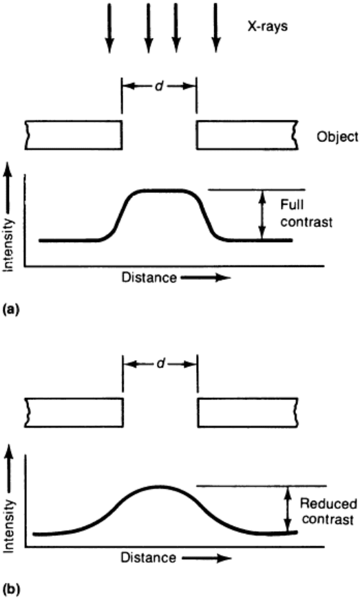
because of unsharpness. If the unsharpness is much smaller than d in Fig. 3, then the contrast is not reduced and the edges
in the image are easily defined. However, if d is much smaller than the unsharpness, then the contrast is reduced. If the
unsharpness is too large, the image cannot be resolved.
Fig. 3 Effect of geometric unsharpness on image contrast. (a) Flaw size, d, is larger t
han the unsharpness, then
full contrast occurs. (b) Flaw size, d, is smaller than the unsharpness, then contrast is reduced.
Subject contrast is the ratio of radiation intensities transmitted by various portions of a testpiece. It depends on the
thickness, shape, and composition of the testpiece; the intensity of the scattered radiation; and the spectrum of the incident
radiation. It does not depend on the detector, the source strength, or the source-to-detector distance. However, it may
depend on the testpiece-to-detector distance because the intensity of scattered radiation (from within the testpiece)
impinging upon a detector is almost eliminated when projective enlargement approaches a magnification factor of three
(see the section "Enlargement" in this article).
In general, a radiograph should be made with the lowest-energy radiation that will transmit adequate radiation intensities
to the detector, because long wavelengths tend to improve contrast. However, radiation energies that are too low produce
excessive amounts of scattered radiation that washes out fine detail. On the other hand, energies that are too high,
although they reduce scattered radiation, may produce images having contrast that is too low to resolve small flaws. For
each situation, there is an optimum radiation energy that produces the best combination of radiographic contrast and
definition, or the greatest sensitivity.
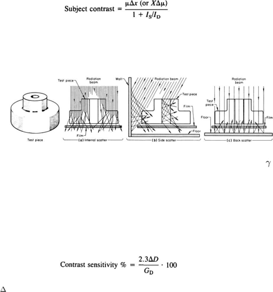
The amount of subject contrast can be modeled as follows. If there is a change in thickness, Δx, of a testpiece with a
uniform attenuation coefficient, μ, then the subject contrast (without scattering) is equal to μΔx. Similarly, if there is a
change in the attenuation coefficient, Δμ, over a distance X, then the subject contrast (without scatter) is equal to XΔμ. If
there is scattering, the subject contrast is reduced such that:
(Eq 1)
where I
S
is the intensity of the scattered radiation and I
D
is the intensity of the direct (or primary) radiation passing
through the testpiece. The scattering may occur from within the testpiece (Fig. 4a) or from the surroundings (Fig. 4b and
c). The various mechanisms of scattering and attenuation are described in the section "Attenuation of Electromagnetic
Radiation" in this article.
Fig. 4 Schematic of the three types of scattered radiation encountered when using x-rays and -rays
Contrast sensitivity refers to the ability of responding to and displaying small variations in subject contrast. Contrast
sensitivity depends on the characteristics of the image detector and on the level of radiation being detected (or on the
amount of exposure for films). The relationship between the contrast sensitivity and the level of radiation intensity (or
film exposure) can be illustrated by considering two extremes. At low levels of radiation intensity, the contrast sensitivity
of the detectors is reduced by a smaller signal-to-noise ratio, while at high levels, the detectors become saturated.
Consequently, contrast sensitivity is a function of dynamic range.
In film radiography, the contrast sensitivity is:
(Eq 2)
where D is the smallest change in photographic density that can be observed when the film is placed on an illuminated
screen. The factor G
D
is called the film gradient or film contrast. The film gradient is the inherent ability of a film to
record a difference in the intensity of the transmitted radiation as a difference in photographic density. It depends on film
type, development procedure, and film density. For all practical purposes, it is independent of the quality and distribution
of the transmitted radiation (see the section "Film Radiography" in this article).
Contrast sensitivity in real-time systems is determined by the number of bits (if the image is digitized) and the signal-to-
noise ratio (which is affected by the intensity of the radiation and the efficiency of the detector). The factors affecting the
contrast sensitivity and signal-to-noise ratio of real-time systems are discussed in the section "Modern Image Intensifiers"
in this article. The best contrast sensitivity of digitized images from fluorescent screens is about 8 bits (or 256 gray
levels).
Another way of specifying the contrast sensitivity of fluorescent screens is with a gamma factor, which is defined by the
fractional unit change in screen brightness, ΔB/B, for a given fractional change in the radiation intensity, ΔI/I. Most
fluorescent screens have a gamma factor of about one, which is not a limiting factor. At low levels of intensity, however,
the contrast is reduced because of quantum mottle (see the section "Screen Unsharpness" in this article) just as
unsharpness reduced the contrast in the example illustrated in Fig. 3.
Dynamic range, or latitude, describes the ability of the imaging system to produce a suitable signal over a range of
radiation intensities. The dynamic range is given as the ratio of the largest signal that can be measured to the precision of
the smallest signal that can be discriminated. A large dynamic range allows the system to maintain contrast sensitivity
over a wide range of radiation intensities or testpiece thicknesses. Film radiography has a dynamic range of up to 1000:1,
while digital radiography with discrete detectors can achieve 100,000:1. The latitude, or dynamic range, of film
techniques is discussed in the section "Exposure Factors" in this article.
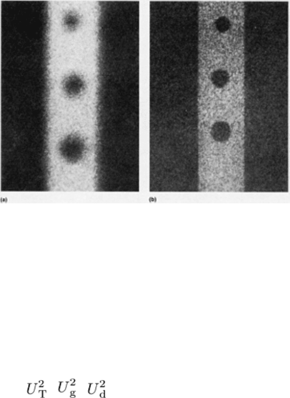
Radiographic definition refers to the sharpness of a radiograph. In Fig. 5(a), for example, the radiograph exhibits
poor radiographic definition even when the contrast is high. Better radiographic definition is achieved in Fig. 5(b), even
though the contrast is lower.
Fig. 5
Radiographs with poor and improved radiographic definition. (a) Advantage of a higher radiographic
contrast is offset by poor definition. (b) Despite lower contrast, better detail is obtained with improved
definition. Source: Ref 1
The degree of radiographic definition is usually specified in line pairs per millimeter or by the minimum distance by
which two features can be physically distinguished. The factors affecting radiographic definition are geometric
unsharpness and the unsharpness (or spatial resolution) of the imaging system. The net effect of these two unsharpnesses
is not directly additive, because the combination of the two unsharpnesses is complex. For most practical radiography, the
total unsharpness, U
T
, is:
= +
(Eq 3)
where U
g
is the geometric unsharpness and U
d
is the unsharpness or spatial resolution of the image-detecting system.
Another important type of unsharpness occurs when in-motion radiography is performed. This type of unsharpness,
termed motion unsharpness, is discussed in the section "In-Motion Radiography" in this article. Geometric unsharpness is
usually the largest contributor to maximum unsharpness in film radiography, while the unsharpness of fluorescent screens
in real-time radiography often overshadows the effect of geometric unsharpness.
Distortion and Geometric Unsharpness. Because a radiograph is a two-dimensional representation of a three-
dimensional object, the radiographic images of most testpieces are somewhat distorted in size and shape compared to
actual testpiece size and shape. The mechanisms of geometric unsharpness and distortion are described in the section
"Principles of Shadow Formation" in this article. The severity of unsharpness and distortion depends primarily on the
source size (focal-spot size for x-ray sources), source-to-object and source-to-detector distances, and position and
orientation of the testpiece with respect to source and detector.
Film unsharpness specifies the spatial resolution of the film and must not be confused with film graininess. Film
unsharpness and film graininess must be treated as two separate parameters.
Film unsharpness depends not only on the film grain size and the development process but also on the energy of the
radiation and the thickness and material of the screens. When a quantum of radiation sensitizes a silver halide crystal in a
film, adjacent crystals may also be sensitized if the quantum has sufficient energy to release electrons during the
absorption by the first crystal. Consequently, a small volume of crystals would be sensitized, thus producing a small disk
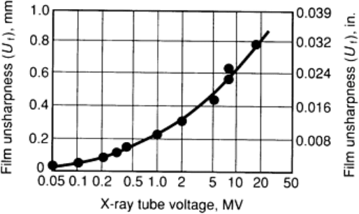
instead of a point image. Figure 6 illustrates an experimental curve of film unsharpness versus radiation energy. The
screens used will also affect the unsharpness with the production of electrons.
Fig. 6 Experimental curve of film unsharpness, U
f
, against x-ray energy (filtered sources). Source: Ref 2
Screen Unsharpness. In addition to the spatial resolution established by the video or image-processing equipment, the
spatial resolution of real-time systems is also affected by screen unsharpness. Screen unsharpness depends not only on the
grain size and thickness of the screen but also on the detection efficiency and the energy and intensity of the radiation.
Statistical fluctuations (quantum mottle) of screen brightness becomes more significant at low levels of screen brightness,
and this can occur when either the radiation intensity or the detection efficiency is lowered. Nevertheless, screen
unsharpness due to statistical fluctuation can be eliminated with the image-processing operation of frame summation,
which makes an image representing the amount of radiation exposure during the frame-summing interval.
Projective Magnification With Microfocus X-Ray Sources. Even though the imaging systems have limits in
their spatial resolution, detail of the testpiece can be enlarged by projective magnification (see the section "Enlargement"
in this article). This process of image magnification minimizes the limits of graininess and unsharpness caused by the
imaging system. With microfocus x-ray sources, a resolution greater than 20 line pairs per millimeter (or a spatial
resolution of 0.002 in.) can be achieved.
Detail Perceptibility of Images. Studies have shown that the perceptibility of image details depends principally on
the product: MTF
2
· DQE; where MTF is the modulation transfer function and DQE is the detective quantum efficiency of
the imaging system. These terms, as previously mentioned in this section, are described in the article "Industrial
Computed Tomography" in this Volume.
Radiation Safety
