Журнал - Проблемы криобиологии 2010 №4
Подождите немного. Документ загружается.


379
* Автор, которому необходимо направлять корреспонденцию:
ул. Переяславская, 23, г. Харьков, Украина 61015; тел.:+38
(057) 373-30-07, факс: +38 (057) 373-30-84, электронная почта:
dudetska@mail.ru
* To whom correspondence should be addressed: 23,
Pereyaslavskaya str., Kharkov, Ukraine 61015; tel.:+380 57 373
3007, fax: +380 57 373 3084, e-mail: dudetska@mail.ru
Institute for Problems of Cryobiology and Cryomedicine of the Na-
tional Academy of Sciences of Ukraine, Kharkov, Ukraine
Институт проблем криобиологии и криомедицины
НАН Украины, г. Харьков
УДК 612.451.014.3.085:612.592
Г.В. ДУДЕЦКАЯ*, Г.А. БОЖОК, Т.П. БОНДАРЕНКО
Влияние скорости охлаждения на сохранность зонально-
дифференцированных популяций клеток надпочечников крыс
UDC 612.451.014.3.085:612.592
G.V. DUDETSKAYA*, G.A. BOZHOK, T.P. BONDARENKO
Effect of Cooling Rate on Integrity of Zone-Differentiated
Adrenal Cell Populations of Rats
С помощью методов флуоресцентной микроскопии и гистохимического окрашивания были изучены клетки надпочечников
взрослых крыс до и после криоконсервирования. Низкие скорости охлаждения позволяют сохранить наибольшее количество
клеток в суспензии после замораживания-отогрева. Скорости охлаждения оказывают селективное действие на сохранность
клеток коркового и мозгового вещества надпочечников.
Ключевые слова: флуоресцентные красители, 3β -гидроксистероиддегидрогеназа, хромогранин А, криоконсервирование,
скорость охлаждения, суспензия клеток надпочечников.
За допомогою методів флуоресцентної мікроскопії та гістохімічного забарвлювання досліджено клітини наднирників дорослих
щурів до та після кріоконсервування. Низькі швидкості охолодження дозволяють зберегти найбільшу кількість клітин у
суспензії після заморожування-відігрівання. Швидкості охолодження чинять селективну дію на збереженість клітин кіркової
і мозкової речовини наднирників.
Ключові слова: флуоресцентні барвники, 3β -гідроксистероїддегідрогеназа, хромогранін А, кріоконсервування, швидкість
охолодження, суспензія клітин наднирників.
By means of the fluorescent microscopy and histochemical staining the adult rat adrenal cells prior to and after cryopreservation
were studied. Low cooling rates enable the preservation of the highest number of cells in suspension after freeze-thawing. Cooling rates
cause a selective effect on preservation of the cells of cortical and medullar substances of adrenal glands.
Key words: fluorescent dyes, 3β-hydroxysteroid dehydrogenase, chromogranin A, cryopreservation, cooling rate, adrenal cell
suspension.
problems
of
cryobiology
Vol. 20, 2010, №4
проблемы
криобиологии
Т. 20, 2010, №4
Применение медикаментозной терапии для ле-
чения надпочечниковой недостаточности не всегда
приводит к положительному эффекту. Это опреде-
лило необходимость поиска новых методов лече-
ния данной патологии. Одним из таких методов
является трансплантация адренокортикальных кле-
ток [2, 9, 10, 14, 17, 21, 24]. Стероидные гормоны,
продуцируемые клетками коркового вещества над-
почечников, оказывают заместительный эффект в
отношении отсутствующих эндогенных гормонов
собственной железы реципиента [19, 24]. Была по-
казана возможность лечения паркинсонизма у экс-
периментальных животных путем трансплантации
хромаффинных клеток мозгового слоя надпочеч-
ников [8]. Поэтому разработка способа криоконсер-
вирования адренокортикоцитов с целью создания
запасов материала для последующей трансплан-
тации актуальна.
В литературе приведены данные о криоконсер-
вировании суспензии хромаффинных клеток, выде-
Application of drug therapy to cure adrenal insuf-
ficiency not always leads to a positive effect. This
preconditioned the necessity of searching the new treat-
ment methods for this pathology. One of these methods
is transplantation of adrenocortical cells [2, 9, 10, 14,
17, 21, 24]. Steroid hormones, produced by the adrenal
cells of cortical substance render a substitutive effect
in respect of missing endogenous hormones of own
recipient’s gland [19, 24]. There has been shown the
possibility of Parkinsonism treatment in experimental
animals by means of transplantation of adrenomedullary
chromaffin cells [8]. Therefore the development of
cryopreservation method for adrenocorticocytes to
create the stocks of the material for following trans-
plantation is an actual one.
There are reported data about cryopreservation of
the suspension of chromaffin cells, isolated from human
embryonic adrenal glands [27]. However, the question
about the effect of cooling rate on the survival of all
cell populations in the suspension has not been studied.

380
problems
of cryobiology
Vol. 20, 2010, №4
проблемы
криобиологии
Т. 20, 2010, №4
ленных из эмбриональных надпочечных желез че-
ловека [27]. Однако вопрос о влиянии скорости
охлаждения на сохранность всех популяций клеток
в суспензии не изучен. В связи с этим необходимы
исследования, направленные на оценку влияния
скорости охлаждения на сохранность клеток над-
почечников при криоконсервировании.
Цель работы – определить сохранность зональ-
но-дифференцированных популяций клеток надпо-
чечников крыс при использовании различных ско-
ростей охлаждения.
Материалы и методы
Объектом исследования служили надпочечники
взрослых крыс. Для получения суспензии клеток
железы измельчали на фрагменты 2–3 мм
3
и в 3
эта-па (30, 10, 10 мин) инкубировали при 37°С в
фер-ментативном растворе, содержащем среду
199, коллагеназу (1 мг/мл) и ДНКазу (0,2 мг/мл)
[16]. Суспензии клеток, полученные на всех этапах
коллагенизации, объединяли и удаляли фермен-
тативный раствор двукратным центрифугиро-
ванием в среде 199, содержащей 0,2% бычьего
сывороточного альбумина (БСА), при 225g в тече-
ние 3 мин. Затем суспензию фильтровали через
нейлоновый фильтр с диаметром пор 100 мкм, и
снова центрифугировали в среде 199 с 10%-й
сывороткой крупного рогатого скота (КРС).
В качестве криопротектора использовали диме-
тилсульфоксид (ДМСО). Криозащитный раствор,
содержащий 14% ДМСО и 10% сыворотки КРС
готовили на среде 199. Поэтапное добавление
криопротектора к суспензии клеток позволяет по-
высить их сохранность при криоконсервировании
[20, 22, 23]. К 500 мкл суспензии клеток надпочеч-
ников с интервалом 1 мин при температуре 0–4°С
добавляли по 100 мкл раствора ДМСО (в 5 этапов).
Конечная концентрация ДМСО в образце составила
7%. Образцы замораживали в криоампулах фирмы
“Nunc” (США) объемом 1,8 мл на программном за-
мораживателе ”Cryoson” (Германия).
При замораживании образцов использовали
постоянные скорости охлаждения: 1; 5; 10; 15; 20;
40 градусов/мин (до –40°С) и затем погружали в
жидкий азот (–196°С). Также замораживали образ-
цы с неконтролируемой скоростью охлаждения
прямым погружением в жидкий азот (↓LN
2
). Ото-
гревали на водяной бане (37°С) до исчезновения
твердой фазы при визуальной оценке.
Криопротектор после размораживания удаляли
поэтапно [1] с последующим центрифугированием
в течение 3 мин при 225g. Отмывочная среда со-
держала 10% сыворотки КРС и 2,5% БСА. Про-
цент жизнеспособных клеток определяли с помо-
In this connection the studies targeted to the estimation
of the cooling rate effect on the survival of adrenal
cells during cryopreservation is necessary.
The research aim is to examine the survival of zone-
differentiated cell populations of rat adrenal glands
when using different cooling rates.
Materials and methods
The research object was adrenal glands of adult
rats. To obtain the cell suspension the glands were
fragmented by 2–3 mm
3
and incubated in 3 stages (30,
10, 10 min) at 37°C in enzyme solution, containing the
medium 199, collagenase (1 mg/ml) and DNase
(0.2 mg/ml) [16]. Cell suspensions obtained at all the
stages of collagenization were combined and enzyme
solution was removed with two-fold centrifugation in
the medium 199, containing 0.2% bovine serum albumin
(BSA), at 225g for 3 min. Then the suspension was
filtered through nylon filter with 100 mm pore diameter
and again centrifugated in the medium 199 with 10%
bovine serum (BS).
Dimethyl sulfoxide (DMSO) was used as the cryo-
protectant. Cryoprotective solution was 14% DMSO
in medium 199 supplemented with 10% BS. Stepwise
adding of cryoprotective solution into cell suspension
allows the increase of cell survival during cryopreser-
vation [20, 22, 23]. 500 µl of DMSO solution was added
to 500 µl of adrenal cell suspension in 5 steps (100 µl
of DMSO solution on each one) with 1 min interval at
the temperature of 0–4°C. The final DMSO concentra-
tion in the sample was 7%. The samples were frozen
in 1.8 ml cryovials (Nunc, USA) using the program-
mable freezer (Cryoson, Germany).
During freezing of the samples the constant cooling
rates, 1; 5; 10; 15; 20 and 40 degrees/min (down to
–40°C) were used and then the vials were plunged
into liquid nitrogen (–196°C). As well the samples were
frozen with non-controlled rate cooling by a direct
plunging into liquid nitrogen (↓LN
2
). The thawing was
performed in water bath (37°C) up to the solid phase
disappearance under visual assessment.
After thawing the cryoprotectant was stepwise re-
moved [1] with following 3 min centrifugation at 225g.
The washing medium contained 10% BS and 2.5%
BSA. The percentage of viable cells was examined
using fluorescent dyes [18]: fluorescein diacetate
(FDA) and propidium iodide (PI). Afterwards the
samples were twice washed with the medium 199 and
the cell fluorescence was assessed with luminescent
microscope Olympus IX-71 (Japan) at the fluo-
rescence excitation wavelength of 488 nm.
Significant number of lipid inclusions and the activity
of 3β-hydroxysteroid dehydrogenase (3β-HSD) are in-
tegral parameters of steroid producing cells in adrenal

381
problems
of cryobiology
Vol. 20, 2010, №4
проблемы
криобиологии
Т. 20, 2010, №4
щью флуоресцентных красителей [18]: флуорес-
цеина диацетата (ФДА) и пропидиум йодида (ПИ).
Далее образцы дважды отмывали средой 199 и
оценивали флуоресценцию клеток на люминес-
центном микроскопе Olympus IX-71 (Япония) при
длине волны возбуждения флуоресценции 488 нм.
Значительное количество липидных включений
и активность 3β-гидроксистероиддегидрогеназы
(3β-ГСД) являются неотъемлемой характерис-
тикой стероидпродуцирующих клеток в надпочеч-
никах. Для выявления липидных включений клетки
окрашивали нильским красным (НК) по методу,
описанному в [29]. В 1 мл ДМСО растворяли 1 мг
красителя. Перед окрашиванием раствор красите-
ля разводили 1:100 физиологическим раствором на
фосфатном буфере (PBS) (рН 7,4) и добавляли
15 мкл данного раствора к 1 мл клеточной суспен-
зии. Клетки окрашивали 10 мин при 37°C и затем
отмывали от избытка красителя физиологическим
раствором на фосфатном буфере. Для регистрации
флуоресценции использовали люминесцентный
микроскоп Olympus IX-71 (при длине волны воз-
буждения 455–500 нм). Сохранность НК
+
-клеток
после криоконсервирования определяли по содер-
жанию в их цитоплазме окрашенных включений и
выражали в процентах по отношению к общему
количеству клеток в образце после отогрева.
Для выявления в клетках активности 3β-ГСД
проводили гистохимическое окрашивание по мето-
ду, предложенному в работе [6]. Для этого суспен-
зию клеток инкубировали в 2,5 мл забуференного
физиологического раствора (рН 7,4), содержащего
0,2 мг/мл нитросинего тетразолия, 1 мг/мл нико-
тинамид-адениндинуклеотид и 0,12 мг/мл дегидро-
эпиандростерона в течение 90 мин при 37°C. Пози-
тивно окрашенные клетки (3β-ГСД
+
– клетки) имели
фиолетовую окраску восстановленного тетразо-
лия. Подсчет 3β-ГСД
+
– клеток осуществляли в
поле зрения микроскопа и выражали в процентном
соотношении их к общему количеству клеток в
образце.
Нативные и криоконсервированные клетки окра-
шивали иммуногистохимическим методом для
выявления белка хромогранина А, специфического
для клеток мозгового вещества надпочечников
[15]. Клетки фиксировали 4%-м параформальде-
гидом, приготовленном на PBS, в течение 15 мин
при температуре 22°С. Пермеабилизацию клеток
проводили в течение 2 мин в абсолютном этиловом
спирте, после чего клетки инкубировали 1 час в
1%-м растворе БСA, приготовленном на PBS, со-
держащем 0,1% тритона Х-100. Затем клетки инку-
бировали с первичными антителами к хромогра-
нину А (антитела кроличьи поликлональные к
glands. To reveal lipid inclusions the cells were stained
with Nile red (NR) according to the method reported
[29]. 1 mg of the dye was diluted in 1 ml DMSO. Prior
to the staining the dye solution was diluted 1:100 with
phosphate buffered physiological solution (PBS)
(pH 7.4) and 15 µl of this solution was added to 1 ml
of cell suspension. The cells were stained for 10 min
at 37°C and then washed with PBS from the surplus
dye. To record the fluorescence the luminescent micro-
scope Olympus IX-71 was used (excitation wavelength
of 455–500 nm). NR
+
cells post-thaw integrity was
examined by the cytoplasm content of stained inclusions
and expressed in the percentage in respect to total cell
number in the sample after thawing.
To reveal 3β-HSD activity in the cells the histo-
chemical staining was performed according to the me-
thod proposed in the paper [6]. For this aim the cell
suspension was incubated in 2.5 ml buffered physio-
logical solution (pH 7.4), supplemented with 0.2 mg/ml
nitroblue tetrazolium, 1 mg/ml nicotinamide adenine
nucleotide and 0.12 mg/ml dehydroepiandrosterone for
90 min at 37°C. Positively stained cells (3β-HSD
+
-
cells) were violet because of the presence of reduced
tetrazolium. The 3β-HSD
+
-cells were counted in the
vision field of microscope and expressed in the percen-
tage vs. total cell number in the sample.
Native and frozen-thawed cells were stained with
immunohistochemical method to reveal the chromo-
granin A protein specific for the cells of adrenomedullar
substances [15]. The cells were fixed with 4% parafor-
maldehyde in PBS during 15 min at 22°C. Permea-
bilization of the cells was performed during 2 min in an
absolute ethyl alcohol, then the cells were incubated
for 1 hr in PBS supplemented with 1% BSA and 0.1%
Triton X-100. Then the cells were incubated with pri-
mary antibodies to chromogranin A (rabbit polyclonal
antibodies to chromogranin A, Abcam, 1:100) for 12
hrs at 4°C, and then with secondary antibodies (sheep
polyclonal antibodies to rabbit IgG, conjugated with
Texas Red, Abcam, 1:200) for 1 hour at 22°C in the
darkness. The fluorescence was recorded using lumi-
nescent microscope Olympus IX-71 at excitation wave
length of 620 nm .
Statistical processing of the results was performed
using the single factor analysis of variance and Stu-
dent’s t-criterion with Excel software. The differences
of indices were considered as statistically significant
at p < 0.05.
Results and discussion
For the analysis of cryopreservation efficiency the
estimation of the number of viable cells is important.
Existing methods of assessing the viability of nucleated
cells are based on the examining the presence of the
382
problems
of cryobiology
Vol. 20, 2010, №4
проблемы
криобиологии
Т. 20, 2010, №4
хромогранину А, Abcam, 1:100) в течение 12 ч при
4°С, и со вторичными (овечьи поликлональные
антитела к кроличьему IgG, конъюгированные с
Texas Red, Abcam,1:200) – в течение часа при тем-
пературе 22°С в темноте. Регистрировали флуо-
ресценцию при помощи люминесцентного микро-
скопа Olympus IX-71 при возбуждении световым
пучком с длиной волны 620 нм.
При статистической обработке результатов ис-
пользовали однофакторный дисперсионный анализ
и t-критерий Стьюдента с помощью пакета прог-
рамм Excel. Разницу показателей считали досто-
верной при значении р < 0,05.
Результаты и обсуждение
Для анализа эффективности криоконсервиро-
вания важна оценка количества жизнеспособных
клеток. Существующие методы оценки жизнеспо-
собности ядерных клеток основаны на определении
наличия ядра, целостности клеточной мембраны
и уровня функциональной активности. В данной ра-
боте для оценки жизнеспособности клеток исполь-
зовали двойное окрашивание ФДА и ПИ. Накоп-
ление ФДА в цитоплазме является показателем
целостности клеточной мембраны и метаболи-
ческой активности клеток [4], а окрашивание ПИ
свидетельствует о наличии ядра. Известно, что
ПИ обладает способностью специфически окра-
шивать нуклеиновые кислоты [18], а использование
глютарового альдегида облегчает проникновение
ПИ и обеспечивает его связывание с ДНК/РНК в
клетках, сохранивших ядра. Исходя из этого, окра-
шивание ПИ проводили в присутствии фиксатора
глютарового альдегида. Формирующаяся при этом
красная и зеленая флуоресценция может визуали-
зироваться при помощи флуоресцентного микрос-
копа [5, 7]. Таким образом, при окрашивании клеток
данным способом наблюдается одновременное
включение ФДА (зеленая флуоресценция) и ПИ
(красная флуоресценция), что является признаком
жизнеспособности ядерных клеток. Наличие толь-
ко красной флуоресценции свидетельствует об от-
сутствии активности внутриклеточных эстераз
ядерной клетки, то есть о ее нежизнеспособности.
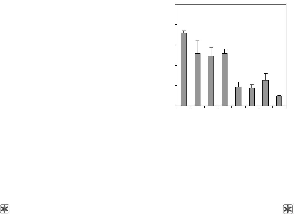
Как видно из рис. 1, жизнеспособность клеток
снижалась после криоконсервирования по всем
программам. Однако наиболее выраженное паде-
ние жизнеспособности суспензии клеток было
выявлено при использовании скоростей охлаждения
свыше 10 градусов/мин и непосредственном погру-
жении в жидкий азот. Наибольшее количество жиз-
неспособных клеток сохранилось при использова-
нии скоростей охлаждения 1; 5 и 10 градусов/мин.
Следовательно, при криоконсервировании суспен-
зии клеток надпочечников крыс необходимо ис-
nucleus, integrity of cell membrane and functional
activity level. In this research to estimate the cell viability
the double staining with FDA and PI was used. FDA
accumulation in cytoplasm is the index of cell mem-
brane integrity and metabolic activity of cells [4] and
staining with PI testifies to the nucleus presence. It is
known that PI has an ability to specifically stain nucleic
acids [18], and the use of glutaraldehyde facilitates
the PI penetration and provides its binding with DNA/
RNA in the cells preserving the nuclei. Therefore PI
staining was done in the presence of glutaraldehyde
fixative agent. Forming herewith red and green fluores-
cence may be visualized by means of fluorescent
microscope [5, 7]. Thus during cell staining with this
method there is observed simultaneous inclusion of
FDA (green fluorescence) and PI (red fluorescence),
testifying to viability of nucleated cells. The presence
of red fluorescence solely means the absence of intra-
cellular esterases in nucleated cell, i. e. its non-viability.
As Fig. 1 shows the cell viability reduced after
freeze-thawing using with all the cryopreservation
protocols. However, the most manifested fall in viability
of cell suspensions was found when using the cooling
rates above 10 degrees/min and after direct plunging
into liquid nitrogen. The highest number of viable cells
was preserved when using the cooling rates of 1, 5
and 10 degrees/min. Consequently during cryopreser-
vation of rat adrenal cell suspensions the low cooling
rates (below 10 degrees/min) should be applied.
0
20
40
60
80
100
Рис. 1. Жизнеспособность клеток надпочечников взрос-
лых крыс (по включению ФДА/ПИ) до (контроль) и после
криоконсервирования с разными скоростями охлажде-
ния; * – различия достоверны по отношению к контролю,
р < 0,05.
Fig. 1. Adrenal cell viability of adult rats (on the inclusion
of FDA/PI) prior to (the control) and after freeze-thawing
with different cooling rates; * – differences are statistically
significant comparing to the control, p < 0.05.
Скорость охлаждения, градусов/мин
Cooling rate, degrees/min
Жизнеспособность клеток, %
Cell viability, %
*
*
*
*
Контроль
Control
1 5 10 15 20 40 ↓LN
2
383
problems
of cryobiology
Vol. 20, 2010, №4
проблемы
криобиологии
Т. 20, 2010, №4
пользовать низкие (до 10 градусов/мин) скорости
охлаждения.
Формирование цитоплазматических липидных
капель в стероид-продуцирующих клетках надпо-
чечников является одним из основных процессов.
Они представляют собой скопление нейтральных
липидов, обычно триглицеридов или холестери-
новых эфиров, которые используются как источник
запаса энергии в организме или как депо холесте-
рина для синтеза стероидных гормонов в стероидо-
генных тканях [3, 9, 11–13, 28]. Для визуализации в
клетках цитоплазматических липидных капель
использовали флуоресцентный краситель НК.
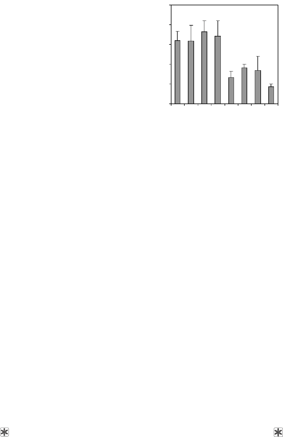
Данные, представленные на рис. 2, свидетель-
ствуют о значительном уменьшении количества
клеток, содержащих липидные капли, после замо-
раживания со скоростями 15; 20, 40 градусов/мин
и неконтролируемой скоростью охлаждения (↓LN
2
).
Образцы, криоконсервированные со скоростями 1;
5 и 10 градусов/мин, сохраняют количество клеток,
окрашенных НК на уровне контрольных значений.
Таким образом, оценивая сохранность липидных
капель в клетках после криоконсервирования, было
установлено, что достоверные отличия между
контрольным значением и данными после замора-
живания с контролируемыми скоростями охлажде-
ния 1, 5, 10 градусов/мин не выявлены. При этом
режимы замораживания с контролируемыми ско-
ростями охлаждения 15, 20 и 40 градусов/мин и
неконтролируемой скоростью охлаждения (↓LN
2
)
снижают этот показатель практически вдвое.
Одним из методов оценки жизнеспособности
стероидпродуцирующих клеток в суспензии явля-
ется определение активности фермента 3β-ГСД,
который обеспечивает конверсию прегненолона в
прогестерон и характеризует функциональную ак-
тивность стероид-продуцирующих клеток. Поэтому
наличие позитивно окрашенных клеток, имеющих
фиолетовую окраску восстановленного тетразолия,
свидетельствует о сохранности стероид-продуци-
рующих клеток в суспензии.
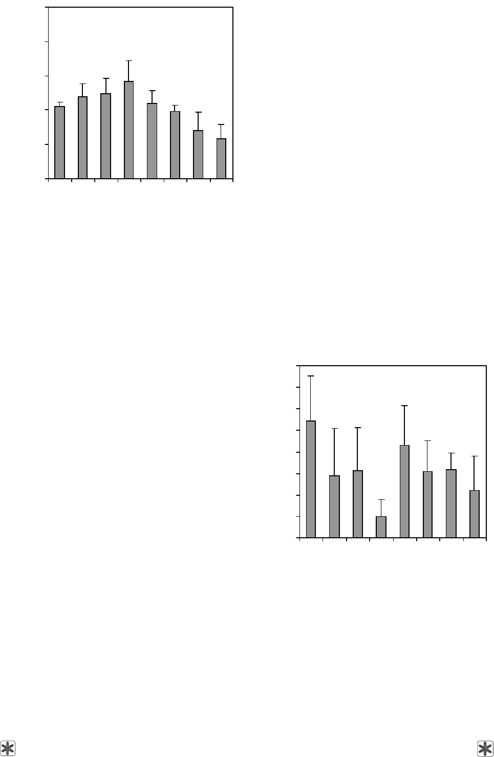
Как видно из рис. 3, сохранность 3β-ГСД
+
–
клеток после криоконсервирования оставалась на
уровне контрольных значений при охлаждении со
скоростями 1; 5, 15 и 20 градусов/мин. Охлаждение
суспензии со скоростью 40 градусов/мин и погру-
жение в жидкий азот привели к снижению коли-
чества 3β-ГСД
+
– клеток. При замораживании сус-
пензии со скоростью 10 градусов/мин сохранность
3β-ГСД
+
– клеток по сравнению с контролем была
достоверно выше. Таким образом, использование
скорости охлаждения 10 градусов/мин для замора-
живания клеток надпочечных желез крыс позво-
ляет получить суспензию клеток с повышенным
содержанием 3β-ГСД
+
– клеток.
The formation of cytoplasm lipid drops in steroid-
producing adrenal cells is one of essential processes.
They represent the cluster of neutral lipids, usually
triglycerides or cholesterol ethers, utilized as the source
of energy in an organism or as the depot of cholesterol
to synthesize steroid hormones in steroidogenic tissues
[3, 9, 11–13, 28]. To visualize the cell cytoplasm lipid
drops we used NR fluorescent dye.
The data demonstrated in Fig. 2 testify to a signifi-
cant decrease in the number of cells, containing lipid drops
after freezing with the rates of 15; 20; 40 degrees/min
and with non-controlled rate cooling (↓LN
2
). In the
samples cooled with the rates of 1, 5 and 10 degrees/min
the post- thaw number of NR
+
cells was at the level of
non-frozen control. Thus when estimating the preserva-
tion of lipid drops in the cells after cryopreservation
no statistically significant differences were revealed
between the non-frozen control values and the data
after freeze-thawing using cooling with controlled rates
of 1; 5 and 10 degrees/min. Herewith the protocols
utilizing cooling with controlled rates of 15; 20 and
40 degrees/min cooling and non-controlled rate (↓LN
2
)
reduce this index almost twice.
One of the methods to assess the viability of steroid-
producing cells in suspension is the examining the
activity of 3β-HSD enzyme, providing the conversion
of pregnenolone into progesterone and characterizes
the functional activity of steroid producing cells. There-
fore the presence of positively stained cells with violet
0
20
40
60
80
100
Рис. 2. Количество клеток надпочечников, включающих
НК до (контроль) и после криоконсервирования с раз-
ными скоростями охлаждения; * – различия достоверны
по отношению к контролю, р < 0,05.
Fig. 2. Adrenal cell number positively stained with NR prior
to (the control) and after freeze-thawing with different cool-
ing rates; * – differences are statistically significant
comparing to the control, p < 0.05.
Количество клеток, %
Cell number, %
*
*
*
*
Скорость охлаждения, градусов/мин
Cooling rate, degrees/min
Контроль
Control
1 5 10 15 20 40 ↓LN
2
384
problems
of cryobiology
Vol. 20, 2010, №4
проблемы
криобиологии
Т. 20, 2010, №4
0
2
4
6
8
10
12
14
16
Рис. 4. Количество клеток надпочечников взрослых крыс,
содержащих хромогранин А до (контроль) и после крио-
консервирования с разными скоростями охлаждения; * –
различия достоверны по отношению к контролю, р < 0,05;
# – различия достоверны по отношению к скорости ох-
лаждения 15 градусов/мин, р < 0,05.
Fig. 4. Amount of adult rats’ adrenal cells, containing chro-
mogranin A prior to (the control) and after cryopreservation
with different cooling rates; * – differences are statistically
significant comparing to the control, p < 0.05; # – differen-
ces are statistically significant comparing to the cooling
rate of 15 degrees/min, p < 0.05.
Поскольку в состав суспензии клеток надпочеч-
ников входят разные популяции клеток, мы оценили
влияние скоростей охлаждения на сохранность кле-
ток мозгового вещества надпочечников.
Как показано на рис. 4, сохранность клеток,
содержащих хромогранин А, после криоконсерви-
рования значительно снижалась относительно
контрольных значений при охлаждении со скорос-
тями 1; 5; 10; 20; 40 градусов/мин и погружении в
жидкий азот. Использование скорости охлаждения
15 градусов/мин для замораживания суспензии
клеток надпочечных желез крыс способствует со-
хранности наибольшего количества клеток, содер-
жащих хромаффинные гранулы, среди исследо-
ванных вариантов.
Таким образом, с учетом данных о влиянии
скорости охлаждения на жизнеспособность адрено-
кортикоцитов взрослых крыс, очевидно, что замо-
раживание с контролируемыми скоростями охлаж-
дения более 10 градусов/мин и использование
неконтролируемых скоростей охлаждения (прямое
погружение в жидкий азот) значительно снижает
показатели жизнеспособности клеток надпочеч-
ных желез крыс. Применение скоростей охлаж-
дения до 10 градусов/мин уменьшает количество
жизнеспособных клеток, однако позволяет сохра-
color due to presence of the reduced tetrazolium testi-
fies to the integrity of steroid producing cells in suspen-
sion.
As Fig. 3 shows the number of 3β-HSD
+
cells after
freeze-thawing remained at the level of non-frozen
control values in the case of cooling with the rates of
1; 5; 15 and 20 degrees/min. Cooling of the suspension
with the rate of 40 degrees/min and plunging into liquid
nitrogen resulted in the decreased number of 3β-HSD
+
cells. When freezing the suspension with the cooling
rate of 10 degrees/min the number of 3β-HSD
+
cells
was quite higher comparing to the control. Thus the
use of cooling rate of 10 degrees/min for freezing the
rat adrenal glands enables the obtaining of suspension
with an increased content of 3β-HSD
+
cells.
Since adrenal cell suspension comprises different
cell populations, we assessed the effect of cooling rates
on the integrity of adrenal medullar substance.
As Fig. 4 shows the number of cells containing
chromogranin A, significantly reduced after freeze-
thawing if compared with the non-frozen control values
when using the cooling rates of 1; 5; 10; 20 and
40 degrees/min and plunging into liquid nitrogen. Use
of cooling rate of 15 degrees/min to freeze the rat adre-
nal cell suspension contributes to the highest amount
Количество клеток, %
Cell number, %
*
*
*#
*
*
*#
0
20
40
60
80
100
Рис. 3. Количество 3β-ГСД
+
– клеток до (контроль) и после
криоконсервирования с разными скоростями охлаж-
дения; * – различия достоверны по отношению к конт-
ролю, р < 0,05; # – различия достоверны по отношению
к скорости охлаждения 10 градусов/мин, р < 0,05.
Fig. 3. The number of 3β-HSD
+
cells prior to (the control)
and after freeze-thawing with different cooling rates; * –
differences are statistically significant comparing to the
control, p < 0.05; # – differences are statistically significant
comparing the cooling rate of 10 degrees/min, p < 0.05.
Количество клеток, %
Cell number, %
*
*#
*#
Скорость охлаждения, градусов/мин
Cooling rate, degrees/min
Контроль
Control
1 5 10 15 20 40 ↓LN
2
Скорость охлаждения, градусов/мин
Cooling rate, degrees/min
Контроль
Control
1 5 10 15 20 40 ↓LN
2

385
problems
of cryobiology
Vol. 20, 2010, №4
проблемы
криобиологии
Т. 20, 2010, №4
нить их наибольшее количество в суспензии. Было
установлено, что при довольно низких значениях
общей сохранности клеток использование скорости
охлаждения 10 градусов/мин дает возможность
сохранить наибольшее количество стероид-про-
дуцирующих клеток в суспензии. При использо-
вании скорости охлаждения 15 градусов/мин можно
сохранить наибольшее количество хромаффинных
клеток. Причем применение скорости охлаждения
15 градусов/мин позволяет сохранить на уровне
контрольных значений количество не только хром-
аффинных, но и стероид-продуцирующих клеток.
Это может быть использовано для последующего
совместного культивирования этих клеток, так как
было показано, что совместное культивирование
клеток коркового и мозгового вещества надпочеч-
ников оказывает стимулирующее влияние на про-
лиферацию стероид-продуцирующих клеток в куль-
туре [25, 26, 30].
Выводы
1. Контролируемые скорости охлаждения до
10 градусов/мин позволяют получить более вы-
сокие показатели жизнеспособности и функцио-
нальной активности клеток надпочечных желез
крыс.
2. Установлено, что скорости охлаждения ока-
зывают селективное действие на сохранность кле-
ток коркового и мозгового вещества надпочечников.
3. Криоконсервирование суспензии клеток над-
почечников крыс со скоростью охлаждения 10 граду-
сов/мин позволяет получить суспензию, обогащен-
ную стероидпродуцирующими клетками.
4. Криоконсервирование суспензии клеток
надпочечников крыс со скоростью охлаждения
15 градусов/мин позволяет получить суспензию,
обогащенную хромаффинными клетками.
of cells, containing chromaffin granules, among the
studied variants.
Thus taking into account the data on the effect of
cooling rate on viability of adult rats’ adrenocortico-
cytes it became evident that freezing with the controlled
cooling rates above 10 degrees/min and use of non-
controlled cooling rates (direct plunging into liquid
nitrogen) significantly reduces the viability indices of
rat adrenal cells. Application of the cooling rates under
10 degrees/min decreases the number of viable cells,
however allows to preserve the highest cell number in
suspension. It has been established that quite low values
of post-thaw total cell integrity after using the cooling
rate of 10 degrees/min is accompanied with the preser-
vation of the highest amount of steroid-producing cells
in the suspension. When using the cooling rate of
15 degrees/min one may preserve the highest number
of chromaffin cells. Herewith the use of the cooling
rate of 15 degrees/min allows the preservation of
number of both chromaffin and steroid-producing cells
at the level of the non-frozen control. This may be
used for the following co-culturing of these cells, since
it has been shown that co-culturing of adrenal cells of
cortical and medullar substances renders the stimu-
lating effect on proliferation of steroid-producing cells
in the culture [25, 26, 30].
Conclusions
1. Controlled cooling rates below 10 degrees/min
enable the obtaining of higher indices of viability and
functional activity of rat adrenal cells.
2. It has been established that the choice of cooling
rates could cause a selective effect on the survival of
the adrenal cells of cortical and medullar substances.
3. Freeze-thawing of rat adrenal cell suspension
using 10 degrees/min cooling rate allowed to obtain
the suspension enriched with steroid-producing cells.
4. Freeze-thawing of rat adrenal cell suspension
using the cooling rate of 15 degrees/min allowed to
obtain the suspension enriched with chromaffin cells.
Литература
Криоконсервирование клеточных суспензий / Под ред.
А. А. Цуцаевой.– Киев: Наукова думка, 1983.– 240 с.
Тронько Н.Д., Рыбаков С.И., Комисаренко И.В. и др.
Лечение хронического гипокортицизма методом трансп-
лантации культур клеток коры надпочечных желез:
Метод. рекомендации.– Киев, 1990.– 24 с.
Renold A.E., Cahill G.F. Adipose tissue. Handbook of
Physiology.– Washington: American Physiological Society,
1965. – 824 p.
Armour A.D., Powell H.M., Boyce S.T. Fluorescein diacetate
for determination of cell viability in tissue-engineered skin //
Tissue Eng. Part C. Methods.– 2008.– Vol. 14, N1.– P. 89–96.
Bachtel N.E., Conaghan I.J., Turek P.J. The relative viability
of human spermatozoa from the vas deferens, epididymis
and testis before and after cryopreservation // Human Repro-
duction.– 1999.– Vol. 14, N12.– P. 3048–3051.
1.
2.
3.
4.
5.
References
Cryopreservation of cell suspensions / Ed. by A.A. Tsutsa-
yeva.– Kiev: Naukova dumka, 1983.– 240 p.
Tronko N.D., Rybakov S.I., Komisarenko I.V. et al. Treatment
of chronic hypocorticism by adrenal cortex cell culture trans-
plantation: Methodical recommendations.– Kiev, 1990.– 24 p.
Renold A.E., Cahill G.F. Adipose tissue. Handbook of
Physiology.– Washington: American Physiological Society,
1965. – 824 p.
Armour A.D., Powell H.M., Boyce S.T. Fluorescein diacetate
for determination of cell viability in tissue-engineered skin //
Tissue Eng. Part C. Methods.– 2008.– Vol. 14, N1.– P. 89–96.
Bachtel N.E., Conaghan I.J., Turek P.J. The relative viability
of human spermatozoa from the vas deferens, epididymis
1.
2.
3.
4.
5.

386
problems
of cryobiology
Vol. 20, 2010, №4
проблемы
криобиологии
Т. 20, 2010, №4
Benton L., Shan L.X, Hardy M.P. Differentiation of adult Leydig
cells // J. Steroid Biochem. Mol. Biol.– 1995.– Vol. 53, N1–6.–
P. 61–68.
Diaz T.M., Pertega S. FDA/PI flow cytometry assay of comple-
ment-mediated cytotoxicity of antibodies generated during
xenotransplantation // Cytometry.– 2004.– Vol. 62, N1.– P. 54–
60.
Drucker-Colin R., Verdugo-Diaz L. Cell transplantation for
Parkinson’s disease: present status // Cell. Mol. Neurobiol.–
2004.– Vol. 24, N3.– P. 301–316.
Dunn J.C., Chu Y., Qin H.H., Zupekan T. Transplantation of
adrenal cortical progenitor cells enriched by Nile red // J.
Surg. Res.– 2009.– Vol. 156, N2.– Р. 317–324.
Dunn J.C., Chu Y., Lam M.M. et al. Adrenal cortical cell
transplantation // J. Pediatr. Surg.– 2004.– Vol. 39, N12.–
P. 1856–1858.
Goldstein J. L., Brown M.S. The low-density lipoprotein
pathway and its relation to atherosclerosis // Annu. Rev.
Biochem.– 1977.– Vol. 46.– Р. 897–930.
Greenspan P., Mayer E.P., Fowler S.D. Nile red: a selective
fluorescent stain for intracellular lipid droplets // J. of Cell
Biol.– 1985.– Vol. 100, N3.– P. 965–973.
Greenspan P., Mayer E.P., Fowler S.D. Spectrofluorometric
studies of the lipid probe, nile red // J. Lipid Res.– 1985.–
Vol. 26, N7.– Р. 781–789.
Grodstein E., Hardy M.A., Goldstein M.J. A case of human
intramuscular adrenal gland transplantation as a cure for chronic
adrenal insufficiency // Am J. Transplant.– 2009.– Vol. 10,
N2.– P. 431–433.
Hawkins K.L., Lloyd R.V., Toy K.A. Immunohistochemical
localization of chromogranin A in normal tissues from labo-
ratory animals // Vet. Pathol.– 1989.– Vol. 26, N6.– P. 488– 498.
Hines G.A., Azziz R. Impact of architectural disruption on
adrenocortical steroidogenesis in vitro // J. Clin. Endocrinol.
Metab.– 1999.– Vol. 84, N3.– P. 1017–1021.
Hornsby P.J. Transplantation of adrenocortical cells // Rev.
Endocr. Metab. Disord.– 2001.– Vol. 2, N3.– P. 313–321.
Jones K.H., Senft J.A. An improved method to determine cell
viability by simultaneous staining with fluorescein diacetate-
propidium iodide // J. Histochem. Cytochem.– 1985.– Vol. 33,
N1.– Р. 77–79.
Kemppainen R.J., Behrend E.N. Adrenal physiology // Vet.
Clin. North Am. Small. Anim. Pract.– 1997.– Vol. 27, N2.– P.173–
186.
Lakey J.R., Anderson T.J., Rajotte R.V. Novel approaches to
cryopreservation of human pancreatic islets // Transplan-
tation.– 2001.– Vol. 27, N72(6).– Р. 1005–1011.
Lee M.K., Bae Y.H. Cell transplantation for endocrine
disorders // Adv. Drug Deliv. Rev.– 2000.– Vol. 42, N1–2.–
P. 103–120.
Maruyama M., Kenmochi T., Sakamoto K. et al. Simplified
method for cryopreservation of islets using hydroxyethyl
starch and dimethyl sulfoxide as cryoprotectants // Transplant.
Proc.– 2004.– Vol. 36, N4.– Р. 1133–1134.
McKay D.B., Karow A.M.Jr. Factors to consider in the asses-
sment of viability of cryopreserved islets of Langerhans //
Cryobiology.– 1983.– Vol. 20, N2.– P. 151–160.
Scheumann G.F., Hiller W.F., Schroder S. et al. Adrenal
cortex transplantation after bilateral total adrenalectomy in the
rat // Henry Ford Hosp Med J.– 1989.– Vol. 37, N3–4.– P. 154–
156.
Shepherd S.P., Holzwarth M.A. Chromaffin-adrenocortical cell
interactions: effects of chromaffin cell activation in adrenal
and testis before and after cryopreservation // Human Repro-
duction.– 1999.– Vol. 14, N12.– P. 3048–3051.
Benton L., Shan L.X, Hardy M.P. Differentiation of adult Leydig
cells // J. Steroid Biochem. Mol. Biol.– 1995.– Vol. 53, N1–6.–
P. 61–68.
Diaz T.M., Pertega S. FDA/PI flow cytometry assay of comple-
ment-mediated cytotoxicity of antibodies generated during
xenotransplantation // Cytometry.– 2004.– Vol. 62, N1.– P. 54–
60.
Drucker-Colin R., Verdugo-Diaz L. Cell transplantation for
Parkinson’s disease: present status // Cell. Mol. Neurobiol.–
2004.– Vol. 24, N3.– P. 301–316.
Dunn J.C., Chu Y., Qin H.H., Zupekan T. Transplantation of
adrenal cortical progenitor cells enriched by Nile red // J.
Surg. Res.– 2009.– Vol. 156, N2.– Р. 317–324.
Dunn J.C., Chu Y., Lam M.M. et al. Adrenal cortical cell
transplantation // J. Pediatr. Surg.– 2004.– Vol. 39, N12.–
P. 1856–1858.
Goldstein J. L., Brown M.S. The low-density lipoprotein
pathway and its relation to atherosclerosis // Annu. Rev.
Biochem.– 1977.– Vol. 46.– Р. 897–930.
Greenspan P., Mayer E.P., Fowler S.D. Nile red: a selective
fluorescent stain for intracellular lipid droplets // J. of Cell
Biol.– 1985.– Vol. 100, N3.– P. 965–973.
Greenspan P., Mayer E.P., Fowler S.D. Spectrofluorometric
studies of the lipid probe, nile red // J. Lipid Res.– 1985.–
Vol. 26, N7.– Р. 781–789.
Grodstein E., Hardy M.A., Goldstein M.J. A case of human
intramuscular adrenal gland transplantation as a cure for chronic
adrenal insufficiency // Am J. Transplant.– 2009.– Vol. 10,
N2.– P. 431–433.
Hawkins K.L., Lloyd R.V., Toy K.A. Immunohistochemical
localization of chromogranin A in normal tissues from labo-
ratory animals // Vet. Pathol.– 1989.– Vol. 26, N6.– P. 488– 498.
Hines G.A., Azziz R. Impact of architectural disruption on
adrenocortical steroidogenesis in vitro // J. Clin. Endocrinol.
Metab.– 1999.– Vol. 84, N3.– P. 1017–1021.
Hornsby P.J. Transplantation of adrenocortical cells // Rev.
Endocr. Metab. Disord.– 2001.– Vol. 2, N3.– P. 313–321.
Jones K.H., Senft J.A. An improved method to determine cell
viability by simultaneous staining with fluorescein diacetate-
propidium iodide // J. Histochem. Cytochem.– 1985.– Vol. 33,
N1.– Р. 77–79.
Kemppainen R.J., Behrend E.N. Adrenal physiology // Vet. Clin.
North Am. Small. Anim. Pract.– 1997.– Vol. 27, N2.– P.173–186.
Lakey J.R., Anderson T.J., Rajotte R.V. Novel approaches to
cryopreservation of human pancreatic islets // Transplan-
tation.– 2001.– Vol. 27, N72(6).– Р. 1005–1011.
Lee M.K., Bae Y.H. Cell transplantation for endocrine
disorders // Adv. Drug Deliv. Rev.– 2000.– Vol. 42, N1–2.–
P. 103–120.
Maruyama M., Kenmochi T., Sakamoto K. et al. Simplified
method for cryopreservation of islets using hydroxyethyl
starch and dimethyl sulfoxide as cryoprotectants // Transplant.
Proc.– 2004.– Vol. 36, N4.– Р. 1133–1134.
McKay D.B., Karow A.M.Jr. Factors to consider in the asses-
sment of viability of cryopreserved islets of Langerhans //
Cryobiology.– 1983.– Vol. 20, N2.– P. 151–160.
Scheumann G.F., Hiller W.F., Schroder S. et al. Adrenal cortex
transplantation after bilateral total adrenalectomy in the rat // Henry
Ford Hosp Med J.– 1989.– Vol. 37, N3–4.– P. 154–156.
Shepherd S.P., Holzwarth M.A. Chromaffin-adrenocortical cell
interactions: effects of chromaffin cell activation in adrenal
6.
7.
8.
9.
10.
11.
12.
13.
14.
15.
16.
17.
18.
19.
20.
21.
22.
23.
24.
25.
6.
7.
8.
9.
10.
11.
12.
13.
14.
15.
16.
17.
18.
19.
20.
21.
22.
23.
24.
25.

387
problems
of cryobiology
Vol. 20, 2010, №4
проблемы
криобиологии
Т. 20, 2010, №4
cell co-cultures // Am. J. Physiol Cell Physiol.– 2001.– Vol.280,
N1.– P. 61–71.
Shepherd S.P., Holzwarth M.A. Frog chromaffin and adre-
nocortical cell co-cultures: a model for the study of medullary
control of corticosteroidogenesis // J. Neuroendocrinol.– 1998.–
Vol. 10, N7.– P. 539–549.
Silani V., Pizzuti A., Strada O., Falini A. et al. Cryopreserva-
tion of human fetal adrenal medullary cells // Brain Res.–
1988.– Vol. 28, N454(1–2).– P. 383–386.
Stanley D.F., Greenspan P. Application of nile red, a fluo-
rescent hydrophobic probe, for the detection of neutral lipid
deposits in tissue sections: comparison with oil red O / /J. Histo-
chem. Cytochem.– 1985.– Vol. 33, N8.– Р. 833–836.
Tchoukalova Y.D., Harteneck D.A., Karwoski R.A. et al. A
quick, reliable, and automated method for fat cell sizing // J.
Lipid. Res.– 2003.– Vol. 44, N9.– P. 1795–1801.
Walker S.W., Lightly E.R.T., Milner S.W., Williams B.C. Cate-
cholamine stimulation of cortisol secretion by 3-day primary
cultures of purified zona fasciculate/reticularis cells isolated
from bovine adrenal cortex // Mol. Cell Endocrinol.– 1988.–
Vol. 57, N3.– P. 139–147.
Поступила 28.09.2010
Рецензент Е.И. Смольянинова
cell co-cultures // Am. J. Physiol Cell Physiol.– 2001.– Vol.280,
N1.– P. 61–71.
Shepherd S.P., Holzwarth M.A. Frog chromaffin and adre-
nocortical cell co-cultures: a model for the study of medullary
control of corticosteroidogenesis // J. Neuroendocrinol.– 1998.–
Vol. 10, N7.– P. 539–549.
Silani V., Pizzuti A., Strada O., Falini A. et al. Cryopreserva-
tion of human fetal adrenal medullary cells // Brain Res.–
1988.– Vol. 28, N454(1–2).– P. 383–386.
Stanley D.F., Greenspan P. Application of nile red, a fluo-
rescent hydrophobic probe, for the detection of neutral lipid
deposits in tissue sections: comparison with oil red O / /J. Histo-
chem. Cytochem.– 1985.– Vol. 33, N8.– Р. 833–836.
Tchoukalova Y.D., Harteneck D.A., Karwoski R.A. et al. A
quick, reliable, and automated method for fat cell sizing // J.
Lipid. Res.– 2003.– Vol. 44, N9.– P. 1795–1801.
Walker S.W., Lightly E.R.T., Milner S.W., Williams B.C. Cate-
cholamine stimulation of cortisol secretion by 3-day primary
cultures of purified zona fasciculate/reticularis cells isolated
from bovine adrenal cortex // Mol. Cell Endocrinol.– 1988.–
Vol. 57, N3.– P. 139–147.
Accepted in 28.09.2010
26.
27.
28.
29.
30.
26.
27.
28.
29.
30.

388
* Автор, которому необходимо направлять корреспонденцию:
ул. Переяславская, 23, г. Харьков, Украина 61015; тел.:+38
(057) 373-41-35, факс: +38 (057) 373-30-84, электронная почта:
cryo@online.kharkov.ua
* To whom correspondence should be addressed: 23,
Pereyaslavskaya str., Kharkov, Ukraine 61015; tel.:+380 57 373
4135, fax: +380 57 373 3084, e-mail: cryo@online.kharkov.ua
Institute for Problems of Cryobiology and Cryomedicine of the Na-
tional Academy of Sciences of Ukraine, Kharkov, Ukraine
Институт проблем криобиологии и криомедицины
НАН Украины, г. Харьков
УДК 57.043:612.111:352.462
Н.А. ПИСАРЕНКО, Н.М. ШПАКОВА*
Влияние амфифильных веществ на гипертонический гемолиз
эритроцитов, модифицированных температурой 49°С
UDC 57.043:612.111:352.462
N.A. PISARENKO, N.M. SHPAKOVA*
Effect of Amphiphiles on Hypertonic Hemolysis of Erythrocytes,
Modified with Temperature of 49°C
Исследовали влияние тепловой предобработки эритроцитов человека и быка на их чувствительность к гипертоническому
стрессу (4,0 моль/л NaCl) при температуре 37 и 0°С. Показано, что инкубация клеток при 49°С, вызывающая денатурацию
спектрина, снижает уровень гипертонического повреждения эритроцитов человека и не влияет на сохранность эритроцитов
быка при 37°С. При 0°С гипертонический гемолиз модифицированных эритроцитов млекопитающих не изменяется. Денатурация
спектрина цитоскелета эритроцитов человека и быка приводит к снижению антигемолитического действия амфифильных
веществ (децилсульфата натрия, додецил-β,D-мальтозида, трифторперазина) при температуре 37 и 0°С.
Ключевые слова: гипертонический стресс, эритроциты, спектрин, амфифильные соединения.
Досліджували вплив теплової передобробки еритроцитів людини і бика на їх чутливість до гіпертонічного стресу (4,0 моль/л
NaCl) при температурі 37 і 0°С. Показано, що інкубування клітин при 49°С, яка викликає денатурацію спектрину, знижує
рівень гіпертонічного пошкодження еритроцитів людини і не впливає на стійкість еритроцитів бика при 37°С. При 0°С
гіпертонічний гемоліз модифікованих еритроцитів ссавців не змінюється. Денатурація спектрину цитоскелета еритроцитів
людини і бика знижує антигемолітичну дію амфіфільних сполук (децилсульфату натрію, додецил-β,D-мальтозиду,
трифторперазину) при температурі 37 і 0°С.
Ключові слова: гіпертонічний стрес, еритроцити, спектрин, амфіфільні сполуки.
The effect of thermal pre-treatment of human and bovine erythrocytes on their sensitivity to hypertonic stress (4.0 mol/l NaCl)
at 37 and 0°C was studied. It has been shown that cell incubation at 49°C, causing spectrin denaturation, decreases the level of human
erythrocyte hypertonic damage and does not affect the integrity of bovine erythrocytes at 37°C. Hypertonic hemolysis of modified
mammalian erythrocytes does not change at 0°C. Cytoskeletal spectrin denaturation of bovine and human erythrocytes results in
reduction of antihemolytic activity of amphiphilic substances (sodium decyl sulfate, dodecyl-β,D-maltoside, trifluoroperazine) at 37
and 0°C.
Key words: hypertonic stress, erythrocytes, spectrin, amphiphilic compounds.
problems
of cryobiology
Vol. 20, 2010, №4
проблемы
криобиологии
Т. 20, 2010, №4
One of the main reasons of cell damage during
freezing is believed to be concentrating of extracellular
solution, associated with water freezing [2, 15]. A
hypertonic stress is the experimental model for studying
this cryodamage factor.
Aggregative and structural state of cytoskeletal
proteins affects the degree of their association with
anchor proteins of membrane, maintaining viscous-
elastic properties of membrane and cell resistance to
stress effect [2]. Therefore modification of cytoskeletal
proteins is important for understanding the mechanisms
of erythrocyte resistance under hypertonic stress.
Whereas amphiphilic compounds, which effect is
realized on membrane, significantly decrease the level
of cell damage under hypertonic conditions [5] there
was of interest to investigate their antihemolytic activity
for erythrocytes with cytoskeleton-membrane complex,
modified with the temperature of 49°C.
Предполагают, что одна из основных причин
повреждения клеток при замораживании – концент-
рирование внеклеточного раствора, связанное с
вымерзанием воды [2, 15]. Экспериментальной мо-
делью для изучения данного фактора криоповреж-
дения является гипертонический стресс.
Агрегатное и структурное состояние белков ци-
тоскелета влияет на степень их ассоциации с якор-
ными белками мембраны, поддерживает вязко-
эластические свойства мембраны и устойчивость
клетки к действию стресса [2]. Поэтому модифи-
кация цитоскелетных белков имеет важное значе-
ние для понимания механизмов устойчивости эрит-
роцитов в условиях гипертонического стресса. Ис-
ходя из того, что амфифильные соединения, дейст-
вие которых реализуется на мембране, значительно
снижают уровень повреждения клеток в гиперто-
нических условиях [5], представлял интерес ис-