Yang J. (ed.) Biometrics
Подождите немного. Документ загружается.


Retinal Identification 11
1. Severe astigmatism: An astigmatic eye’s optics image dots as lines. This results in
problems with focusing and also bad optical quality of measurements.
2. Cataracts: A cataract is an eye condition in which clouding develops in the crystallinen
lens. The lens becomes opaque so seeing becomes compromised. Obviously any optical
measurement in the eye, including the RI, becomes increasingly difficult.
3. Severe eye diseases, such as the age-related macula degeneration (AMD), can change the
structure of the retina, either by destroying retinal tissue or by stimulating growth of new
blood vessels.
6.2 RI using birefringence
Eight eyes of four volunteer subjects were measured. Both absorption- and birefringence-
based signals were recorded two times for each eye. For verifying purposes, fundus
photographs were taken from all the eyes. The measured peaks and the blood vessels on
the fundus photos were compared as follows:
A20
◦
diameter circle was drawn on a transparent overlay, on which the measured peaks were
marked at corresponding angles on the perimeter of the circle. The transparency was then
placed on the fundus photo to compare the marked signal peaks with the blood vessels on
the photo. Only the vessels larger than a certain threshold size (set for each eye individually)
were taken into account, i.e. the smallest vessels were ignored. In this way, the numbers
of blood vessels corresponding to the peaks in the measured absorption/reflectance and
birefringence-based tracings were calculated. If a confirmed peak did not correspond to a
vessel above the threshold size on the fundus photo, it was considered a false positive.
Altogether 55 blood vessels were located on the fundus photos of the ’better’ eyes (the ones
which yielded clearer signals) of the four volunteers, of which 34 could be correlated with the
’peaks’ in the measured signal. The calculated sensitivities (number of vessels identified /
number of vessels altogether) and specificities (number of positive recognitions / number
of positive + false positive recognitions) - are presented in Table 1. The columns in the
tables represent total percentage of blood vessel ’peaks’ correlating with vessels in the fundus
photo (Total), from the two reflection/absorption measurements (Sum) and from the two
birefringence measurements (Diff).
Total Sum Diff
Average sensitivity 69% 51% 33%
Average specificity 78% 76% 60%
Table 1. Summarized results of the measurements taken from the eye with a better signal of
each subject.
6.2.1 Limitations
Our system was of ’proof-of-principle’ -nature and - unlike the conventional RI technique -
had no inherent defocus compensation; the test subjects were required to have a refractive
error of less than about
±2 diopters or a good corrected vision using contact lenses. The eye
conditions disturbing the measurement include cataracts and astigmatism as well as a severe
glaucoma, which damages the RNFL by killing the nerve fibers going through the optic nerve.
109
Retinal Identification

12 Will-be-set-by-IN-TECH
6.3 Other techniques
The author in unaware of any scientific studies on the accuracy of the other techniques
mentioned here.
7. Discussion
Retinal Identification remains the most reliable and secure biometric. Falsifying an image of
a retinal blood vessel pattern appears impossible. The eye scans are generally considered
invasive or even harmful, especially if a l aser (even if the light is weak intensity and harmless
to the eye, as in our system). However, the enrollees should be able to overcome their fear of
these scans once they acquire more user experience.
The original RI technique uses the choroidal blood vessel pattern for identification; however,
several newer RI techniques center the scan around the optic disc. The main drawback in
such a device is that the eye has to fixate 15 degrees off-axis while being measured. However,
if this is achieved, the blood vessels around the optic disc are easier to detect - in addition, the
- as the authors and his co-workers proved - birefringence-based blood vessel detection can
help detecting more blood vessels, only at a small cost of specificity. The measured signal was
also directly linked to blood vessels and not to other retinal structures, as in the original RI
technique.
The newer techniques - imaging the optic disc using an SLO, or the combined retina- and
iris scan - appear very promising. However, the author is unaware either of any commercial
devices or of any scientific studies on the accuracy of these identifying method.
The use of retinal identification is not limited to humans. In 2004, a patent was filed on
identifying various animals using their retinal blood vessel pattern [Golden et al. (2004)]. To
develop the technique for animals, a company OptiBrand was started (Ft. Collins, Colorado,
USA). The company produces and develops hand-held video camera -based devices which
provide an acceptable image an animal’s eye fundus to allow identification. This method is
certainly preferable over the traditional hot iron branding, which is not only painful to the
animal, but also costly in the lost hide value. When a false identification is not disastrous (as
opposed to some high security installations), a simple video camera based device provides an
accurate enough identification.
8. Appendix: The Stokes parameters
The modern treatment of polarization was first suggested by Stokes in the mid-1800’s. The
polarization state of the light can be completely represented with four quantities, the Stokes
parameters:
S
0
= E
2
x
T
+ E
2
y
T
S
1
= E
2
x
T
−E
2
y
T
S
2
= 2E
x
E
y
cos
T
S
3
= 2E
x
E
y
sin
T
,(1)
where
=
y
−
x
is the phase difference between x- and y-polarization and the
T
denote
time averages.
For totally unpolarized light S
0
> 0andS
1
= S
2
= S
3
= 0. For completely polarized light, on
110
Biometrics

Retinal Identification 13
the other hand, S
2
0
= S
2
1
+ S
2
2
+ S
2
3
. The polarization state of the light can be defined as
V
=
S
2
1
+ S
2
2
+ S
2
3
S
0
.(2)
The importance of the Stokes parameters lies in the fact that they are connected to easily
measurable intensities:
S
0
(θ, ρ)=I(0
◦
,0)+I(90
◦
,0)
S
1
(θ, ρ)=I(0
◦
,0) − I(90
◦
,0)
S
2
(θ, ρ)=I(45
◦
,0) − I(135
◦
,0)
S
3
(θ, ρ)=I(45
◦
, π/2) − I(135
◦
, π/2),(3)
where θ is the angle of the azimuth vector measured from the x-plane and ρ is the
birefringence. These can be easily measured: for example, S
1
can be measured with polarizing
beam splitter and two detectors, which are placed so that they measure the intensities coming
out of the beam splitter, and S
3
can be measured by adding a quarter wave plate before the
aforementioned system.
9. References
Agopov, M.; Gramatikov, B.I.; Wu, Y.K.; Irsch, K; Guyton, D.L. (2008). Use of retinal nerve
fiber layer birefringence as an addition to absorption in retinal scanning for biometric
purposes. Applied Optics 47: 1048-1053
Bettelheim, F. (1975). On optical anisotrophy of the lens fiber cells. Exp. Eye Res. 21: 231-234
Cope, W.; Wolbarsht; Yamanashi, B. (1978). The corneal polarization cross. J. Opt Soc Am 68:
1149-1140
Dreher, A.; Reiter, K. (1992). Scanning laser polarimetry of the retinal nerve fiber layer. SPIE
1746:34-41
Fernandez, E. J.; Unterhuber, A; Pieto, P.M.; Hermann, B.; Drexler, W.; Artal, P. (2005).
Ocular aberrations as a function of wavelength in the near infrared measured with a
femtosecond laser. Optics Expr ess 13:400-409
Hill, R. (1978). Apparatus and method for identifying individuals through their retinal
vasculature patterns, United States Patent 4109237
Hill, R. (1981). Rotating beam ocular identification apparatus and method. United S tates Patent
4,393,366
Hill, R. (1986). Fovea-centered eye fundus scanner. United States Patent 4620318
Johnson, J.C.; Hill, R. (1996). Eye fundus optical scanner system and method, United States
Patent 5532771
Golden, B .L.; Rollin, B.E.; Switzer, R.; Comstock, C.R. (2004). Retinal vasculature image
acquisition apparatus and method, United States Patent 6766041
Arndt, J.H. (1990). Optical alignment system. United States Patent 4923297
Klein Brink, H. B.; Van Blockland, G.J. (1988). Birefringence of the human foveal area assessed
in vivo with the Mueller-Matrix ellipsometry. J. Opt. Soc. Amer. A 5:49-57
Marshall, J.; Usher, D. (2004). Method for generating a unique and consistent signal pattern
for identification of an individual, United States Paten t 6757409
111
Retinal Identification

14 Will-be-set-by-IN-TECH
Muller, D. F.; Heacock G. L.; Usher D. B. (2007). Method and system for generating a combined
retina/iris patter biometric, United States Patent 7248720
Sandia National Laboratories (1991). Performance Evaluation of Biometric Identification
Devices, Technical Report SAND91-0276, UC-906
Simon C.; Goldstein I. (1935). A New Scientific Method of Identification, New York State Journal
of Medicine, Vol. 35, No. 18, pp. 901-906
Tower, P. (1955). The fundus Oculi in monozygotic twins: Report of six pairs of identical twins,
Archives of Ophthalmology, Vol. 54, pp. 225-239
Weinreb, R.; Dreher A.; Coleman, A.; Quigley H.; Shaw, B.; Reiter, K. (1990). Histopatologic
validation of Fourier-ellipsometry measurements of retinal nerve fiber layer
thickness, Arch Opthalmol 108:557-60
112
Biometrics

0
Retinal Vessel Tree as Biometric Pattern
Marcos Ortega and Manuel G. Penedo
University of Coruña
Department of Computer Science
Spain
1. Introduction
In current society, reliable authentication and authorization of individuals are becoming
more and more necessary tasks for everyday activities or applications. Just for instance,
common situations such as accessing to a building restricted to authorized people (members,
workers,...), taking a flight or performing a money transfer require the verification of the
identity of the individual trying to perform these tasks. When considering automation of
the identity verification, the most challenging aspect is the need of high accuracy, in terms
of avoiding incorrect authorizations or rejections. While the user should not be denied to
perform a task if authorized, he/she should be also ideally inconvenienced to a minimum
which further complicates the whole verification process Siguenza Pizarro & Tapiador Mateos
(2005).
With this scope in mind, the term biometrics refers to identifying an individual based
on his/her distinguished intrinsic characteristics. Particularly, this characteristics usually
consist of physiological or behavioral features. Physiological features, such as fingerprints,
are physical characteristics usually measured at a particular point of time. Behavioral
characteristics, such as speech or handwriting, make reference to the way some action
is performed by every individual. As they characterize a particular activity, behavioral
biometrics are usually measured over time and are more dependant on the individual’s
state of mind or deliberated alteration. To reinforce the active versus passive idea of both
paradigms, physiological biometrics are also usually referred to as static biometrics while
behavioral ones are referred to as dynamic biometrics.
The traditional authentication systems based on possessions or knowledge are widely spread
in the society but they have many drawbacks that biometrics try to overcome. For instance, in
the scope of the knowledge-based authentication, it is well known that password systems are
vulnerable mainly due to the wrong use of users and administrators. It is not rare to find some
administrators sharing the same password, or users giving away their own to other people.
One of the most common problems is the use of easily discovered passwords (child names,
birth dates, car plate,...). On the other hand, the use of sophisticated passwords consisting
of numbers, upper and lower case letters and even punctuation marks makes it harder to
remember them for an user.
Nevertheless, the password systems are easily broken by the use of brute force where powerful
computers generate all the possible combinations and test it against the authentication system.
In the scope of the possession-based authentication, it is obvious that the main concerns are
6

2 Will-be-set-by-IN-TECH
related to the loss of the identification token. If the token was stolen or found by another
individual, the privacy and/or security would be compromised. Biometrics overcome most
of these concerns while they also allow an easy entry to computer systems to non expert
users with no need to recall complex passwords. Additionally, commercial webs on the
Internet are favored not only by the increasing trust being transmitted to the user but also
by the possibility of offering a customizable environment for every individual along with the
valuable information on personal preferences for each of them.
Many different human biometrics have been used to build a valid template for verification
and identification tasks. Among the most common biometrics, we can find the fingerprint
Bolle et al. (2002); Maio & Maltoni (1997); Seung-Hyun et al. (1995); Venkataramani & Kumar
(2003), iris Chou et al. (2006); He et al. (2008); Kim, Cho, Choi & Marks (2004); Ma et al. (2002);
Nabti & Bouridane (2007) or face Kim, Kim, Bang & Lee (2004); Kisku et al. (2008); Mian et al.
(2008); Moghaddam & Pentland (1997); Yang et al. (2000) or hand geometry Jain et al. (1999);
Sidlauskas (1988); systems Lab (n.d.); Zunkel (1999). However, there exist other emerging
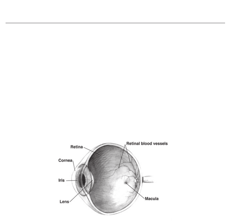
biometrics where we can find retina biometrics. Identity verification based on retina uses the
blood vessels pattern present in the retina (Figure1).
Fig. 1. Schema of the retina in the human eye. Blood vessels are used as biometric
characteristic.
Retinal blood vessel pattern is unique for each human being even in the case of identical
twins. Moreover, it is a highly stable pattern over time and totally independent of genetic
factors. Also, it is one of the hardest biometric to forge as the identification relies on the blood
circulation along the vessels. These property make it one of the best biometric characteristic
in high security environments. Its main drawback is the acquisition process which requires
collaboration from the user and it is sometimes perceived as intrusive. As it will be further
discussed, some advances have been done in this field but, in any case, this continues to be
the weak point in retinal based authentication.
Robert Hill introduced the first identification system based on retina Hill (1999). The general
idea was that of taking advantage of the inherent properties of the retinal vessel pattern to
build a secure system. The system acquired the data via a scanner that required the user to be
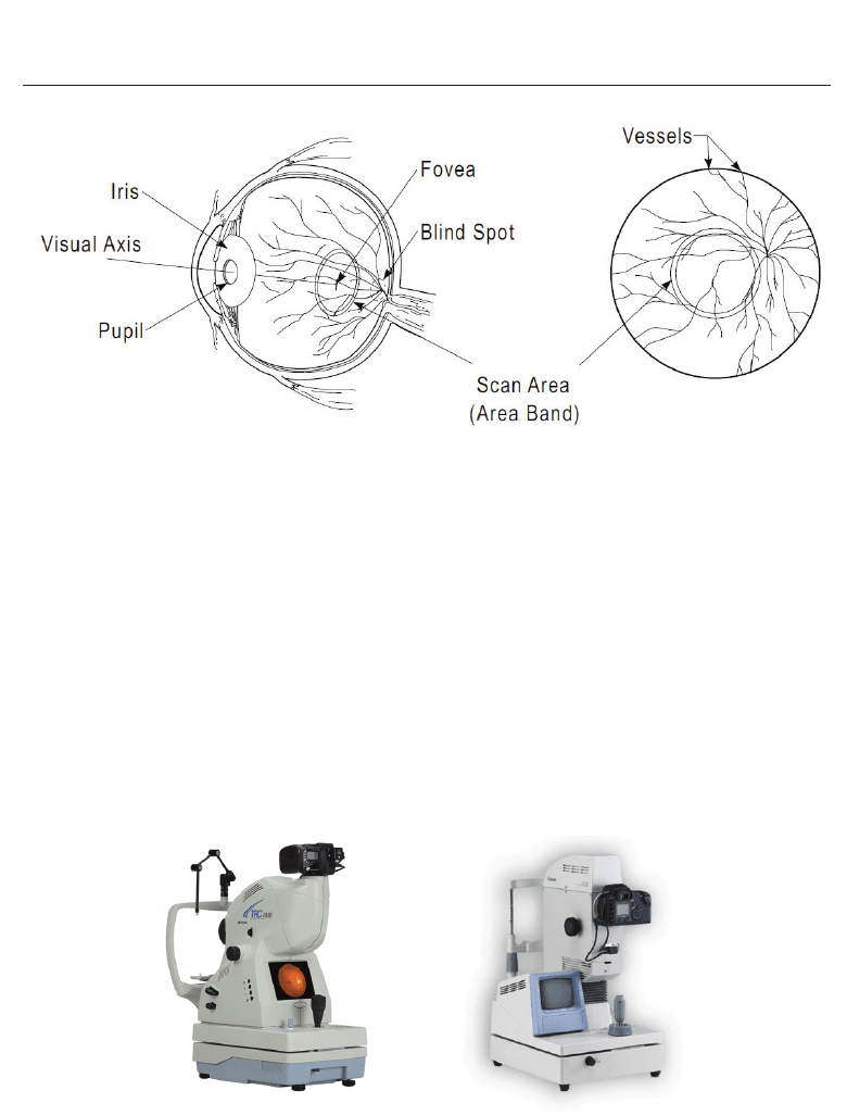
still for a few seconds. The scanner captured a band in the blood vessels area similar to the
one employed in the iris recognition as shown in Figure 2.
The scanned area is a circular band around blood vessels. This contrast information of this area
is processed via fast Fourier transform. The transformed data forms the final biometric pattern
114
Biometrics

Retinal Vessel Tree as Biometric Pattern 3
Fig. 2. Illustration of the scan area in the retina used in the system of Robert Hill.
considered in this system. This pattern worked good enough as the acquisition environment
was very controlled. Of course, this is also the source of the major drawbacks present in
the device: the data acquisition process. This process was both slow and uncomfortable
for the user. Moreover, the hardware was very expensive and, therefore, it rendered the
system hardly appealing. Finally, the result was that the use of retinal pattern as a biometric
characteristic, despite all its convenient properties, was discontinued.
Nowadays, retinal image cameras (Figure 3) are capable of taking a photograph of the retina
area in a human eye without any intrusive or dangerous scanning. Also, currently, the devices
are cheaper and more accessible in general. This technology reduces the perception of danger
by the user during the retina acquisition process but also brings more freedom producing
a more heterogeneous type of retinal images to work with. The lighting conditions and the
movement of the user’s eye vary between acquisitions. This produces as a result that previous
systems based on contrast information of reduced areas may lack the required precision in
some cases, increasing the false rejection rate.
Fig. 3. Two retinal image cameras. The retinal image is acquired by taking an instant
photograph.
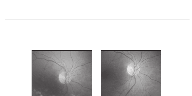
In Figure 4 it can be observed two images from the same person acquired at different times by
the same retinograph. There are some zones in the retinal vessels that can not be compared
115
Retinal Vessel Tree as Biometric Pattern

4 Will-be-set-by-IN-TECH
because of the lack of information in one of the images. Thus, to allow the retinal biometrics
to keep and increase the acquisition comfortability, it is necessary to implement a more robust
methodology that, maintaining the extremely low error rates, is capable to cope with a more
heterogeneous range of retinal images.
Fig. 4. Example of two digital retina images from the same individual acquired by the same
retinal camera at different times.
This work is focused on the proposal of a novel personal authentication system based on the
retinal vessel tree. This system deals with the new challenges in the retinal field where a more
robust pattern has to be designed in order to increase the usability for the acquisition stage.
In this sense, the approach presented here to the retinal recognition is closer to the fingerprint
developments than to the iris ones as the own structure of the retinal vessel tree suggests.
Briefly, the objectives of this work are enumerated:
• Empirical evaluation of the retinal vessel tree as biometric pattern
• Design a robust, easy to store and process biometric pattern making use of the whole retinal
vessel tree information
• Development of an efficient and effective methodology to compare and match such retinal
patterns
• Analysis on similarity metrics performance to establish reliable thresholds in the
authentication process
To deal with the suggested goals, the rest of this document is organized as follows. Second
section introduces previous works and research on the retinal vessel tree as biometric pattern.
Section 3 presents the methodology developed to build the authentication system, including
biometric template construction and template matching algorithms. Section 4 discusses the
experiments aimed to test the proposed methodologies, including an analysis of similarity
measures. Finally, Section 5 offers some conclusions and final discussion.
2. Related work
Awareness of the uniqueness of the retinal vascular pattern dates back to 1935 when two
ophthalmologists, Drs. Carleton Simon and Isodore Goldstein, while studying eye disease,
realized that every eye has its own unique pattern of blood vessels. They subsequently
published a paper on the use of retinal photographs for identifying people based on their
blood vessel patterns Simon & Goldstein (1935). Later in the 1950s, their conclusions were
supported by Dr. Paul Tower in the course of his study of identical twins. He noted that,
of any two persons, identical twins would be the most likely to have similar retinal vascular
116
Biometrics

Retinal Vessel Tree as Biometric Pattern 5
patterns. However, Tower showed that, of all the factors compared between twins, retinal
vascular patterns showed the least similarities Tower (1955).
Blood vessels are among the first organs to develop and are entirely derived from the
mesoderm. Vascular development occurs via two processes termed vasculogenesis and
angiogenesis. Vasculogenesis, this is, the blood vessel assembly during embryogenesis, begins
with the clustering of primitive vascular cells or hemangioblasts into tube-like endothelial
structures, which define the pattern of the vasculature. In angiogenesis, new vessels arise by
sprouting of budlike and fine endothelial extensions from preexisting vessels Noden (1989).
In a more recent study Whittier et al. (2003), retinal vascular pattern images from livestock
were digitally acquired in order to evaluate their pattern uniqueness. To evaluate each retinal
vessel pattern, the dominate trunk vessel of bovine retinal images was positioned vertically
and branches on the right and left of the trunk and other branching points were evaluated.
Branches from the left (mean 6.4 and variance 2.2) and the right (mean 6.4 and variance 1.5) of
the vascular trunk; total branches from the vascular trunk (mean 12.8 and variance 4.3), and
total branching points (mean 20.0 and variance 13.2) showed differences across all animals
(52). A paired comparison of the retinal vessel patterns from both eyes of 30 other animals
confirmed that eyes from the same animal differ. Retinal images of 4 cloned sheep from the
same parent line were evaluated to confirm the uniqueness of the retinal vessel patterns in
genetically identical animals. This would be confirming the uniqueness of animal retinal
vascular pattern suggested earlier in the 1980s also by De Schaepdrijver et al. (1989).
In general, retinal vessel tree his is a unique pattern in each individual and it is almost
impossible to forge that pattern in a false individual. Of course, the pattern does not change
through the individual’s life, unless a serious pathology appears in the eye. Most common
diseases like diabetes do not change the pattern in a way that its topology is affected.
Some lesions (points or small regions) can appear but they are easily avoided in the vessels
extraction method that will be discussed later. Thus, retinal vessel tree pattern has been
proved a valid biometric trait for personal authentication as it is unique, time invariant and
very hard to forge, as showed by Mariño et al. C.Mariño et al. (2003); Mariño et al. (2006),
who introduced a novel authentication system based on this trait. In that work, the whole
arterial-venous tree structure was used as the feature pattern for individuals. The results
showed a high confidence band in the authentication process but the database included only
6 individuals with 2 images for each of them. One of the weak points of the proposed system
was the necessity of storing and handling a whole image as the biometric pattern. This
greatly difficults the storing of the pattern in databases and even in different devices with
memory restrictions like cards or mobile devices. In Farzin et al. (2008) a pattern is defined
using the optic disc as reference structure and using multi scale analysis to compute a feature
vector around it. Good results were obtained using an artificial scenario created by randomly
rotating one image per user for different users. The dataset size is 60 images, rotated 5 times
each. The performance of the system is about a 99% accuracy. However, the experimental
results do not offer error measures in a real case scenario where different images from the
same individual are compared.
Based on the idea of fingerprint minutiae, a robust pattern is introduced where a set of
landmarks (bifurcations and crossovers of retinal vessel tree) were extracted and used as
feature points. In this scenario, the pattern matching problem is reduced to a point pattern
matching problem and the similarity metric has to be defined in terms of matched points. A
common problem in previous approaches is that the optic disc is used as a reference structure
in the image. The detection of the optic disc is a complex problem and in some individuals
117
Retinal Vessel Tree as Biometric Pattern

6 Will-be-set-by-IN-TECH
with eye diseases this cannot be achieved correctly. In this work, the use of reference structures
is avoided to allow the system to cope with a wider range of images and users.
3. Retinal verification based on feature points
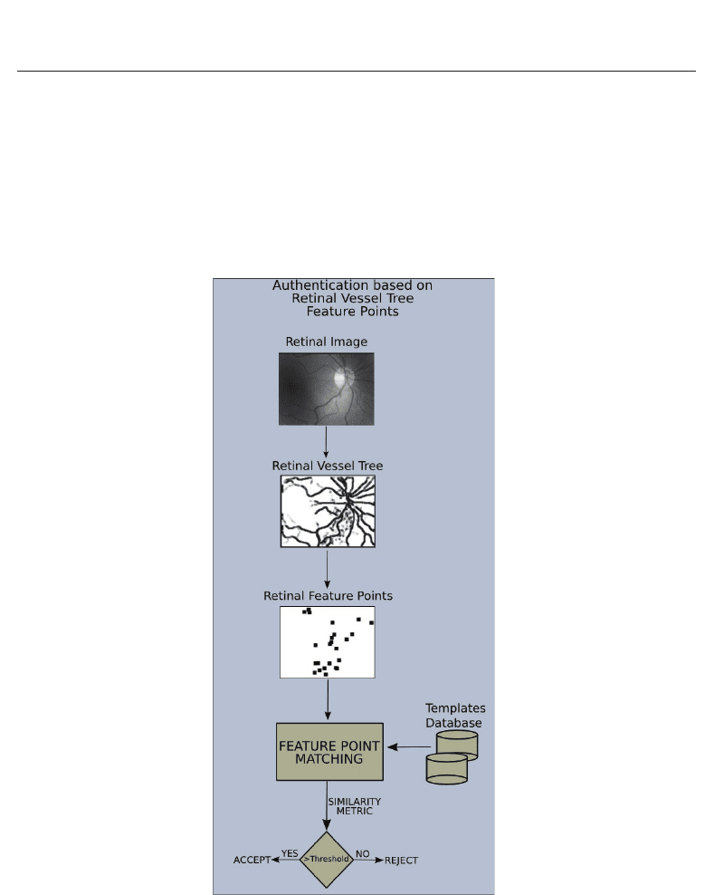
Figure 5 illustrates the general schema for the new feature point based authentication
approach. The newly introduced stages are the feature point extraction and the feature point
matching. The following chapter sections will discuss the methodology on these new stages
of the system.
Fig. 5. Schema of the main stages for the authentication system based in the retinal vessel tree
structure.
3.1 Feature points extraction
Following the idea that vessels can be thought of as creases (ridges or valleys) when images are
seen as landscapes (see Figure 6), curvature level curves will be used to calculate the creases
118
Biometrics
