Yang J. (ed.) Biometrics
Подождите немного. Документ загружается.

13
The Use of Saliva Protein Profiling as a
Biometric Tool to Determine the Presence
of Carcinoma among Women
Charles F. Streckfus and Cynthia Guajardo-Edwards
University of Texas Dental Branch at Houston,
United States of America
1. Introduction
1.1 Background information
Biometrics is the science and technology of measuring and analyzing biological data. It also
refers to technologies that measures and analyzes human body characteristics for
identification purposes. In the context of this book chapter, identification will refer to the
recognition of those individuals in a disease state i.e., carcinoma of the breast. Using “start-
of-the-art” mass spectrometry protein analysis, the author will demonstrate the use of
salivary protein profiles to recognize individuals at risk for carcinoma of the breast.
Proteomic analyses of varying body fluids are propelling the field of medical research
forward at unprecedented rates due to its consistent ability to identify proteins that are at
the fentomole level in concentration. These advancements have also benefited biometric
research to the point where saliva is currently recognized as an excellent diagnostic medium
for biometric authentication of human body characteristics. The saliva microbiome, for
example, is reputed to be biometrically as accurate as a fingerprint. Collectively, these
efforts are in the area of biological verification; however, biometric can be applied to identify
the biological characteristics of a diseased individual.
1.2 Why Saliva as a diagnostic media?
1.2.1 Analytical advantages of Saliva
Saliva as a diagnostic fluid has significant biochemical and logistical advantages when
compared to blood. Bio-chemically, saliva is a clear liquid with an average protein
concentration of 1.5 to 2.0 mg/ml. As a consequence of this low protein concentration, it was
once assumed that this was a major drawback for using saliva as a diagnostic fluid;
however, current ultra sensitive analyte detection techniques have eliminated this barrier.
Saliva specimen preparation is simple involving centrifugation prior to storage and the
addition of a cocktail of protease inhibitors to reduce protein degradation for long-term
storage.
Blood is a far more complex medium. A decision has to be made as to whether to use serum
or plasma. Serum has a total protein concentration of approximately 60-80 mg/ml. Since
serum possesses more proteins than saliva, assaying trace amounts of “factors” (e.g.,
oncogenes, etc.), may result in a greater risk of non-specific interference and a greater chance

Biometrics
250
for hydrostatic (and other) interactions between the factors and the abundant serum
proteins. Serum also possesses numerous carrier proteins, e.g., albumin, which must either
be removed or treated prior to being assayed for protein content. Additionally, it has been
demonstrated that clotting removes many background proteins, which may be altered in the
presence of disease. It has been demonstrated that enzymatic activity continues during this
process, which may cleave proteins from many relevant pathways (Koomen et al., 2005).
It would be ideal if all enzymatic activity in serum would cease at the time of collection;
however, proteomic analyses of serum has shown that this is not the case. As a consequence,
plasma is also being explored as a diagnostic fluid. The main consideration in using plasma
is the selection of a proper anticoagulant (Koomen et al., 2005; Teisner et al., 1983). Heparin
for example can be used as an anti-clotting agent; however, current research has found that
heparin has a relatively short half life (3 to 4 hours) and can produce products of
coagulation which are abundantly comparable to those assayed in serum. Based on these
observations, it is recommended that blood specimens be collected with ethylenediamine
tetraacetic acid (EDTA).
1.2.2 Collection advantages of Saliva
From a logistical perspective, the collection of saliva is safe (e.g., no needle punctures), non-
invasive and relatively simple, and may be collected repeatedly without discomfort to the
patient [4]. Consequently it may be possible to develop a simplified method for “home-
testing”, testing in a “health fair” setting or in dental clinics where individuals are available
for periodic oral examinations. This diagnostic potential could reach many individuals who
for personal, logistical or economical reasons lack access to preventive care.
Blood is a more complicated medium to collect. It requires highly trained personnel to
collect it and if collected incorrectly, can lead to misinterpretations which can result in
patient mismanagement (Ernest & Balance, 2006). Blood specimens need to be collected in a
specific sequence and under-filling tubes with additives may possibly alter protein analyses.
Additionally, if specimens are collected during hospital or clinical settings, there may be a
lapse of time before being processed.
1.2.3 Saliva collection
The oral cavity receives secretions from three pairs of major salivary glands and numerous
minor salivary glands that are located on the oral buccal mucosa, palate, and tongue each
producing a unique type of secretion with varying protein constituents (Birkhed & Heintze,
1989). For example the parotid and Von Ebner glands (located on the tongue) produce
serous secretions while the minor salivary glands produce mucinous secretions. The
submandibular and sublingual glands, however, produce mixed secretions which are both
serous and mucinous. As a consequence, composite or “whole” saliva is preferred as it
enhances the chances of finding a biomarker due to the variety of sources from which it
derives and because of its simplicity to collect.
There are basically two types of saliva to collect. One type is “resting” or unstimulated
whole saliva and the other is stimulated whole saliva. There are several methods for
collecting unstimulated whole saliva. These include the draining or drool method, spitting
method, suction method and the swab method. These methods will yield 0.47, 0.47, 0.54 and
0.52 ml/minute of saliva respectively. Of the four methods, the most reliable is the suction
method with a reliability coefficient of r = 0.93. It also revealed a within subject variance of

The Use of Saliva Protein Profiling as a Biometric Tool
to Determine the Presence of Carcinoma among Women
251
0.14. This is a very reliable method; however, a vacuum pump is required to collect the
specimens (Birkhed & Heintze, 1989).
There are several drawbacks when using unstimulated saliva. The major problem is the
small amount of saliva derived from collection. The 0.47-0.54 ml/minute is the range for
healthy individuals (0.25–0.35 ml/minute normal range) under ideal conditions using those
aforementioned collection methodologies. If the subject is taking medications that decrease
flow rates (e.g., anti-hypertensive medications) the amount collected will be significantly
reduced. Additionally, if the subject has autoimmune disorders (e.g., Sjögren’s syndrome),
has under gone head and neck radiation, or is very elderly, it will be difficult to obtain 0.5
ml over a five minute period. Unstimulated saliva flow rates are also influenced by
circadian and circannual rhythms. Therefore, for consistency, individuals will need to be
serially assessed at approximately the same time of day that the baseline specimen was
collected. All other participants will need to be collected at approximately the same time in
order to reduce inter-variability among the participating subjects. In conclusion, due to the
small quantity of specimen obtained from these techniques and the large within subject
variance, one can conclude that using unstimulated saliva is not the ideal medium for cancer
biomarker discover.
The alternative to using unstimulated whole saliva is obviously to use stimulated whole
saliva. Stimulated secretions produce about three times the volume of unstimulated
secretions and are not subjected to the effects of circadian rhythm. Additionally, you will be
able to collect sufficient quantities of saliva despite health status and medication usage. The
flow rate range is 1 – 3 ml/minute for healthy individuals (Birkhed & Heintze, 1989; Gu et
al., 2004).
There are two methods for collecting stimulated whole saliva. One method of collection is
the gustatory method and the other is the reflexive or “masticatory” technique. The
gustatory technique requires the use of an oral based secretory stimulant. Citric acid is the
most widely used stimulant. Five drops of a 1-6% citric acid solution is applied to the
dorsum of the tongue every 30 seconds. The saliva accumulates in the mouth and is
expectorated intermittently for a period of five minutes. This technique produces copious
amounts of saliva; however, the reliability is only r = 0.76 and has a within subject variance
of 0.49.
The reflexive method is based on the reflex response occurring during the mastication of a
bolus of food. Usually, a standardized bolus (1 gram) of paraffin or a gum base (Wrigley
Co., Peoria, IL) is given to the test subject and they chew the substance at a regular rate. The
subject expectorates intermittently during the collection period for duration of five minutes.
This is an accurate technique as it has a reliability coefficient of r = 0.95 and a within subject
variability of 0.11. The authors recommend this salivary collection method for biomarker
discovery.
The procedure for collecting Stimulated Whole Salivary Gland Secretions is as follows: A
standard piece of unflavored gum base (1.0 - 1.5 g.) is placed in the subject's mouth. The
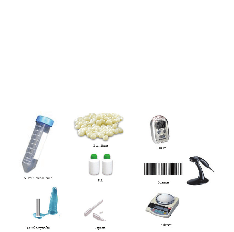
armamentarium used for this procedure is illustrated in Figure 1. The patient is asked to
swallow any accumulated saliva and then instructed to chew the gum at a regular rate
(using a metronome). The subject, upon sufficient accumulation of saliva in the oral cavity,
expectorates periodically into a preweighed disposable plastic cup. This procedure is
continued for a period of five minutes. The cup with the saliva specimen is reweighed and
the flow rate determined gravimetrically. The volume and flow rate is then recorded along
with a brief description of the specimen’s physical appearance (Gu et al., 2004).

Biometrics
252
1.2.4. Long-term Saliva specimen banking
Roughly 2 - 5 ml of whole saliva will be obtained from the individual. In order to minimize
the degradation of the proteins, protease inhibitor cocktail (Sigma, 1 mg/ml whole saliva)
and 1 mM of sodium orthovanadate are added immediately after sample collection
(Shevchenko et al., 2002). All samples are kept on ice during the process. The specimen is
next divided into 0.5 ml aliquots, placed into bar code labeled cryotubes, and frozen (-80°C).
To assess specimen degradation, ten healthy subjects were serially sampled for saliva over a
five-year period. We used c-erbB-2 to test for specimen stability as this is a large 185-kDa
protein, which would be susceptible to degradation by proteases and other biochemical
activity. The results are shown in Figure 1 and illustrate protein stability when frozen at -
80°C.These results are consistent with Wu et al, 1993 where they assayed serially sampled
salivary specimens which were collected over a ten year period for total protein, lactoferrin
(77 kDa) and histidine rich proteins concentrations. In their study, they found no
concentration differences due to specimen aging.
Fig. 1. Armamentarium for the collection and storage of stimulated whole saliva
1.3 Studies using Saliva protein profiling for disease state detection
The majority of the literature concerning human saliva biometrics is associated with the oral
cavity and its associated maladies. An example of this statement is demonstrated in a
manuscript assessing salivary proteins associated with burning mouth syndrome (Moura et
al., 2007). The principle objective the present study was to analyze the characteristics of
salivary production and its composition in individuals with burning mouth syndrome. The
investigators compared salivary flow rates, potassium, iron, chloride, thiocyanate,
magnesium, calcium, phosphorus, glucose, total protein and urea concentrations, as well as
the expression profile of salivary proteins by SDS-PAGE among healthy individuals and
those diagnosed with burning mouth syndrome. The results of the study showed that mean
salivary flow rates among control patients were lower than that of burning mouth syndrome
patients. Chloride, phosphorus and potassium levels were elevated in patients with burning

The Use of Saliva Protein Profiling as a Biometric Tool
to Determine the Presence of Carcinoma among Women
253
mouth syndrome (p = 0.041, 0.001 and 0.034, respectively). Total salivary protein
concentrations were reduced in individuals with burning mouth syndrome (p = 0.223).
Additionally, the analysis of the expression of salivary proteins by Coomassie blue SDS-
PAGE revealed a lower expression of low molecular weight proteins in individuals with
burning mouth syndrome compared to healthy controls. The results suggested that the
identification and characterization of low molecular weight salivary proteins in burning
mouth syndrome may be important in understanding BMS pathogenesis, thus contributing
to its diagnosis and treatment.
Another study using salivary protein profiles investigated the modification of the salivary
proteome occurring in type 1 diabetes and to highlight potential biomarkers of the disorder.
High-resolution two-dimensional gel electrophoresis and matrix-assisted laser
desorption/ionization time-of-flight mass spectrometry was combined to perform a large
scale analysis of the salivary specimens. The proteomic comparison of saliva samples from
healthy subjects and poorly controlled type-1 diabetes patients revealed a modulation of 23
proteins. Fourteen isoforms of α-amylase, one prolactin inducible protein, three isoforms of
salivary acidic protein-1, and three isoforms of salivary cystatins SA-1 were detected as
under expressed proteins, whereas two isoforms of serotransferrin were over expressed
secondary to type-1 diabetes. The proteins under expressed were all known to be implicated
in the oral anti-inflammatory process, suggesting that the pathology induced a decrease of
non-immunological defense of oral cavity. As only particular isoforms of proteins were
modulated, type-1 diabetes seemed to differentially affect posttranslational modification of
the proteins (Hirtz et al., 2006).
An additional study (Delaleu et al., 2008) investigated the involvement of 87 proteins
measured in serum and 75 proteins analyzed in saliva in spontaneous experimental
Sjögren's syndrome. In addition, they intended to compute a model of the immunological
situation representing the overt disease stage of Sjögren's syndrome. In this animal study,
they used non-diabetic, non-obese diabetic mice for salivary gland dysfunction. The mice
aged 21 weeks and were evaluated for salivary gland function, salivary gland inflammation
and extra-glandular disease manifestations. The analytes, comprising chemokines,
cytokines, growth factors, autoantibodies and other biomarkers, were quantified using
multi-analyte profile technology and fluorescence-activated cell sorting. Age-matched and
sex-matched Balb/c mice served as a reference. The investigators found non-diabetic, non-
obese diabetic mice tended to exhibit impaired salivary flow, glandular inflammation and
increased secretory SSB (anti-La) levels. Thirty-eight biomarkers in serum and 34 in saliva
obtained from non-diabetic, non-obese diabetic mice were significantly different from those
in Balb/c mice. Eighteen biomarkers in serum and three chemokines measured in saliva
could predict strain membership with 80% to 100% accuracy. Factor analyses identified
principal components mostly correlating with one clinical aspect of Sjögren's syndrome and
having distinct associations with components extracted from other families of proteins. They
concluded that the autoimmune manifestations of Sjögren's syndrome are greatly
independent and associated with various immunological processes; however, CD40, CD40
ligand, IL-18, granulocyte chemotactic protein-2 and anti-muscarinic M3 receptor IgG3 may
connect the different aspects of Sjögren's syndrome. Processes related to the adaptive
immune system appear to promote Sjögren's syndrome with a strong involvement of T-
helper-2 related proteins in hyposalivation. This approach further established saliva as an
attractive biofluid for biomarker analyses in Sjögren's syndrome and provides a basis for the
comparison and selection of potential drug targets and diagnostic markers (Delaleu et al.,
2008).

Biometrics
254
2. Current research in Salivary protein profiling for cancer detection
2.1 Methods
2.1.1 Study design
The purpose of this study was to determine if individuals could be protein profiled and
potentially classified as having cancer. The investigator also wanted to ascertain if there
were alterations of the protein profiles due to the primary tissue site and the varying degree
of tumor staging. In order to achieve this objective, the investigators collected saliva from
women that were healthy and from those diagnosed with carcinoma breast in the following
stages: Stage 0, Stage I, Stage IIa and Stage IIb. All the tumors were adenocarcinomas.
Additionally, specimens were collected from women diagnosed with varying gynecological
carcinomas. These included women diagnosed with moderate cervical dysplasia, severe
cervical dysplasia and cervical carcinoma in situ. These tumors were all squamous cell
carcinomas. Women diagnosed with ovarian and endometrial carcinomas were also
included in the study. These malignancies were identified as adenocarcinomas. The final
group consisted of women diagnosed with head and neck squamous cell carcinomas ten
women with varying stages of development. Due to the difficulty in obtaining early stage
tumors for ovarian, endometrial and head/neck carcinomas, a composite of varying staged
patients formed these saliva pools.
This study was performed under the UTHSC IRB approved protocol number HSC-DB-05-
0394. All procedures were in accordance with the ethical standards of the UTHSC IRB and
with the Helsinki Declaration of 1975, as revised in 1983. The specimens were banked at the
University of Texas Dental Branch Saliva repository, which stores the specimens at -80°C.
Ten saliva specimens were pooled for each type of carcinoma. The saliva samples were
pooled by combining equal volumes of cleared stimulated whole saliva from a set of
archived healthy and cancer subjects. The subjects were matched for age and race and were
non-tobacco users. Previous studies by the investigator have demonstrated that properly
prepared specimens can remain in storage for a long period of time.
2.1.2 Saliva collection and sample preparation
Stimulated whole salivary gland secretion is based on the reflex response occurring during
the mastication of a bolus of food. Usually, a standardized bolus (1 gram) of paraffin or a
gum base (generously provided by the Wrigley Co., Peoria, IL) is given to the subject to
chew at a regular rate. The individual, upon sufficient accumulation of saliva in the oral
cavity, expectorates periodically into a preweighed disposable plastic cup. This procedure is
continued for a period of five minutes. The volume and flow rate is then recorded along
with a brief description of the specimen’s physical appearance (Navazesh &Christensen,
1982). The cup with the saliva specimen is reweighed and the flow rate determined
gravimetrically. The authors recommend this salivary collection method with the following
modifications for consistent protein analyses. A protease inhibitor from Sigma Co (St. Louis,
MI, USA) is added along with enough orthovanadate from a 100mM stock solution to bring
its concentration to 1mM. The treated samples were centrifuged for 10 minutes at top speed
in a table top centrifuge. The supernatant was divided into 1 ml aliquots and frozen at -80°C.
2.1.3 LC-MS/MS mass spectroscopy with isotopic labeling
Recent advances in mass spectrometry, liquid chromatography, analytical software and
bioinformatics have enabled the researchers to analyze complex peptide mixtures with the

The Use of Saliva Protein Profiling as a Biometric Tool
to Determine the Presence of Carcinoma among Women
255
ability to detect proteins differing in abundance by over 8 orders of magnitude (Wilmarth et
al., 2004). One current method is isotopic labeling coupled with liquid chromatography
tandem mass spectrometry (IL-LC-MS/MS) to characterize the salivary proteome (Gu et al.,
2004). The main approach for discovery is a mass spectroscopy based method that uses
isotope coding of complex protein mixtures such as tissue extracts, blood, urine or saliva to
identify differentially expressed proteins (18). The approach readily identifies changes in the
level of expression, thus permitting the analysis of putative regulatory pathways providing
information regarding the pathological disturbances in addition to potential biomarkers of
disease. The analysis was performed on a tandem QqTOF QStar XL mass spectrometer
(Applied Biosystems, Foster City, CA, USA) equipped with a LC Packings (Sunnyvale, CA,
USA) HPLC for capillary chromatography. The HPLC is coupled to the mass spectrometer
by a nanospray ESI head (Protana, Odense, Denmark) for maximal sensitivity (Shevchenko
et al., 2002). The advantage of tandem mass spectrometry combined with LC is enhanced
sensitivity and the peptide separations afforded by chromatography. Thus even in complex
protein mixtures MS/MS data can be used to sequence and identify peptides by sequence
analysis with a high degree of confidence (Birkhed et al., 1989; Gu et al., 2004; Shevchenko et
al., 2002; Wilmarth et al., 2004).
Isotopic labeling of protein mixtures has proven to be a useful technique for the analysis of
relative expression levels of proteins in complex protein mixtures such as plasma, saliva
urine or cell extracts. There are numerous methods that are based on isotopically labeled
protein modifying reagents to label or tag proteins to determine relative or absolute
concentrations in complex mixtures. The higher resolution offered by the tandem Qq-TOF
mass spectrometer is ideally suited to isotopically labeled applications (Gu et al., 2004;
Koomen et al 2004; Ward et al., 1990).
Applied Biosystems recently introduced iTRAQ reagents (Gu et al., 2004; Koomen et al 2004;
Ward et al., 1990), which are amino reactive compounds that are used to label peptides in a
total protein digest of a fluid such as saliva. The real advantage is that the tag remains intact
through TOF-MS analysis; however, it is revealed during collision induced dissociation by
MSMS analysis. Thus in the MSMS spectrum for each peptide there is a fingerprint
indicating the amount of that peptide from each of the different protein pools. Since
virtually all of the peptides in a mixture are labeled by the reaction, numerous proteins in
complex mixtures are identified and can be compared for their relative concentrations in
each mixture. Thus even in complex mixtures there is a high degree of confidence in the
identification.
2.1.4 Salivary protein analyses with iTRAQ
Briefly, the saliva samples were thawed and immediately centrifuged to remove insoluble
materials. The supernatant was assayed for protein using the Bio-Rad protein assay
(Hercules, CA, USA) and an aliquot containing 100 µg of each specimen was precipitated
with 6 volumes of -20ºC acetone. The precipitate was resuspended and treated according to
the manufacturers instructions. Protein digestion and reaction with iTRAQ labels was
carried out as previously described and according to the manufacturer’s instructions
(Applied Biosystems, Foster City, CA). Briefly, the acetone precipitable protein was
centrifuged in a table top centrifuge at 15,000 x g for 20 minutes. The acetone supernatant
was removed and the pellet resuspended in 20 ųl dissolution buffer. The soluble fraction
was denatured and disulfides reduced by incubation in the presence of 0.1% SDS and 5 mM
TCEP (tris-(2-carboxyethyl)phosphine)) at 60ºC for one hour. Cysteine residues were

Biometrics
256
blocked by incubation at room temperature for 10 minutes with MMTS (methyl methane-
thiosulfonate). Trypsin was added to the mixture to a protein:trypsin ratio of 10:1. The
mixture was incubated overnight at 37ºC.
The protein digests were labeled by mixing with the appropriate iTRAQ reagent and
incubating at room temperature for one hour. On completion of the labeling reaction, the
four separate iTRAQ reaction mixtures were combined. Since there are a number of
components that can interfere with the LC-MS/MS analysis, the labeled peptides are
partially purified by a combination of strong cation exchange followed by reverse phase
chromatography on preparative columns. The combined peptide mixture is diluted 10 fold
with loading buffer (10 mM KH
2
PO
4
in 25% acetonitrile at pH 3.0) and applied by syringe to
an ICAT Cartridge-Cation Exchange column (Applied Biosystems, Foster City, CA) column
that has been equilibrated with the same buffer. The column is washed with 1 ml loading
buffer to remove contaminants.
To improve the resolution of peptides during LCMSMS analysis, the peptide mixture is
partially purified by elution from the cation exchange column in 3 fractions. Stepwise
elution from the column is achieved with sequential 0.5 ml aliquots of 10 mM KH2PO4 at
pH 3.0 in 25% acetonitrile containing 116 mM, 233 mM and 350 mM KCl respectively. The
fractions are evaporated by Speed Vacuum to about 30% of their volume to remove the
acetonitrile and then slowly applied to an Opti-Lynx Trap C18 100 ul reverse phase column
(Alltech, Deerfield, IL) with a syringe. The column was washed with 1 ml of 2% acetonitrile
in 0.1% formic acid and eluted in one fraction with 0.3 ml of 30% acetonitrile in 0.1% formic
acid. The fractions were dried by lyophilization and resuspended in 10 ul 0.1% formic acid
in 20% acetonitrile. Each of the three fractions was analyzed by reverse phase LCMSMS. The
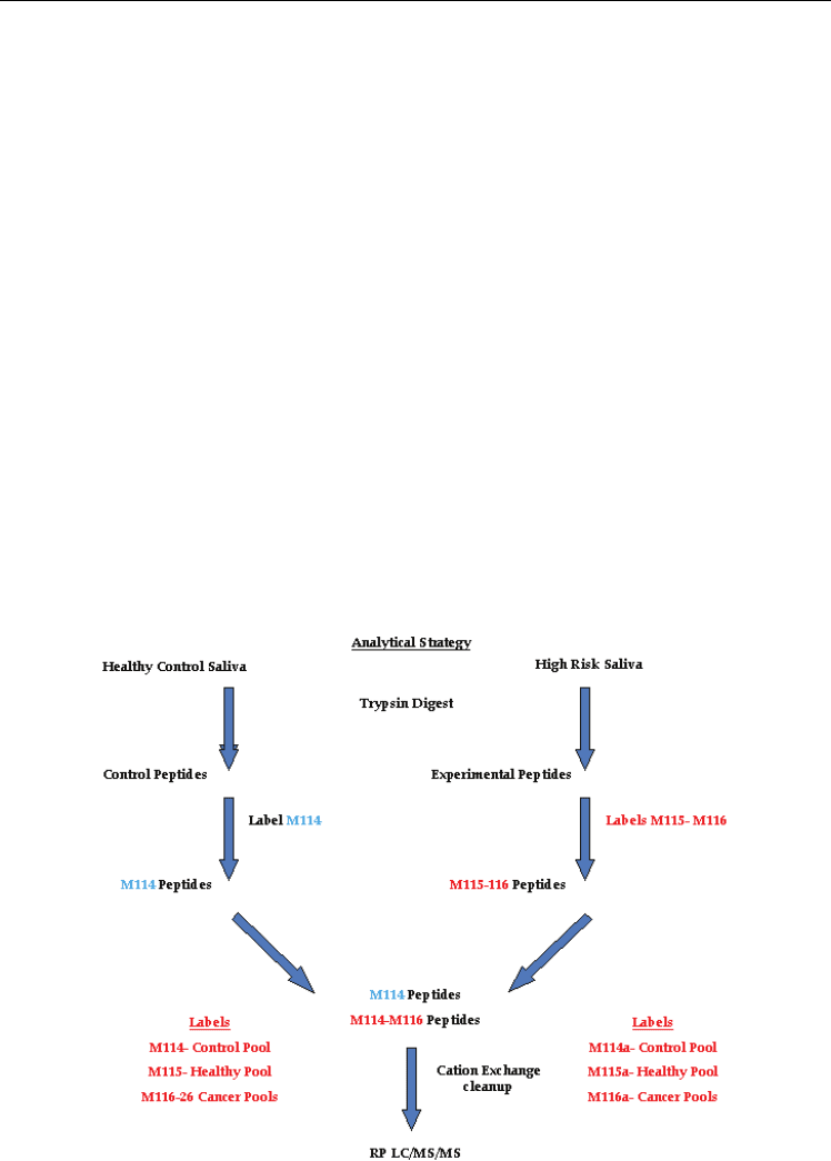
analytical strategy is illustrated in Figure 2.
Fig. 2. Analytical strategy for quantifying peptides using iTRAQ tagging

The Use of Saliva Protein Profiling as a Biometric Tool
to Determine the Presence of Carcinoma among Women
257
2.1.5 Reverse phase LCMSMS
The desalted and concentrated peptide mixtures were quantified and identified by nano-LC-
MS/MS on an API QSTAR XL mass spectrometer (ABS Sciex Instruments) operating in
positive ion mode. The chromatographic system consists of an UltiMate nano-HPLC and
FAMOS auto-sampler (Dionex LC Packings). Peptides were loaded on a 75cm x 10 cm, 3cm
fused silica C18 capillary column, followed by mobile phase elution: buffer (A) 0.1% formic
acid in 2% acetonitrile/98% Milli-Q water and buffer (B): 0.1% formic acid in 98%
acetonitrile/2% Milli-Q water. The peptides were eluted from 2% buffer B to 30% buffer B
over 180 minutes at a flow rate 220 nL/min. The LC eluent was directed to a NanoES source
for ESI/MS/MS analysis. Using information-dependent acquisition, peptides were selected
for collision induced dissociation (CID) by alternating between an MS (1 sec) survey scan
and MS/MS (3 sec) scans. The mass spectrometer automatically chooses the top two ions for
fragmentation with a 60 second dynamic exclusion time. The IDA collision energy
parameters were optimized based upon the charge state and mass value of the precursor
ions. Each saliva sample set there are three separate LCMSMS analyses.
The accumulated MSMS spectra are analyzed by ProQuant and ProGroup software
packages (Applied Biosystems) using the SwissProt database for protein identification. The
ProQuant analysis was carried out with a 75% confidence cutoff with a mass deviation of
0.15 Da for the precursor and 0.1 Da for the fragment ions. The ProGroup reports were
generated with a 95% confidence level for protein identification.
2.1.6 Bioinformatics
The Swiss-Prot database was employed for protein identification while the PathwayStudio
®
bioinformatics software package was used to determine Venn diagrams were also
constructed using the NIH software program (http://ncrr.pnl.gov). Graphic comparisons
with log conversions and error bars for protein expression were produced using the
ProQuant
®
software.
2.1.7 Western blot analysis for marker validation
2.1.7.1 Preparation of samples
We selected the protein profilin-1 for validating the presence of these proteins in saliva. The
profilin-1 antibody was a rabbit polyclonal from the Abcam Co. #Ab10608 diluted 1:1000.
The saliva samples from a healthy individuals, benign tumor subjects and individuals
diagnosed with Stage IIa her2/neu receptor positive breast cancer subjects and Stage IIa
her2/neu receptor negative breast cancer subjects were pooled by combining equal volumes
of cleared stimulated whole saliva from a set of archived specimens.
The pooled saliva was mixed with loading buffer (Laemmli buffer containing BME) in 1:1
ratio. The sample was then incubated at 95°C for 5 minutes and was then loaded onto the 4-
15% Tris-HCl polyacrylamide gel. Four-fifteen percent Tris-HCl polyacrylamide gels were
loaded with molecular weight markers, controls, and the pooled saliva samples.
Electrophoresis was run at 200 Volts, 30 minutes in 1X TGS buffer. The gels were
equilibrated and extra thick blot paper and PVDF membranes were soaked in 1X TGS buffer
for 15 minutes prior to running Western Transfer. Semi-dry transfer apparatus was used.
Transfer conditions were 0.52mA constant, 17 volts, 19 minutes. Polyvinylidene fluoride
(PVDF) membranes were air dried for minimum of 1 hour. Dry PVDF membranes were
activated in methanol for about 10 seconds then transferred to soak in 1X PBS-T for 3 washes

Biometrics
258
of 5 minutes each. The membranes were then incubated for 1 hour in 5% NFDM in PBS-T.
Afterwards the membranes were washed 3 times for 5 minutes in PBS-T. The membranes
were incubated overnight with a primary antibody in PBS-T. The membranes were washed
3 times for 5 minutes in PBS-T and were incubated for 4 hours with secondary antibody
(HRP conjugate) in PBS-T. Again, the membranes were washed 3 times for 5 minutes in PBS-
T. Finally, the membranes were treated with ECL plus detection reagents and photographed
with exposure of 800 seconds.
3. Experimental results in salivary protein profiling for cancer detection
3.1 Mass spectrometry analysis
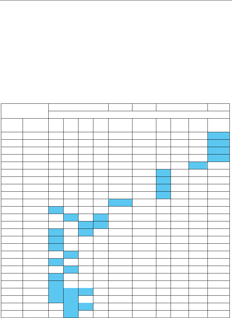
Tables 1-5 summarize the results of the proteomic analysis and illustrates protein
comparisons between breast (Stage 0 – IIb), cervical (moderate, severe dysplasia and in situ)
Proteins
Breast Ovarian Endo. Cervical H & N
Staging
Gene ID Accession
Stage
0
Stage
I
Stage
IIa
Stage
IIb
Variable Variable Mod. Severe Stage 0 Variable
A1AT P01009 4.32
ANXA3 P12429 0.74
CO3 P01024 2.16
HPTR P00739 3.57
K1C16 P08779 5.37
COBA1 P12107 0.57
LUZP1 Q86V48 0.62
ZN248 Q8NDW4 0.84
CYTC P01034 0.77
KAC P01834 1.42
K2C6C P48666 3.40
K1CJ P13645 0.47 0.26
SCOT2 Q9BYC2 0.83 0.50
VEGP P31025 1.36 0.47
PROF1 P07737 0.74
NGAL P80188 0.90
NUCB2 P80303 1.28
HEMO P02790 0.74
CYTD P28325 0.82
CRIS3 P54108 0.65
ACBP P07108 1.42
KLK P06870 0.80 1.46 0.86
PIP P12273 0.88 0.80
PERL P22079 1.31 0.88
PPIB P23284 1.32
Table 1. The table shows which proteins are unique to each type and stage of carcinoma
