Yang J. (ed.) Biometrics
Подождите немного. Документ загружается.

0
Retinal Identification
Mikael Agopov
University of Heidelberg
Germany
1. Introduction
Since the pioneering studies of Drs. Carleton Simon and Isodore Goldstein in
1935 [Simon & Goldstein (1935)], it has been known that every eye has its own unique pattern
of blood vessels, and that retinal photographs can be used for identifying people. In the 1950’s,
this was proven to hold even for identical twins [Tower (1955)]. Hence the idea of using the
retinal blood vessel pattern for identification. Eye fundus photography for the purpose of
identification is impractical, however. An optical device which scans the retinal blood vessel
pattern is required.
Such an optical Retinal Identification (RI) device was originally patented in 1978 [Hill (1978)];
after several subsequent patents, it developed into a commercial product in the 1980s and
1990s. As the patent of retinal identification (opposed to the actual design of the device) has
now worn off, new developments in the field have taken place. In this chapter, the history,
technique and recent developments of RI are discussed.
1.1 The anatomy and optical properties of the human eye
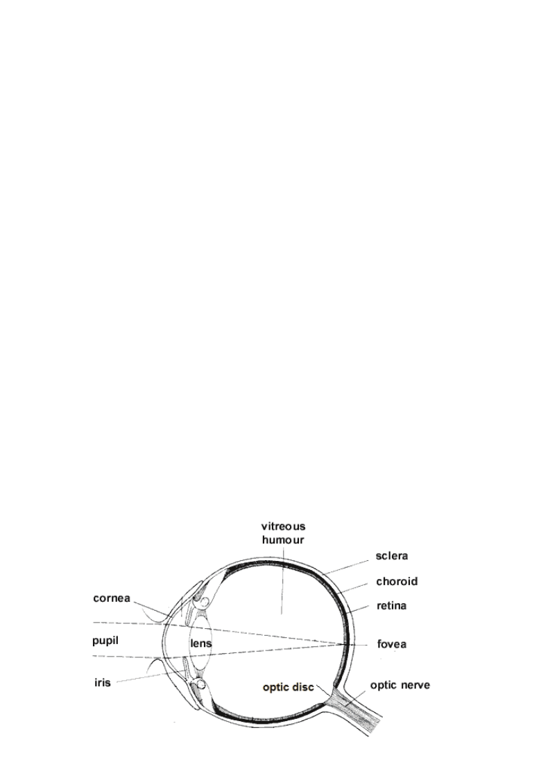
Fig. 1. A schematic picture of the human eye.
5

2 Will-be-set-by-IN-TECH
A schematic picture of the human eye is shown in Figure 1. The eyeball is of about 24 mm
in diameter and filled with vitreous humor, jelly-like substance similar to water; its outer shell,
the sclera, is made of rigid proteins called collagen. The light entering the eye first passes
through the pupil, an aperture-like opening in the iris. The size of the pupil limits the amount
of light entering the eye. The light is focused by the cornea and the crystalline lens onto the
retina. The retina converts the photon energy into an electric signal, which is transferred to the
brain through the optic nerve.
The cornea is about 11 mm in diameter and only 0.5 mm thick. It accounts for most of
the refractive power of the eye (about 45 D). The remaining 18 D come from the crystalline
lens, which is also - through deformation - able to change its refractive power, thus partly
compensating for the refractive error and helping to focus the eye.
The retina is a curved surface in the back of the eye. The point of sharpest vision is called
fovea - here the light-sensing photoreceptor cells are only behind a small number of other cells.
Elsewhere on the retina, the light has to travel through a multi-layered structure of different
cells. These various cells are responsible for the eye’s ’signal processing’, i.e. turning the
incoming photons first into a chemical and then to an electric signal.
After being amplified and pre-processed, the signal is transferred to the nerve fibers, which
reside on the peripheral area of the retina around the optic disc, where they form the retinal
nerve fiber layer (RNFL). The optic disc is an approximately 5
◦
×7
◦
, ellipse-like opening in the
eye fundus, through which the nerve fibers and blood vessels enter the eyeball. It is about 15
◦
away from the fovea in the nasal direction. The choroid is the utmost layer behind the retina
just in front of the sclera. It has a bunch of small blood vessels, and is responsible for the
retina’s metaboly.
1.2 The birefringence properties of the eye
Birefringence is a form of optical anisotrophy in a material, in which the material has different
indices of refraction for p- and s-polarization components of the incoming light beam. The
components are thus refracted differently, which in general results the beam being divided
into two parts. If the parts are then reflected back by a diffuse reflector (such as the
eye fundus), a small portion of the light will travel the same way as it came, joining the
polarization components into one again, but having changed the beam’s polarization state
in process
1
.
The birefringence of the eye is well documented (Cope et al. (1978), Klein Brink et al. (1988),
Weinreb et al. (1990), Dreher et al. (1992)). The birefringence of the corneal collagen fibrils
constitutes the main part of the total birefringence of the eye. Its amount and orientation
changes throughout the cornea. In the retina, the main birefringent component is the retinal
nerve fiber layer (RNFL), which consists of the axons of the nerve fibers. The thickness of
RNFL is not constant over the retina; the amount of birefringence varies according to the RNFL
thickness and also drops steeply if a blood vessel (which is non-birefringent) is encountered.
The most successful application of measuring the RNFL thickness around the optic disc is
probably the GDx glaucoma diagnostic device (Carl Zeiss Meditec, Jena, Germany). It uses
scanning laser polarimetry to topograph the RNFL thickness on the retina. A reduced RNFL
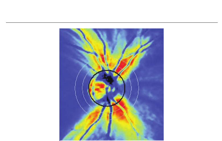
thickness means death of the nerve fibers and thus advancing glaucoma. A typical GDx image
isshowninFigure2.
1
See Appendix about how the polarization change can be measured.
100
Biometrics
Retinal Identification 3
Fig. 2. A typical GDx image of a healthy eye. The birefringent nerve fiber layer is seen
brightly in the image, as well as the blood vessels which displace the nerve fibers, thus
resulting in a weaker measured signal (darker in the image).
2. RI using retinal blood vessel absorption
The first patent of the biometric identification using the retinal blood vessel pattern dates
back to 1978 [Hill (1978)]. Soon afterwards, the author of the patent founded the company
EyeDentity (then Oregon, Portland, USA) and began full-time efforts to develop and
commercialize the technique.
In the original patent, the retinal blood vessel pattern is scanned with the help of two rings of
LEDs. The amount of light reflected back from the retina is measured - when the beam hits
a blood vessel, it is absorbed to a bigger extent than when it hits other tissue. In the original
retina scan, green laser light was used - it was strongly absorbed by the red blood vessels.
However, it was found out that visible light causes discomfort to the identified individual, as
well as pupil constriction, causing loss of signal intensity.
Since the first working prototype RI, patented in 1981 [Hill (1981)], near-infrared (NIR) light
has been used for illumination. The infrared light is not absorbed by the photoreceptors
(the absorption drops steeply above 730 nm); however, the retinal blood vessels are fairly
transparent to the NIR wavelengths as well - the light is absorbed by the smaller choroidal
blood vessels instead (thus, considering this technique, the term Retinal Identification is slightly
misleading) before being reflected back from the eye fundus. The image acquisition technique
has also been changed: the LEDs are given up in favour of scanning optics. A circular scan is
preferred over a raster, which suffers from the problem of reflections from the cornea.
In the patent of 1986 [Hill (1986)], the scan is centered around the fovea instead of the optic
disc. The fovea is on the optical axis of the eye, so this arrangement has the definite advantage
that no fixation outside the normal line of sight is required, unlike when a ring around the
optic disc is scanned, when the subject has to look 15 degrees off-axis. The downside is that
101
Retinal Identification
4 Will-be-set-by-IN-TECH
the choroidal blood vessels are much thinner than around the optic disc, and they don’t form
a clear pattern. Thus the price paid for easier fixation is the quality of the signal.
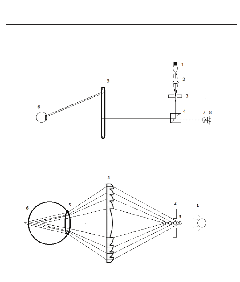
Fig. 3. A schematic drawing of the current Retinal Identification technique, based on the
patent from 1996. A detailed explanation is in the text.
Fig. 4. A schematic drawing fixation-alignment technique using a Fresnel lens. A detailed
explanation is in the text.
Current RI technology is based on an active US patent [Johnson & Hill (1996)]. A schematic
drawing of the measurement setup is shown in Figure 3. An infrared light source, for example
a krypton lamp (1), is focused by a lens (2) through an optical mirror (explained below) via a
pinhole (3). The light enters a beam splitter (4) which reflects it into a Fresnel optical scanner
(5). The rotating optical scanner scans a ring on the cornea, which - if properly focused - will
hit the retina at the same angle. A multifocus Fresnel lens, cemented in the scanner, creates
a fan of nearly-collimated light beams which hit the eye of the tested subject. One of the
102
Biometrics

Retinal Identification 5
beams will be focused on the retina by the eye’s own optical apparatus, thus compensating
for refractive error (explained in detail below).
The light reflected back from the eye fundus travels the same way through the scanner and
into the beam splitter; a part of it is transmitted into a photodetector (8) through a focusing
lens (7). After being measured by the photodetector, the signal is A/D-converted, amplified
and processed. The processed signal is converted into points, which are stored in an array,
which is used for matching. A similar process is used in all RI techniques.
Fixation and alignment of the subject’s eye in a RI measurement is critical; it is almost
impossible to scan a non-willing subject. The fixation system of the RI technique, which was
also patented (Arndt (1990)) , is illustrated in Figure 4. A Light-Emitting Diode (LED) is
situated next to the Krypton lamp. It illuminates the optical double-surface mirror, creating
several reflections, ’ghost images’, of the LED on the optical axis of the system. These images
function as targets for the test subject’s eye. The eye looks at them through a multifocal
Fresnel lens. The lens, which consists of several focusing parts with different focal lengths,
focuses the ghost images on different points on the eye’s optical axis. Regardless of whether
the test subject is emmetropic (normal visual acuity), myopic (near-sighted) or hyperopic
(far-sighted), one of the images will almost certainly end up on the retina and will thus
result a sharp image and effectively compensate for the refractive error of the subject’s eye.
However, this happens at the cost of optical image quality; the measured pattern is a sum of
contributions from choroidal blood vessels and other structures.
2.1 Matching
At first, a reference measurement, which the further measurements will be compared against,
has to be taken from each tested subject. Any further measurement will be compared against
the reference. As the subject’s eye can rotate around the optical axis (due to different head
position, i.e. head tilt, between the measurements), the best possible match is found by
’rotating’ the measurement points in the array. The matching is done using a Fourier-based
correlation; the match is measured on a scale of +1,0 (a perfect match) to -1,0 (a complete
mismatch). User experience has shown that a match above 0,7 can be considered a matching
identification.
3. RI using the RNFL birefringence
The blood vessels emerging into the retina through the optic disc often displace the nerve
fibers in the retinal nerve fiber layer. Unlike the RNFL nerve fiber axons, the blood vessels are
not birefringent - thus, if the birefringence (change in the state of polarization) of the scanning
laser beam is measured around the optic disc, a steep signal drop proportional to the blood
vessel size is measured wherever one is encountered. This can be seen in the GDx-pictures,
where the blood vessels are seen as dark lines on the otherwise bright nerve fiber layer.
The author and his co-workers studied the possibility of using blood vessel-induced RNFL
birefringence changes for biometric purposes [Agopov et al. (2008)]. A measurement device
was built to scan a circle of 20
◦
around the optic disc. The measured birefringence would drop
steeply where a blood vessel is encountered, creating a sharp drop, or ’blip’, in the measured
signal. The scanning angle is big enough to catch the major blood vessels which enter the
fundus through the disc.
103
Retinal Identification
6 Will-be-set-by-IN-TECH
3.1 Apparatus and method
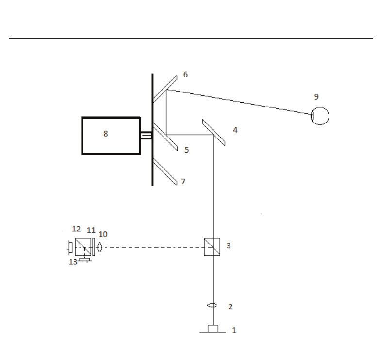
Fig. 5. A schematic drawing of the RI measurement setup using the RNFL birefringence. A
detailed explanation is in the text.
The measurement setup is explained in detail in [Agopov et al. (2008)]; here it is explained
briefly. The apparatus and the light paths within it are shown in Figure 5. The light path
is drawn with a solid line. A 785 nm laser diode (1) was used as a light source. The
beam was collimated by a lens (2); then it passed through a non-polarizing beam splitter (3)
and was reflected by a mirror (4) further into the optical scanner (5-8). The two scanning
mirrors (5 and 6) and a counterweight (7) were cemented on an aluminium plate which
was spun by a DC motor (8). The center mirror was (5) tilted 45
◦
from the disc plane
rotated clockwise and reflected the laser beam onto the edge mirror. The edge mirror (6)
was tilted 50
◦
, creating a circular scan subtending a 10
◦
radius of visual angle (20
◦
diameter)
in the tested subject’s eye (9). The reflection from the ocular fundus traveled back the
same optical path through the scanner; now, however, the beam splitter (3) reflected the
useful half of the beam (drawn with dashed line) into the detection system (10-13). A lens
(10) focused the beam into a polarizing beam splitter (12) through a quarter wave plate
(11) which had its fast axis 45
◦
to the original plane of polarization. The polarizing beam
splitter separated the p- and s-polarization components; two avalanche photodiodes (13) were
placed right after it, measuring the two signals which corresponded the two polarization
components. Amplified by the detection electronics, the signals were added and subtracted
104
Biometrics

Retinal Identification 7
respectively. The polarization was manipulated so that the Stokes parameters S
0
and S
3
were
measured - it was decided to measure S
3
instead of S
1
for birefringence-based changes as it
appeared to suffer less from various amounts and orientations of corneal birefringence in our
computerized model.
As the alignment of the eye is critical, special care was taken to properly align the test subject’s
eye. The measurement apparatus included three eyepieces: the measurement was taken
through the fixed central piece; in addition there were two horizontally movable ones; the
subject could look through the central piece with either eye while having a ’dummy’ eyepiece
available for the other eye. Thus possible head tilt was reduced to almost zero.
Because the fixation point of the retina, the fovea, is approximately 15 degrees away from
the optic disc, the measured eye had to look 15
◦
away in the nasal direction to center the
scan on the optic disc. To achieve this, two fixation LEDs were set at 15
◦
angletothecentral
axis of the scan. Because the human eye is about 0,75 D myopic at the wavelength 785 nm
(see [Fernandez et al. (2005)] for details) the fixation LEDs were placed at 130 cm distance, so
that the eye’s fixation would compensate for this.
4. RI using the optic disc structure
Another interesting RI technology was patented in 2004 [Marshall & Usher (2004)]. The idea
is to use the image of the optic disc - taken by a scanning laser ophthalmoscope (SLO) -
for identification. A company Retinal Technologies (Boston, MA, USA) was founded for
developing the technique.
4.1 The principle of an SLO
The best-known application of the SLO is probably the Heidelberg Retina Tomograph
(Heidelberg Engineering, Heidelberg, Germany), which is used for glaucoma diagnostics. The
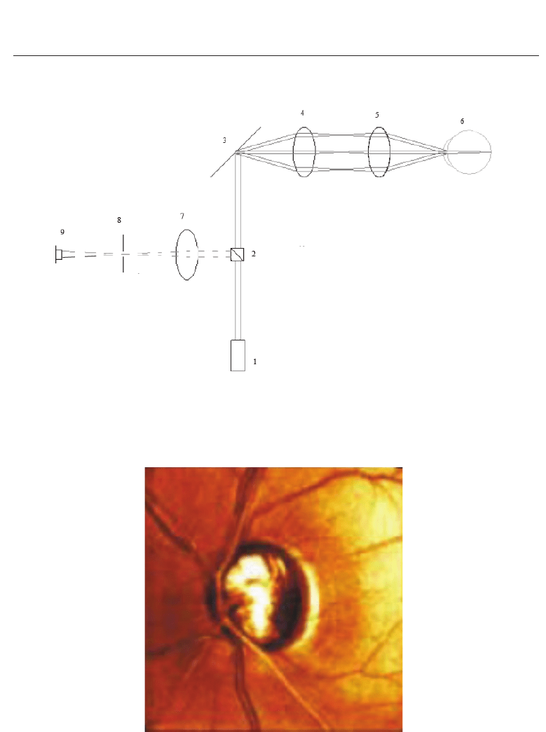
principle of an SLO is illustrated in 6. A low-intensity laser diode (1) is used for illumination.
A collimated laser beam goes through a beam splitter (2) into an optical scanner (3). The
scanner consists of two rotating mirrors, a fast and a slow one, creating a raster scan. The
scanning beam is imaged through two lenses (4 and 5) onto exactly one point called the
conjugated plane. If the imaged subject’s cornea is at this point(6), the eye’s optics focus the
scanning beam onto the retina (7). The reflection from the eye fundus travels back through
the system, but is reflected into the detection system (7-9) by the beam splitter. A lens (7)
focuses the beam through a pinhole (8) onto a photodiode (9). The pinhole is very important -
only the light which comes exactly from the conjugated plane reaches the photodetector. Thus
the SLO creates a high-resolution microscopic image of the retina. The scan is usually centered
around the optic disc (using an off-axis fixation target - as in the previous setup). A typical
SLO image of the optic disc (taken with the HRT) is shown in Figure 7.
4.2 Image analysis
The boundary of the optic disc is found from the image taken by the SLO. There is a clear
boundary between the disc and surrounding tissue (as seen in Figure 7); an ellipse is fit onto
the image by analyzing the average intensity of the pixels around the boundary.
Once the disc is identified, a recognization pattern is created from the its structures. This
fairly complicated procedure is explained in detail in the patent. The patterns of recognized
105
Retinal Identification
8 Will-be-set-by-IN-TECH
Fig. 6. An schematic drawing of a scanning laser ophthalmoscope. A detailed explanation is
in the text.
Fig. 7. A typical SLO image of the optic disc (taken with an HRT).
106
Biometrics
Retinal Identification 9
individuals are stored in a database for comparing with the pattern of a person wishing to be
identified.
5. Combined retina and iris identification
Fig. 8. The measurement setup of a combined retina and iris identification device. The details
are in the text.
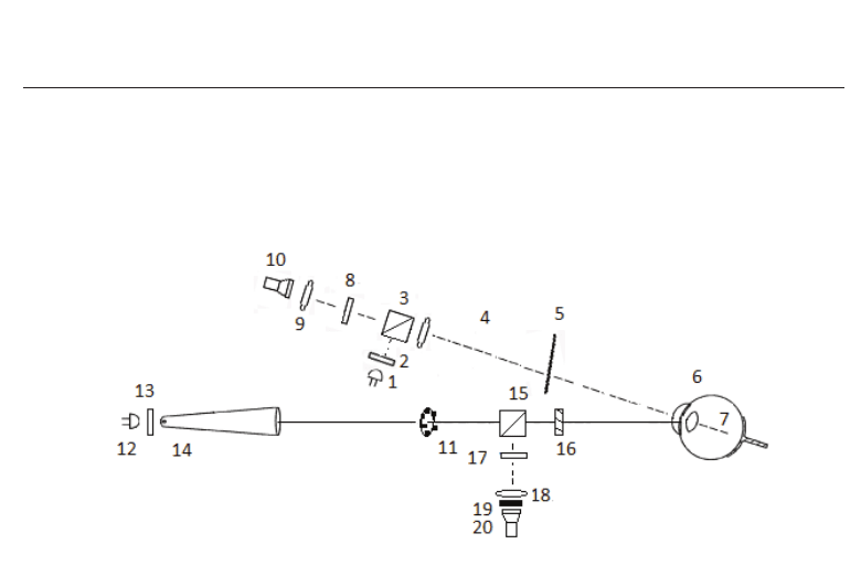
In a fairly recent patent [Muller et al. (2007)], a combination of retina and iris identification
was suggested (about iris identification, see the previous chapter). The measurement system
is constructed so that both biometrics can be recorded with one scan. This is obviously
advantageous, as now two biometrics are available simultaneously. The measurement setup
is shown in Figure 8. The setup has two optical axes: one for the retinal image (dashed line)
and one for alignment and the iris image (solid line). They are 15
◦
apart; as explained earlier,
the incoming light is focused on the area around the optic disc only if the eye is looking 15
◦
in
the nasal direction. To achieve this, the test subject has to be faced towards the dashed line but
look in the direction of the solid line. As a fixation target, a ring of green LED’s (11) is placed
around the optical axis of the iris scanning system.
Two illuminating LED’s are used: One infrared (λ
≈ 800 nm) LED (1), and one red (λ ≈ 660
nm) LED (12). The interference from the ’wrong’ light source is blocked a dichroic mirror (5).
The light from the infrared LED (1) first goes through a polarizer (2), which ensures that the
outgoing light is linearly polarized. The outgoing beam is then divided into two parts by a
beam splitter (3). The transmitted (useless) part crosses the beam splitter and is preferably
absorbed by a light trap. The reflected (useful) beam part is collimated by a lens (4) and
goes through the dichroic mirror (5) into the eye. The beam is focused by the eye’s optics (6)
onto the area around the optic disc; after being reflected from the eye fundus (7), a reflection
of the beam returns back the same way as the beam came; however, coming from the other
direction, a part of the reflected beam now passes through the beam splitter into the detection
system. The polarizer (8) is set so that it absorbs the illuminating LED’s polarization direction.
However - having changed its state of polarization while passing through the eye tissue - a
part of the beam is now able to pass through the polarizer. The beam is focused by a lens (9)
onto the detector (10), which can be for example a CCD camera. It should be noted that this
setup has no optical scanner (nor it is confocal) - a wide-field image around the optic disc is
107
Retinal Identification

10 Will-be-set-by-IN-TECH
captured, thus resulting in lower resolution than a confocal scanning system would achieve.
For the iris scan, the illuminating light from the red LED (12) is first linearly polarized (13)
2
.
and then guided into the eye through an alignment tube (14). The function of the alignment
tube is not explained in the patent; preferably it would consist of at least one lens with a long
focal length, which would focus the light onto the iris (the beam entering the eye should not
be parallel, otherwise it will be focused on the retina). The distance between the lens and the
beam splitter should be much bigger than that between the beam splitter and the eye, so that
the slightly de-focused reflection image of the iris can be caught. On its way to the iris and
back, the light double-passes a quarter wave plate (16). The wave plate’s axes are set so that
both the fast and the slow axis of it are 45
◦
to the original polarization direction - when the
beam passes through it, its polarization becomes circular; the second pass (the reflection from
the iris) turns the circular polarization into linear again, but having turned the polarization
direction by 90
◦
in the process. After entering the detection system (17-20), the light can now
pass through the polarizer (17), which is set to absorb the polarizing direction of the initial
polarizer (13). The beam is focused by a lens onto the detector (a CCD camera); the possible
reflections of the green LED are filtered out by a red band pass filter (19).
The two detection systems are electronically synchronized so that one scan records both
images simultaneously. The recording is triggered by a switch, which is turned on when
the eye is correctly aligned.
The eye is at a crossing of two optical axes - its distance and orientation are critical. In the
setup suggested in the patent, the distance is controlled by an ultrasound transducer. It sends
and receives ultrasound pulses which are reflected back from the surface of the cornea. When
the distance is right, and the optic disc is seen on the CCD (10), the eye is aligned properly,
and the recording can be taken.
6. Results of performance tests and limitations of the RI techniques
6.1 RI using absorption
In a performance test by Sandia National Laboratories [Sandia Laboratories (1991)], the
EyeDentity RI device recognized >99% of the tested subjects in a three-attempt measurement,
with no false positives.
6.1.1 Limitations
As the light has to pass twice through the pupil during the measurement, a constricted
pupil can increase the number of false negative scan results. Thus dim light conditions are
preferable for the RI (of course, this is true for almost any optical measurement); the technique
has difficulties in broad daylight. In addition, various eye conditions can disturb the light’s
passing through the eye, compromising vision; this also affects measuring the eye’s properties
optically, including the RI.
2
In the patent, the device is desribed without the polarizers (13 and 17), the quarter wave plate (16), the
filter (19); instead of a beam splitter (15), a dichroic mirror is suggested. The fixation LEDs are also
placed together with the red illuminating LED. However, this leads to unsurmountable difficulties.
First of all, the IR light and the green light used for fixation cannot both pass through the dichroic
mirror; moreover, the IR light would first have to pass through the mirror - the reflection from the eye
would then have to be reflected by it. Therefore, the author suggests slight modifications in the setup.
108
Biometrics
