Stroscio Michael A., Dutta Mitra. Biological Nanostructures and applications of Nanostructures in biology: electrical, mechanical and optical properties
Подождите немного. Документ загружается.

106
ANJUM ANSARI AND SERGUEI V. KUZNETSOV
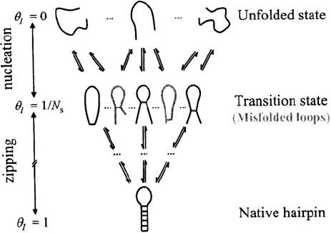
Figure 3. Zipper model (with misfolded conformations). A schematic representation of the ensemble of
microstates in the unfolded, transition state, and the native state. The misfolded conformations are represented
by hairpin loops with mismatched stems, which act as dead-ends in the folding process.
3. REVIEW OF EXPERIMENTAL RESULTS AND PUZZLES
The earliest measurements on the kinetics of duplex and hairpin formation were done
using temperature-jump (T-jump) techniques (Cohen and Crothers, 1971; Coutts, 1971;
Craig et al., 1971; Porschke and Eigen, 1971; Gralla and Crothers, 1973; Porschke,
1974b; 1974a; 1977; Chu and Tinoco, 1983; Xodo et al., 1988). The temperature of the
sample was raised on microsecond time-scales using electrical pulses generated by
discharging a capacitor (Eigen and de Maeyer, 1963). A modified T-jump apparatus,
using a coaxial cable capacitor, has been used for some sub-microsecond measurements
(Hoffman, 1971; Porschke, 1974a). An excellent discussion of some of the results from
the early T-jump measurements can be found in Cantor and Schimmel (1980).
The experimental data on the kinetics of duplex formation from complementary
oligonucleotides showed that the activation enthalpies for the helix formation step, for
sequences containing only A·U base-pairs, are about –4 to –9 kcal/mol (Craig et al.,
1971; Porschke and Eigen, 1971), and for sequences containing G·C base-pairs, the
activation enthalpies are about +6 kcal/mol to +9 kcal/mol (Porschke et al., 1973). As
described in the previous Section, the negative activation enthalpies observed for
sequences with A·U base-pairs indicate that the helix nucleation is not a simple
elementary step, but instead consists of the formation of a critical nucleus that has 2 or 3
HAIRPIN FORMATION IN POLYNUCLEOTIDES
107
base-pairs (Porschke, 1977). The nucleation of the helix is then the rate-determining step,
followed by the rapid zipping of the stem. The positive activation enthalpies for
sequences with G·C base-pairs were explained by assuming that, for these sequences,
only 1 or 2 base-pairs may be sufficient to form the nucleating helix (Porschke et al.,
1973).
Early estimates of the zipping rate, i.e., the rate at which a base-pair is added to an
existing helix, vary widely, from very fast (Spatz and Baldwin, 1965; Wetmur
and Davidson, 1968) to relatively slower, between and (Craig et al., 1971;
Porschke and Eigen, 1971). Porschke (1974a) made direct measurements of the
zipping/unzipping rate by carrying out sub-microsecond T-jump measurements on dimers
of poly(A) and poly(U) oligomers of chain lengths 14 and 18 at temperatures below the
melting transition. The kinetics measurements revealed two distinct processes, one
occurring with a time constant of about independent of the oligomer
concentration, and a much slower relaxation occurring with a time constant of a few
seconds. The slow component is the overall helix-to-coil transition, while the fast
component was assigned to the unzipping at the ends. A kinetic zipper model was used to
estimate the rate coefficient for the zipping step to be at 25°C
(Porschke, 1974a).
Hairpin formation in ssDNA or RNA chains requires the formation of a loop
stabilized by a few base-pairs as the nucleation step, followed by zipping as in duplex
formation. Because of the close proximity of the two ends of the ss-chain, hairpins are
expected to form on time-scales considerably faster than duplex formation. The early T-
jump measurements on short self-complementary oligomers revealed that hairpins with 4-
6 bases in the loop and less than 10 bases in the stem form on time-scales of tens of
microseconds (Coutts, 1971; Gralla and Crothers, 1973; Porschke, 1974b). To form
hairpins, the ss-chain has to overcome an entropic barrier in forming a loop, which is
countered by the stabilizing free energy of a few base-pairs. To understand the time-
scales for forming hairpins requires an estimation of the time-scales for forming the
critical nucleus, which in turn requires reliable estimates of the free energy cost of loop
formation.
It was recognized quite early that the free energy of loop formation in ss-
polynucleotides deviates from the simple estimates of entropic costs expected for a
random coil model, especially for loop sizes smaller than about 10 nucleotides,
presumably from favorable stacking interactions of bases within small loops (Vallone et
al., 1999). However, there is considerable uncertainty in the estimates of the enthalpic
contribution to loop closure, obtained from the thermodynamic analysis of melting
profiles of hairpins, ranging from ~21 kcal/mol (Uhlenbeck et al., 1973) to ~11 kcal/mol
(Porschke, 1974b) for an hairpin, and ~0 kcal/mol for an hairpin
(Gralla and Crothers, 1973). The very large enthalpic cost for loop closure for the
hairpin was explained as arising from the unstacking of cytosine residues in the poly(C)
strand in order to form the loop (Uhlenbeck et al., 1973; Porschke, 1974b). However,

108
ANJUM ANSARI AND SERGUEI V. KUZNETSOV
that conclusion seems inconsistent with essentially no enthalpic cost reported by Gralla
and Crothers ( 1973).
Kinetics measurements on the formation of hairpins impose much more stringent
constraints on the possible estimates of thermodynamic parameters, and are therefore
indispensable for accurate estimates of the free energies that stabilize secondary structure
in ssDNA and RNA. The early T-jump measurements, however, also showed quite large
variation in the activation enthalpies obtained from the temperature dependence of the
measured rates, ranging from –22 kcal/mol for the closing step of a hairpin fragment
from (Coutts, 1971) to ~2.5 kcal/mol for the hairpin
(Porschke, 1974b), suggesting a sequence dependence to the free energy of loop
formation as well as to the size of the critical nucleus. Based on a comparison of the
estimated enthalpy for forming the first base-pair from thermodynamic measurements on
and the measured activation enthalpy for the hairpin formation step from
kinetics measurements on that hairpin, Porschke argued that a stable nucleus that leads to
zipping is formed only after the formation of the fourth A·U base-pair (Porschke, 1974b).
In recent years there has been a surge in the investigation of hairpin kinetics using a
variety of new experimental tools, such as fluctuation correlation spectroscopy (FCS)
(Bonnet et al, 1998; Goddard et al., 2000; Wallace et al., 2000); laser T-jump
measurements (Ansari et al., 2001; Shen et al., 2001), and single-molecule techniques
(Deniz et al., 1999; Grunwell et al., 2001; Liphardt et al., 2001).
Libchaber and co-workers have carried out a series of elegant measurements on the
kinetics of conformational fluctuations in ssDNA hairpin-loops by FCS (Bonnet et al.,
1998; Goddard et al., 2000). They attached a fluorophore and a quencher at either end of
their oligonucleotide sequence, and the state of the molecule, whether hairpin (closed) or
unfolded (open), was monitored by the intensity of the fluorescence. In the open state the
molecule is fluorescent because the fluorophore and the quencher are far apart, whereas
in the closed state the fluorescence is quenched. They monitored the time-scales for
fluctuations between the open and the closed states by analyzing the autocorrelation
function of the fluctuations in the fluorescent signal. The sequences of DNA hairpins
investigated by Libchaber and co-workers were where X
was either T or A, and the size of the loop (N) varied from N=12 to N=30 for the
poly(dT) loop and from N=8 to N=30 for the poly(dA) loop.
The primary results from Libchaber’s group are: (i) closing times depend on the
sequence and length of the loop, whereas the opening times are insensitive to the loop
composition; (ii) the closing times scale with the length of the loops as for poly(dT)
loops and as for poly(dA) loops; (iii) closing times for poly(dA) loops are about 10
times slower than for poly(dT) loops at 20°C; and (iv) the activation enthalpies for the
closing step increase nearly linearly for poly(dA) loops, from ~5 kcal/mol for loops with
8 bases to >15 kcal/mol for loops with 30 bases, whereas for the poly(dT) loops the
activation enthalpies decrease slightly with the loop size (Bonnet et al., 1998; Goddard et
al., 2000).
Kinetics measurements on another DNA hairpin whose
stem sequence is complementary to that of one of Libchaber’s sequence, have been
HAIRPIN FORMATION IN POLYNUCLEOTIDES
109
performed by Klenerman and co-workers, also using FCS techniques (Wallace et al.,
2000; 2001). One difference between the two sets of FCS measurements is that in
Libchaber’s set-up the fluorescence of the excited label is quenched upon contact with
the second label, whereas in Klenerman’s set-up, the fluorescence labels attached at the
two ends of the hairpin stem are donor-acceptor pair for fluorescence resonance energy
transfer (FRET), and the intensity of the donor changes as the two ends come closer, but
without necessarily making direct contact. Another difference is the method by which
they subtract the contribution from the diffusion of the DNA molecules in and out of their
observation volume to their intensity fluctuation measurements (Wallace et al., 2000).
However, the results of their measurements are quite strikingly different and as yet
unresolved. Libchaber’s group reports single-exponential kinetics for temperatures
ranging from ~10-50°C, as observed previously in T-jump measurements, whereas
Klenerman’s group observes highly nonexponential relaxation kinetics at ~20°C that they
describe in terms of stretched exponentials (Wallace et al., 2000; 2001). The Klenerman
group also reports non-Arrhenius temperature dependence for the opening and closing
rates, and a viscosity dependence for the rates that scales nearly inversely with the
solvent viscosity (Wallace et al., 2001).
Bustamante and co-workers (Liphardt et al., 2001) have used mechanical force to
induce the unfolding and refolding of single RNA molecules, including a simple RNA
hairpin, a molecule containing a three-helix junction, and a domain of a ribozyme. For
their hairpin, which has ~22 base-pairs in the stem, approximately half of which are G·C
base-pairs, and 4 bases in the loop, they find that the hairpin unfolds at a force of ~15 pN,
similar to forces required to unzip DNA helices (Essevaz-Roulet et al., 1997; Rief et al.,
1999; Bockelmann et al., 2002; Thomen et al., 2002). By imposing a constant force on
the molecule, they were able to monitor the end-to-end distance between the two ends of
the hairpin and to watch the distance hop back and forth between two values
characteristic of the fully unfolded and the fully folded hairpin, with no evidence of any
intermediate states. They determined the folding and unfolding rates from the average
lifetimes in the two states, and found that, at the critical force for which the opening and
closing rates are the same (~14 pN in the presence of the folding times are
~1s. The very slow folding times observed in these measurements, compared to the
folding times of tens of microseconds observed in FCS and T-jump measurements for
hairpins with similar loop sizes, but smaller stems 5-7 base-pairs long, has been
explained as arising from the very large free energy barrier for folding in the presence of
the applied force, which has been estimated to be (or ~6 kcal/mol) for their
hairpin, and because of its long stem (Liphardt et al., 2001; Cocco et al., 2003a).
Schultz and co-workers (Deniz et al., 1999; Grunwell et al., 2001) have developed a
single-molecule FRET measurement technique to monitor the conformational
fluctuations of ssDNA hairpins immobilized on a glass surface. For their hairpin with 40
poly(dA) bases in the loop, they report closing times that are ~140ms, i.e. more than
about 30-100 times longer than is predicted from scaling the measured closing times from
the Libchaber group by The long closing times in the single-molecule FRET
measurements may be a result of the interactions of the hairpin with the derivatized glass
110
ANJUM ANSARI AND SERGUEI V. KUZNETSOV
surface, or perhaps another manifestation of the anomalous dependence of the dynamics
of poly(dA) loops with increasing loop-size.
FCS and single-molecule measurements are limited in their time-resolution to
microseconds and milliseconds, respectively. The rapid development of nanosecond laser
T-jump techniques has opened up the field to investigate the dynamics of biomolecules
with ~10 ns time-resolution (Williams et al., 1989; Hofrichter, 2001), while overcoming
the limitation of the earlier T-jump setups that required the use of solutions of high
conductivity and thus high ionic strength. Laser T-jump has been used extensively by
several groups to investigate rapid events in the protein folding process such as the
kinetics of formation of elementary secondary structures, and (Munoz
et al., 1997; Dyer et al., 1998; Eaton et al., 1998; Gruebele et al., 1998; Jager et al.,
2001). In our laboratory, we have used laser T-jump to investigate hairpin dynamics in
ssDNA (Ansari et al., 2001; Kuznetsov et al., 2001; Shen et al., 2001), as well as to
investigate the dynamics of wrapping and unwrapping of ssDNA on a single-stranded
binding protein (Kuznetsov et al., 2004). Our T-jump measurements on hairpin formation
are consistent with single-exponential relaxation dynamics, although the current time-
resolution is not sufficient to determine whether there is any missing amplitude on the
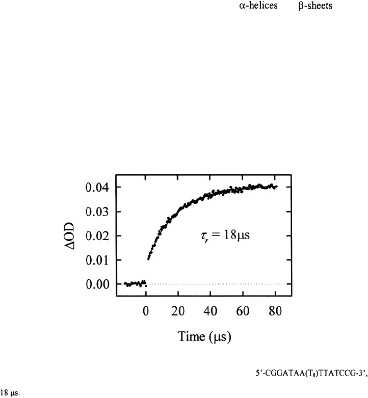
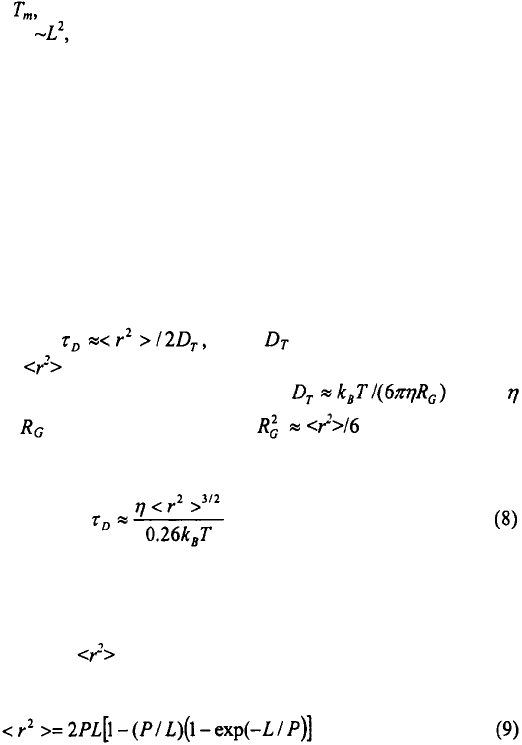
sub-microsecond time-scale, see Figure 4. The rapid change in absorbance in the laser T-
jump measurements has contributions from any unresolved relaxations and from an
apparent change in the optical density of the sample from thermal lensing effects, which
occurs on the time-scale of the T-jump (Hofrichter, 2001).
Figure 4. The change in absorbance as a function of time, for the hairpin
after a T-jump from 42°C to 51°C. The kinetics are described as a single-exponential with a relaxation time of
HAIRPIN FORMATION IN POLYNUCLEOTIDES
111
The primary results from our T-jump measurements are: (i) the free energy of the
hairpin relative to the unfolded state scales with the loop size with an apparent exponent
of ~7, much larger than the exponent of ~1.8 expected from the entropic cost of loop
formation for a semiflexible polymer; (ii) the equilibrium zipper model, which was used
to calculate free energy profiles along an effective reaction coordinate, suggests that the
transition state ensemble consists of looped conformations stabilized by one base-pair
closing the loop; (iii) the equilibrium model predicts negative activation enthalpies of
~–9 kcal/mol for the closing step, and which are confirmed in kinetics measurements;
(iv) at temperatures near the closing times for both poly(dT) loops and poly(dA)
loops scale with loop size as consistent with the scaling expected for a semiflexible
polymer; (v) the opening and closing times exhibit an apparent viscosity independence, a
conclusion that is contradictory to an earlier study on viscosity dependence by the
Klenerman group (Wallace et al., 2001)
3.1. Why is hairpin formation so slow?
The nucleation step in hairpin formation requires the ss-polynucleotide to form a
loop with one or more base-pairs to stabilize the loop. Models describing the
characteristic time for two ends of a polymer chain to come into contact have been
proposed in several theoretical studies (Wilemski and Fixman, 1974; Doi, 1975; Szabo et
al., 1980; Friedman and O’Shaughnessy, 1989; Guo and Thirumalai, 1995;
Podtelezhnikov and Vologodskii, 1997). An order-of-magnitude estimate for the end-to-
end contact time is estimated as where is the translational diffusion
coefficient of the chain and is the mean-square end-to-end distance (Winnik, 1986).
We can estimate the translational diffusion coefficient from where
is the solvent viscosity and is the radius of gyration, (DeGennes, 1979).
Therefore, the end-to-end contact time becomes
The result in Eq. (8) is nearly identical to the more rigorous calculation of Szabo et al.
(1980) who model the dynamics of the end-to-end contact of a flexible (Gaussian) chain
as diffusion in a harmonic potential well.
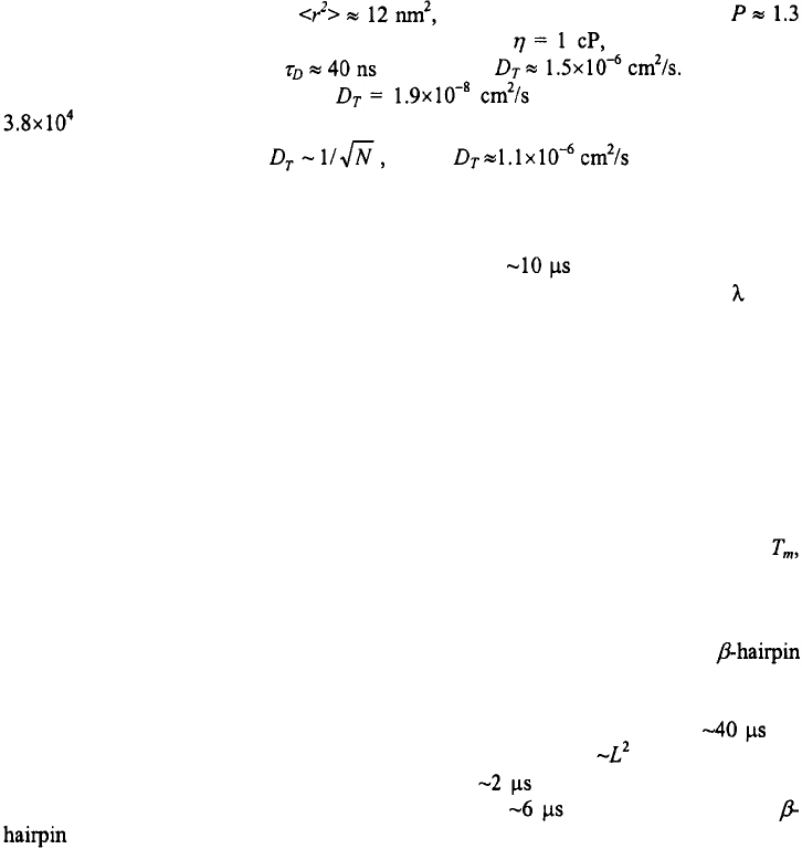
For a semiflexible polymer, can be written as (Landau and Lifshitz, 1980;
Rivetti et al., 1998)
112
ANJUM ANSARI AND SERGUEI V. KUZNETSOV
where P is the persistence length of the chain, and L is the contour length. Note that this
formula predicts a stiff-rod behavior for L<P and random-coil behavior for L>>P. For a
ss-polynucleotide chain ~10 nucleotides long, and assuming an internucleotide distance
of ~0.6 nm, yields L ~6nm, and where we have used a value of
nm (Rivetti et al., 1998). Therefore, at T = 25°C and the diffusion-limited
contact time is estimated to be at 25°C and Wetmur and
Davidson (1968) report a value of for a ss-polynucleotide with
nucleotides. If we scale the experimentally measured value for a long ss-chain
down to shorter chains using we get for a strand of ~10
nucleotides, in close agreement with our crude estimate.
If the contact time between the two ends of the polymer is indeed ~40 ns, then
formation of the loop cannot be the rate-determining step in hairpin formation, which
occurs ~250 times slower, with hairpin closing times of at 25°C for hairpins with
about 10 poly(dT) bases in the loop. It is well known that cyclization times for DNA
molecule with cohesive ends are also much longer than the end-to-end contact times for a
semiflexible polymer (Wang and Davidson, 1966a; 1966b; 1968). One explanation for
this discrepancy was first proposed by Wang and Davidson (1966b), who argued that the
rate-determining step in the joining of the two ends is the very slow chemical step of
base-pair formation and not the diffusion-limited time for contact formation. They based
their arguments on two observations: first, that the temperature dependence of the
measured cyclization times exhibited a very large (~24 kcal/mol) activation energy;
second, that the viscosity dependence of the cyclization times did not follow a simple
scaling with solvent viscosity as expected for a diffusion-controlled reaction. Hairpin
closing times, on the other hand, exhibit negative activation energies, especially near
and therefore the chemical step of base-pair formation cannot be the rate-determining
step. The viscosity dependence of the opening and closing times of a DNA hairpin is still
an open question. This point is discussed further in Section 3.4.
It is of interest to compare hairpin formation in ss-polynucleotides with
formation in polypeptides, which are also found to occur on time-scales of several
microseconds (Munoz et al., 1997). Early measurements of the time-scales for loop
formation in polypeptide chain under strongly denaturing conditions yielded for
loops of ~50 residues (Hagen et al., 1996). Using a scaling of for a semiflexible
polymer of length L yields loop formation times of for ~10 residues long loops,
which is close to the experimentally measured time of for the formation of a
(Munoz et al., 1997), thus suggesting that the initiation of the loop could set the
time-scale for hairpin formation.
Subsequent measurements of first contact time between two ends of Gly-rich
polypeptide sequences designed to have little or no secondary structure have yielded
values of ~30-100 ns for ~10 residues long loops (Bieri et al., 1999; Lapidus et al., 2000;
Hudgins et al., 2002). The origin of the discrepancy between these and the earlier
measurements is not clear. One suggestion is that the persistence length of the chain,
which is known to be highly sequence dependent (Miller et al., 1967), was ~5 times
HAIRPIN FORMATION IN POLYNUCLEOTIDES
113
bigger in the polypeptide chain of the denatured protein that was used in the first set of
measurements compared to the designed Gly-rich sequences of the subsequent
measurements (Lapidus et al., 2000). Another explanation is that the slow contact times
in the early set of measurements is a result of high concentration of GdnHCl in the
solution, which binds to the protein in the denatured state, and could slow down the
effective diffusion coefficient of the chain (Hagen et al., 2001).
Several theoretical and computational studies of protein folding have postulated
another source for the decrease in the effective diffusion coefficient of the polypeptide
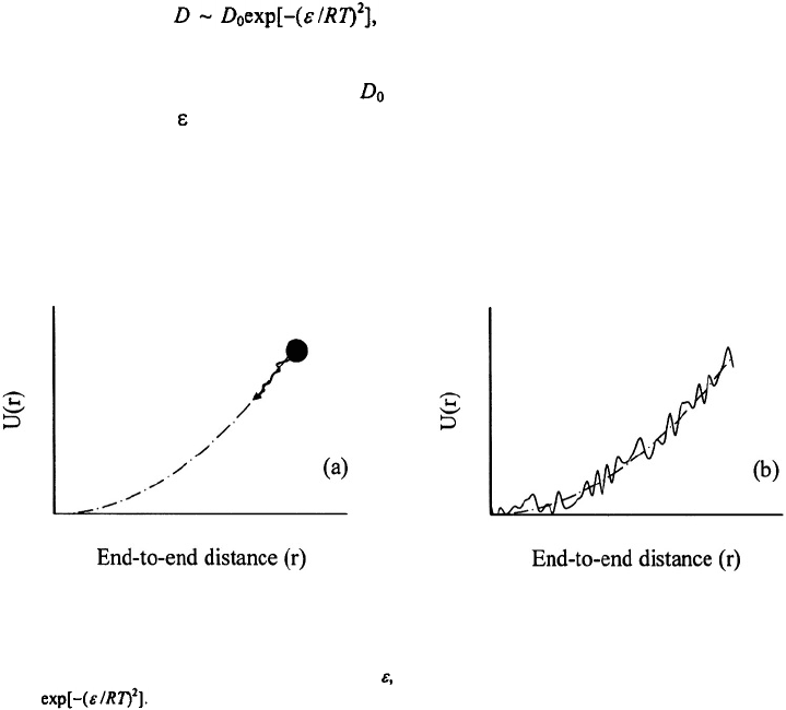
chain, of the form as a result of interactions within the chain,
especially under folding conditions, which give rise to a “roughness”in the energy
surface of the polypeptide (Zwanzig, 1988; Bryngelson and Wolynes, 1989; Bryngelson
et al., 1995; Socci et al., 1996). Here is the intrinsic diffusion coefficient of the
polymer chain and is the amplitude of the roughness; see Figure 5. Eaton and co-
workers have postulated that even for their Gly-rich sequence especially designed to have
no secondary structure, there seems to be a ~16-fold decrease in the effective diffusion
coefficient of the probes attached to the two ends of the polypeptide chain, and they
attributed this decrease to transient intrachain interactions (Lapidus et al., 2000).
Figur
e
5. Diffusion in a harmonic potential. (a) For an ideal gaussian chain, the diffusion coefficient is
characteristic of the relative diffusion of the two ends of the chain. (b) For chains with intrachain interactions,
the harmonic potential has a roughness of amplitude and the effective diffusion coefficient is reduced by a
factor
In a series of recent papers, we proposed that such transient intrachain interactions in
the unfolded state of ss-polynucleotides could lead to the slow formation of the critical
nucleus for forming hairpins (Ansari et al., 2001; Kuznetsov et al., 2001; Shen et al.,
2001; Ansari et al., 2002). This slowing down could arise from (i) non-native base-pairs
that don’t lead to a complete hairpin and that act as dead-ends during the folding process,
114
ANJUM ANSARI AND SERGUEI V. KUZNETSOV
or (ii) non-native hydrogen bonds or non-native stacking interactions (mis-stacked bases)
as was suggested to explain the anomalous loop-size dependence of the hairpin closing
times for hairpins with long poly(dA) loops (Ansari et al., 2002). Such a mechanism
would increase the nucleation time by decreasing the effective intrachain diffusion
coefficient. A characteristic roughness of only would decrease
the effective diffusion coefficient and increase the characteristic first contact time by a
factor of ~250 at 25°C.
Two computational studies of ss-polynucleotide conformational dynamics support
some of the ideas postulated above. The first study, by Zhang and Chen (2002), presents
a detailed folding kinetic analysis of a 21 -nucleotide RNA hairpin (9 base-pairs in the
stem and 3 bases in the loop), using a statistical mechanical model that enumerates all
conformations of the RNA chain with two or more contiguous (stacked) base-pairs,
including all misfolded conformations. They calculate the free energy of each
conformation using a statistical mechanical model for RNA thermodynamics (Chen and
Dill, 2000), and using the base-pairing and stacking interactions from the RNA
thermodynamics literature (Serra and Turner, 1995). The various conformations are
coupled via elementary transition steps in which only one base-pair is formed or broken
in any single kinetic step. The rates of transitions between the conformations are
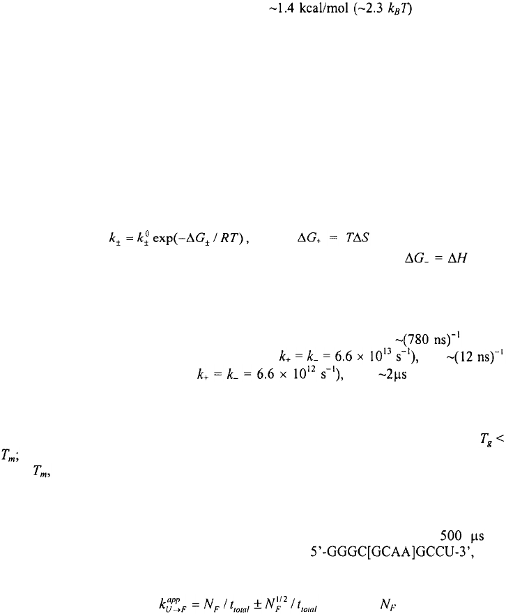
parameterized as where is the barrier for the
formation of a base-pair and is assumed to be entirely entropic, and is the
barrier for the disruption of a base-pair, and is assumed to be the enthalpic cost of
breaking the hydrogen bonding and stacking interactions. They use this model to
calculate in detail the folding pathways, the relaxation kinetics, and the temperature
dependence of the relaxation rates. In their model, the parameters that best describe the
experimentally measured folding rates of small RNA hairpins yield for the
elementary step of forming G·C base-pairs (with and
for forming A·U base-pairs (with with obtained for the
closing time of a hairpin with 9 base-pairs in the stem and 3 bases in the loop (Shi-Jie
Chen, private communication). Important results from their study include (i) a rugged
energy landscape for RNA folding; (ii) folding pathways that lead to dead-ends or traps,
especially at temperatures below what they define as the glass transition temperature
and (iii) a distinctly non-Arrhenius temperature dependence for the closing rates.
Near these traps are not deep; nevertheless, they could lead to slowing down of the
chain dynamics.
A second study on ss-polynucleotide dynamics comes from large-scale, parallel,
molecular dynamics simulation of Pande and co-workers, which involves sampling a
large number of constant temperature trajectories that total more than of
simulations for an all-atom model of an RNA hairpin with
continuum representation of solvent effects (Sorin et al., 2002; Sorin et al., 2003). From
their simulated trajectories, they calculate the apparent transition rates for folding by
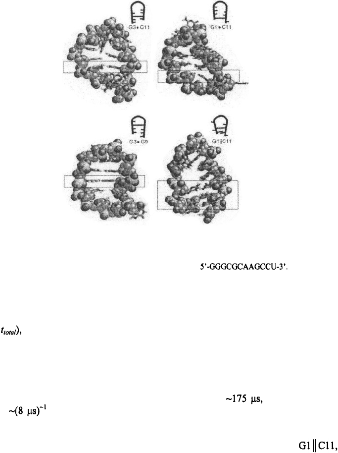
using the approximation, (where is the number of
HAIRPIN FORMATION IN POLYNUCLEOTIDES
115
Figure 6. Members of the misfolded trap ensemble for the hairpin The figure is
adapted from Sorin et al. (2003) and shows the atomistic (and schematic) pictures of the non-native interactions
found in the collapsed state in their simulations.
transitions that occur from the unfolded state U to the folded state F in a total simulation
time valid for all processes that exhibit single-exponential kinetics (Shirts and
Pande, 2001; Zagrovic et al., 2001). To investigate the folding process at 300K, the
simulations were started from the fully extended, denatured state. They observe at least
two dominant mechanisms by which the hairpin folds, the first is a loop formation
followed by zipping, and the other is a nonspecific collapse mechanism, similar to the
hydrophobic collapse in proteins (Dill, 1990; Thirumalai et al., 2001). They find that a
total of 21 trajectories undergo a nativelike collapse within giving a collapse
rate of at ~300K, which is very close to the experimentally observed hairpin
closing rates. The individual conformations observed in their collapsed state show an
ensemble of misfolded traps with base-pairing interactions between G3·C11 or G1·C11,
hydrogen bonding interactions between G3·G9, and base-stacking interactions
as in Figure 6. Thus, the simulations of Pande and co-workers support the notion that
transient trapping can result not only from non-native base-pairing interactions, as
explicitly included in the model of Zhang and Chen (2002), but also from non-native
