Stroscio Michael A., Dutta Mitra. Biological Nanostructures and applications of Nanostructures in biology: electrical, mechanical and optical properties
Подождите немного. Документ загружается.

G.YOUREK
ET AL
The final AFM image is virtually a composite or “convolution” between the geomet-
ric properties of the tip and the sample surface.
93
Undoubtedly, one of the most impor-
tant controllable parameter for AFM imaging, either in ambient air or fluid, is proper
selection of scanning tips. The AFM scanning “tip” typically consists of a sharp micro-
fabricated barb or spike mounted at the end of a “cantilever” to form a unified “probe”.
Although these terms are sometimes used interchangeably, only the microstructured com-
ponent at the end of the cantilever is the part that actually indents the sample surface dur-
ing the AFM scanning. A wide variety of AFM scanning tips are currently available,
differing in geometry, material properties, and chemical composition.
Tips are broadly defined by their aspect ratio (length to width), opening angle and/or
radius of curvature. Relatively rough sample surfaces, in the range of micrometers,
should be scanned using high-aspect-ratio tips, which combine small opening angles with
a long tip. However, low-aspect-ratio tips, corresponding with high opening angles, are
more suitable for scanning relatively flat specimens.
94, 95
A special type of low-aspect-
ratio tips exist where the very end is shaped into a high-aspect-ratio peak with overall
low-aspect-ratio configuration of the tip. These tips are referred to as “sharpened tips”
and ensure greater scan depth with improved resolution when scanning relatively flat
samples. Radius of curvature of the AFM tip reflects the nanometric sharpness of the
tip’s peak. Typical radius of curvature of sharpened tips is less than 20 nm, while that of
unsharpened tips ranges from 20-50 nm. Oxide sharpened silicone nitride tips are widely
utilized in the AFM scanning of living cells and various other biological structures
3, 96, 97
due to their high versatility and ability to combine high resolving power with physical
tolerance on soft sample surface.
The tip-cantilever assembly is most commonly made of crystal silicone or silicone
nitride, which are both suitable for microfabrication due to their stiffness and wear resis-
tance. Silicone nitride tips are more suitable for contact mode imaging due to their flexi-
bility and “forgiveness” on the sample surface compared to the stiffer crystal silicone
probes. Another distinctive characteristic of silicone nitride probes is the greater ten-
dency of the silicone nitride tips to be trapped by the surface tension attractive forces
during interactions with the sample surface than the crystal silicon probes. Such forces,
although micro- or nano-scale in nature, might be strong enough to deform the surface of
soft samples. Therefore, considerable attention to the selection of the scanning tip should
be taken, especially when imaging delicate samples. By contrast, when scanning harder
samples or using tapping mode AFM, stiffer crystal silicone probes are likely more ap-
propriate. However, increased brittleness of the crystal silicone tips due to their greater
stiffness mandates considerable care during tip handling and preparation for the scanning
session.
Several recent efforts are directed toward substituting the silicone and the silicone ni-
tride with more characterized materials for the fabrication of enhanced AFM probes.
Carbon nanotubes
98-101
are gaining rising popularity to be the backbone structural mate-
rial for the second generation of AFM probes. Additional advantages offered by the car-
bon nanotubes include their well-characterized structure, mechanical robustness, and
unique chemical properties that allow well-defined surface modification without jeopard-
izing the AFM scanning resolution. For example, utilizing this last feature of feasibility
of carbon nanotubes’ surface modification under high controllability, many aspects of
structural and thermodynamic properties of protein-protein and protein-nucleic acid com-
76
Based on the nature of the interactions between the scanning tip and sample surface,
AFM scanning can be in contact mode, tapping mode, or error-signal mode. Mode selec-
tion largely depends on the nature of the sample and the desirable images to be obtained
by the
AFM.
In contact mode, the scanning tip makes a direct contact with the surface of the sam-
ple throughout the scanning period with the topographic features of the sample’s surface
dictating the degree of cantilever reflection. However, since the amount of cantilever
reflection is pre-adjusted through the control system to a certain value (the operating set-
point), this imaging mode is also known as “constant-force mode”. In addition to the fact
that this mode is the original AFM imaging mode and can be readily accomplished for a
large variety of samples, the most advantageous feature of contact-mode AFM is its abil-
ity to perform the scanning under both air and fluid conditions. Fluid imaging with con-
tact mode AFM is necessary for imaging living cells in an appropriate fluidic medium so
that cell viability can be maintained in quasi-physiologic conditions.
93
Contact-mode
AFM imaging in fluid is also beneficial in eliminating the capillary action forces, adding
to the precision of the scanning force.
94
On the other hand, a disadvantage associated
with contact-mode AFM scanning is the lateral frictional forces due to direct contact be-
tween the tip and the sample surface. In the case of soft samples, such as the cell surface,
this direct contact may damage the structures of the imaged surface.
105
In contact-mode
AFM, the deflection signals from the cantilever in the z direction are plotted against x and
y to produce informative height images that reflect the nanometric height variations of the
scanned surface.
Also known as “oscillating mode” or “non-contact mode”, in tapping mode the scan-
ning tip is literally bouncing up and down or “tapping” as it travels across the sample
surface. The driving principle behind implementation of tapping mode is to eliminate the
lateral shear forces associated with imaging in contact mode.
105
However, similar to con-
tact-mode AFM, the vertical movement of the cantilever is maintained at constant oscilla-
tion amplitude throughout the scanning period. Similarly, tapping-mode AFM can be
performed in air or fluid environment. The imaging of living cells in aqueous environ-
ment using tapping mode may actually have the potential of minimizing the frictional
forces between the tip and the relatively soft surface of the cell membrane and thus can
reduce the accumulation of membrane structures on the scanning tip that might hinder the
scanning resolution.
90
However, for AFM topographic imaging, the use of the tapping
scanning mode may result in less accurate duplication of surface topographic features
since the deflection of the scanning tip is predetermined and is not dependent on the
height variations of the scanned surface.
Error signal mode is the scanning mode of choice to get the most accurate reproduc-
tion of the surface topographic features, especially when scanning relatively rough and
rigid sample surfaces such as that of cells or bacteria.
94
Error-signal mode or “deflection
mode” derives its name from the fact that the operating set-point (the scanning force) is
reduced to the lowest value and therefore the input signals that are translated into the re-
THE CYTOSKELETON
77
plexes interactions have been revealed.
102, 103
Moreover, besides their use for AFM imag-
ing, a recent attempt introduced a novel chromosomal dissection method using carbon
nanotube probes under direct AFM imaging.
104
4.5. Scanning Modes
To date, many cell types have been imaged successfully with AFM under quasi-
physiologic conditions (for reviews: 93, 97, 105, 108-111). Topographic images are
probably the most commonly acquired AFM images to analyze the sub-micron structural
complexity of various biological surfaces, including cell membrane structures.
l03, 112, 113
Topographic images represent the output data for the vertical deflection of the cantilever
tip in response to encountering surface height variations, where the input signals are am-
plified and digitally translated into topographic images by the AFM control system. The
extended ability to derive and analyze the surface roughness of the sample utilizing rou-
tine AFM height images has significantly supplemented the physical characterization
process of the cellular membrane. Since early utilization of AFM for imaging living
cells, the subsurface cytoskeletal structures have been observed and described in the
nanometric-scale range.
114, 115
The cytoskeleton most readily resolved by the AFM is
actin filaments.
116
The conjunction of the AFM with other imaging techniques has also
confirmed the ability to study microtubules and intermediate filaments with the AFM.
117-
120
Tightly adherent cells are stiffer than cells that are loosely attached,
118
suggesting a
dynamic reorganization of the cytoskeletal elements induced by the cellular attachment to
the substrate. Also, the portion of the plasma membrane overlying the nucleus is 10
times softer than the rest of the membrane in living fibroblasts.
117
Upon study of the
three cytoskeletal elements with immunofluorescent dyes using confocal laser scanning
microscopy, the elasticity of the cell membrane is related to the distribution of actin and
intermediate filaments, but much less to microtubules,
117
Similar observations in two
fibroblast cell lines confirmed the crucial importance of the actin filament network for the
mechanical stability of living cells,
31
Whereas the disaggregation of actin filaments
causes a decrease in the average elastic modulus of the cell membrane, induced disas-
sembly of microtubules has little effect on cell membrane elasticity,
31
The relative con-
tribution of the cytoskeleton to the elasticity of the cell membrane is also demonstrated in
many other cell types, including epithelial cells,
106
cardiocytes,
121
astrocytes,
122
liver si-
nusoidal endothelial cells,
123
cancer cells and macrophages,
124
erythrocytes,
125
plate-
lets,
126, 127
articular chondrocytes,
92
osteoblasts,
128
and a variety of other cell lines.
129-132
78
G.YOUREK
ET AL
suiting surface image are virtually registered by the amount of deviation, or “error”, of
the scanning tip and the cantilever in response to the surface features. This cantilever
deflection as a result of encountering height variations on the sample surface with mini-
mal “interference” or control from the feedback loop system, translates into scanning
forces that are largely dictated by the sample surface roughness and hence more accurate
reproduction of the surface topographic features are produced. The resulting topographic
image is then literally a surface force map where high spots on the surface are repre-
sented by areas of high force on the image as a result of greater deflection of the cantile-
ver and the similarly minimal cantilever deflection will be recorded as a region low force.
Although more detailed topographic images can be obtained by utilizing the error-signal
mode, one must be cautious when scanning living cells of any cell membrane disintegra-
tion as a result of high “poking” force in response to greater deflection from the scanning
tip. A number of cell types and fine structural details of the cell membrane have been
exposed by successfully employing the error-signal AFM scanning mode.
96, l06-108
5.
TOPOGRAPHIC IMAGING OF LIVING CELLS WITH AFM
Not only has the AFM provided a tool for observing the cytoskeletal elements with
nanometric-scale resolution and analyzing their corresponding involvement in cell mem-
brane elasticity, but it also has become a versatile instrument to assess the effect of dif-
ferent pharmacological agents on the cytoskeletal elements and cell membrane.
31, 133-135
In addition to the cytoskeleton, the AFM is gaining popularity for the biostructural analy-
sis of microdomains and junctions that constitute the plasma membrane.
136-139
In particu-
lar, a promising approach is the constitutive amalgamation of selected molecules to the
AFM tip to control the local interaction and recognition events between the AFM tip and
certain molecules or receptors on the cell surface.
140-142
The implications of this approach
extend beyond the structural analysis of local distribution of selected molecules to the
specific mechanical characterization of molecules in the nanometric-scale range.
142
In-
deed, the AFM’s capability to characterize both the structural and mechanical properties
of biological structures at the cellular and subcellular levels with nanometric resolution
has provided an unprecedented tool for biological science.
The early use of the AFM to visualize living cells has been complemented by inno-
vative incorporation of the AFM to derive the elastic properties of cells.
106, 143, l44
The
elastic properties of cells can be determined by generating force images or “force-volume
images” that represent the elastic behavior of the cell’s surface or intracellular structures
to well-controlled vertical probing forces applied to the surface by the AFM scanning tip.
The cantilever deflection, as the tip probes areas with varying degrees of elasticity on the
scanned surface, is reflected on the photodiode detector, where these signals are then
transformed digitally into force image, representing the elastic response of various points
within the scanned field of the sample surface in response to the introduced probing
force. Force curves are generated automatically by selecting particular points on the
force-volume image within each scanning field. Force curves typically consist of ap-
proaching and retraction phases, representing the arrival and departure motions of the
AFM scanning tip over the sample surface. As the AFM probing tip approaches the sam-
ple surface, a number of interactive events take place before the tip makes an actual con-
tact with the surface. Electrostatic forces between the scanning tip and the sample sur-
face can be either repulsive or attractive, depending upon the electric charge of both the
tip and sample surface. These repulsive or attractive forces are the first interactive forces
between the material surface of the scanning tip and the outermost layer the sample sur-
face, When scanning in fluid, as is the case when scanning living cells, the effect of ten-
sile forces on the fluid surface should be taken into consideration as the liquid medium
forms a thin layer between the approaching tip and the sample surface. As the tip moves
closer to the sample surface, the Van Der Waals’ attractive forces start to take effect be-
tween the molecules and atoms of the tip and the scanned surface. Finally, when the tip
makes its way down to the sample surface, a nanoindentation on the surface is produced.
The amount of the nanoindentation depends on several factors including the elastic be-
havior of the sample surface, nanoindentation force, and the type of the AFM scanning
tip. Nanoindentation can be derived from force-displacement plots that are recorded by
the AFM each time the tip approaches and retracts from the sample surface. The force
displacement curve is simply a systematic translation for the deflection of the cantilever
THE CYTOSKELETON
79
6.
FORCE IMAGING OF LIVING CELLS WITH AFM
tip versus the z-direction displacement of the piezo-scanner as a result of the interaction
between the AFM tip and the sample surface.
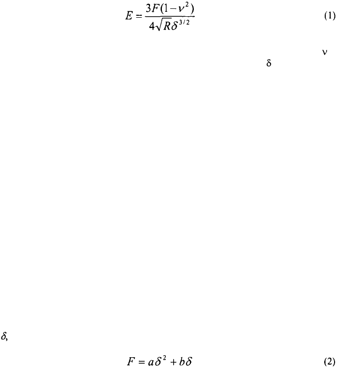
The Hertz model
145
is the most straightforward mathematical derivation for describ-
ing the elastic responses of an indented sample by the tip of the AFM.
146, 147
The Hertz
model is described as:
where E is the Young’s modulus of elasticity, F is the applied nanomechanical load, is
the Poisson’s ratio, R is the radius of the curvature of the AFM tip, and is the amount of
sample indentation. The amount of applied load and the indentation depth can be derived
from force versus distance plots that are recorded by the AFM each time the tip ap-
proaches and retracts from the sample surface. The force versus distance curve is simply
a systematic translation for the deflection of the cantilever tip versus the z-direction dis-
placement of the piezo-scanner as a result of the interaction between the AFM tip and the
sample surface.
The Poisson’s ratio is a material property that describes the ratio of transverse de-
formation to the axial deformation of the material under axial loading.
148
Typical values
for Poisson’s ratio are in the range of 0.3 to 0.5.
97, l49
Thus, by substituting obtainable
values in the Hertz model, it is possible to estimate the Young’s modulus of elasticity of
the studied sample. In the case of soft samples, such as living cells, one frequent limita-
tion with mathematical fitting of force curves using the Hertz model is the difficulty in
determining the tip-sample contact point due to (i) the softness of the cell surface and (ii)
the lack of sharpness in curve deviation as the AFM tip contacts the sample surface as is
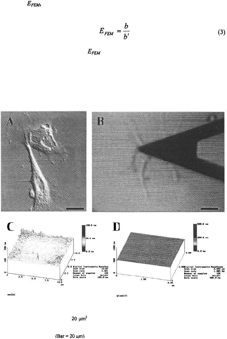
the situation when the tip contacts a glass surface (Figure 1). Also, the legitimacy of the
Hertz model for application to living cells is further depreciated by the highly anisotropic
behavior of cellular systems
143
. Nonetheless, with a lack of more sophisticated analytical
formulas to translate the AFM data into mechanical values, the Hertz model still presents
a workable framework with valuable application to mechanical mapping of living cells,
especially when a comparison of mechanical properties rather than absolute measurement
of a mechanical value to be pursued.
Recently, the quadratic equation has proven to be a useful tool for modeling force-
indentation curves.
32, 150
Applied forces, F, and resulting indentation depths up to 500
nm, have been expressed by the quadratic equation:
where a and b are the parameters expressing the nonlinearity and the initial stiffness of
the force-indentation curve, respectively. Significant correlations (r > 0.99) have been
found for the quadratically defined curves when compared with experimentally obtained
force-indentation curves.
32, 150
As the thickness of a specimen increases, the parameter b
decreases monotonically until a constant value is reached while an increase in the b pa-
rameter indicates an increase in mechanical stiffnes.
32
Both a and b parameters increase
for sheared endothelial cells which may indicate a remodeling of the cytoskeletal struc-
ture since these cells exhibited thick stress fibers of F-actin bundles.
32
An elastic
80
G.YOUREK
ET AL
THE CYTOSKELETON
81
modulus, was determined using Finite Element Modeling (FEM). It’s relationship
to the quadratic equation variables is as follows:
AFM has made the structural study of the nanosized cytoskeleton elements possible.
However, the study of the cytoskeleton comes at a cost. Low loading forces result in
smooth versions of the cell’s membrane, while the high forces uncover the true structure
of the cell’s cytoskeleton.
121
The high loading forces necessary to view the cytoskeletal
Figure 1. AFM imaging of living mesenchymal stem cells (MSCs) on glass. A. A single MSC during nanoin-
dentation with V-shaped AFM cantilever. B. The height image representing the cellular extensions of two
MSCs collected at a scan area of showing height variation within the scanning field. C. The height
image of the glass substrate showingabsence of height variation when cells are not present on the glass surface.
D. The height image of the glass substrate with the fluid imaging medium present but without cells showing
minimal height variation.
where b’ is the linear coefficient. increases in sheared endothelial cells.
32
6.1. AFM in Cytoskeleton Imaging

structures cause a large lateral drag which damages the cell and make any preceding
evaluation difficult. The importance of the actin cytoskeleton becomes readily apparent
with its disruption with cytochalasin B. Chicken cardiomyocytes appear more rounded
and a large decrease in the elastic modulus by about a factor of 3 occurred. This directly
demonstrated that the elastic response of the chicken cardiomyocyte is due in a large part
to the actin network.
Mechanical properties for multiple cell types have been reported including glial
cells,
116
epithelial cells,
106, 151
cardiocytes,
121
myocytes,
152
platelets,
126
erythrocytes,
141, 153
macrophages,
154
endothelial cells,
32, 150, 155-157
fibroblasts,
31, 118, 130, l58, 159
osteoblasts,
128, 160.
161
chondrocytes,
92
hair cells,
162, 163
F9 carcinoma cell line,
164
and bone marrow-derived
mesenchymal stem cells.
3
Mapping of mechanical properties of living cells can be
viewed as an indicator of the cytoskeleton structure and function.
165
Multiple vital proc-
esses of the cell are dependent on the dynamics of the cytoskeleton, such as cell migra-
tion,
34, 166, 167
cell division,
168, 169
cell adhesion,
170, 171
cellular transport system,
172, l73
phagocytosis,
174, 175
and maintenance of overall stability and mechanical integrity of the
cell.
176, 177
In addition to the cytoskeleton, numerous other molecules may also contribute
to the overall mechanical map of the cell membrane and have an active role in determin-
ing the elastic properties of living cells.
140, 142, l78
Wang et al.
179
used actin staining with palloidin to demonstrate changes in the ac-
tin cytoskeleton upon differentiation of human MSCs with osteogenic (bone inducing)
supplements (ascorbate, dexamethasone, and vitamin In addi-
tion to expressing markers specific to bone cells such as alkaline phosphatase, osteocal-
cin, and bone sialoprotein (BSP), production of collagen type I and bone sialoprotein and
extracellular matrix mineralization, the differentiated cells displayed highly organized
actin fibers alongside abundant bone sialoprotein complexes. In contrast to the latter, the
undifferentiated MSCs had long actin filaments and sparse amounts of bone sialoprotein.
When the actin cytoskeleton of chick limb bud mesenchymal cells (CLBMCs) is dis-
rupted chondrogenesis occurs. CLBMCs were isolated from stage 22-24 White Leghorn
chick embryos and the cytoskeleton disrupted using cytochalasin D (see section 3.2).
181
The cells rounded up, lost their actin cables, and underwent chondrogenesis, as demon-
strated by type II collagen and matrix production. These characteristics were amplified
with increasing concentrations of cytochalasin as well as with introduction of the agent at
earlier time points. However, the cells did not react this way with microtubule disrupting
drugs, leading to the conclusion that actin filaments, rather than microtubules, control the
shape of cultured limb bud cells and that these filaments play an important role in the
early development of cartilage. Furthermore, the activation of the cell signaling molecule
protein kinase C and the inhibition of the extracellular signal regulated kinase-1 has been
shown to be involved in the regulation of chondrogenesis by actin cytoskeleton disruption
by cytochalasin.
182, 183
82
7.
CELL NANOSTRUCTURAL CHANGES INDUCED BY DIFFERENTIA-
TION OR MECHANICAL FORCES
7.1.
Cell Differentiation
G.YOUREK
ET AL
180
THE CYTOSKELETON
Recent work in our laboratory has focused on the contribution of the cytoskeleton
during the process of osteogenic (bone forming cell) differentiation. We have exploited
the effect of cytochalasin on the actin cytoskeleton of human mesenchymal stem cells
(hMSCs) to study the changes of the cytoskeleton as a stem cell changes from a some-
what “generic” cell to a cell with a specific purpose, namely to form bone tissue. Fluo-
rescent rhodamine-phalloidin staining (specific for F-actin
180
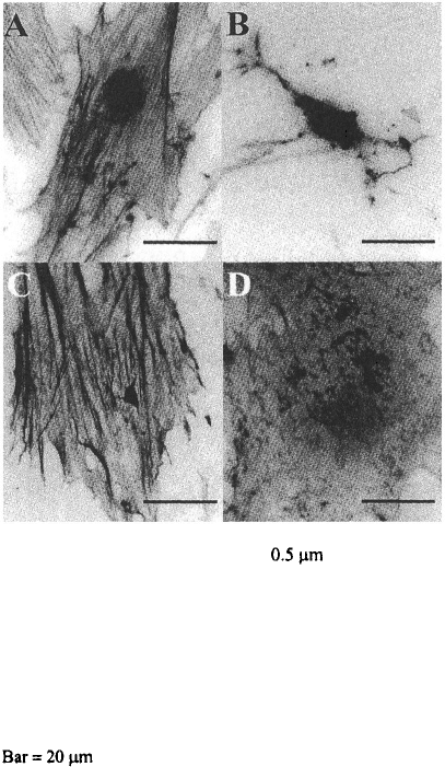
) reveals the intense trans-
formation of the actin cytoskeleton upon osteogenic differentiation; see Figure 2. The
actin cytoskeleton becomes robust and ordered upon osteogenic differentiation. It can be
speculated that the natural forces that MSCs encounter in a physiological environment do
not necessitate a strong cytoskeleton. However, upon osteogenic differentiation, in which
the stem cells become a part of a larger bone structure that functions to provide both form
and strength, the supporting structure of the cells becomes enhanced to better enable
function. The hMSCs also abandon their long, fibroblast-like, spindle shape for more of
a circular shape which may better fit their role as bone forming cell. A greater effect of
cytochalasin D on the actin cytoskeleton of undifferentiated hMSCs than on hMSCs dif-
ferentiated down the osteogenic lineage also supports the idea that the cytoskeleton of
83
Figure 2. Human BMSCs in control (Con) or Osteogenic
(OG) media 2 hr. after treatment with Cytochalasin-D
(cytD) or DMSO (Con). Actin network stained with rhoda-
mine-labeled phalloidin (pictures have been converted to
black and white so phalloidin shows dark in the images).
A. Con-Con B. Con-CytD C. OG-Con D. OG-CytD
Cells grown in osteogenic medium are more differentiated
and have a more defined cytoskeleton which is less easily
disrupted by cytD than that of undifferentiated hBMSCs.
From this we can predict differences in nanomechanical
properties of these cells caused by cytoskeletal disruption.
It has been proposed that mechanical loading of bone causes fluid flow around bone
cells and ultimately deformation (strain) at the cell membrane.
1
Blood in the circulation
system constantly exposes endothelial cells to shear and strain forces by a fluid shear
stress and endothelial cells have been proposed as sensors and regulators of vessel struc-
ture and morphogenesis.
184, 185
Erythrocytes, or red blood cells (RBCs), are constantly
exposed to a shear stress by the fluid portion of the blood as they move through the circu-
latory system.
Slides are placed in a parallel-plate flow chamber at 80% confluence (this is the op-
timum cell density to allow cells to change shape, to better visualize cell morphology and
for more precise measurement with AFM). The apparatus used in our laboratory has
been described previously.
76
In brief, the system consists of two cylindrical glass reser-
voirs, one above the other, with a parallel-plate flow chamber connected to them. The
distance between the upper reservoir and the return outlet of the flow chamber drives the
fluid flow through the chamber by the hydrostatic pressure head created. A constant
pressure is created by continuous pumping of culture medium between the lower to upper
reservoir. The flow chamber is a machine-milled polycarbonate plate, a rectangular gas-
ket on top of the bottom polycarbonate plate, a polycarbonate slide with the attached cell
layer on top of the gasket, and a top machine-milled polycarbonate plate. These will be
held together by ten nuts and bolts spaced around the outside of the plates. The polycar-
bonate plate has two holes through which medium enters and exits the channel. All parts
of the flow loop apparatus are washed, dried, assembled, and autoclaved prior to the ex-
periment.
The wall shear stress on the cell monolayer can be calculated assuming Newtonian
fluid and parallel-plate geometry:
84
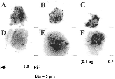
osteogenic hMSCs is robust as compared to undifferentiated hMSCs; see Figure 2. At
different concentrations of cytochalasin D, the cytoskeleton of osteogenic MSCs was less
disrupted than that of undifferentiated MSCs; see Figure 3.
7.2.
Fluid Flow Forces
7.2.1.
Parallel-Plate Flow Chamber Methodology
Figure 3. At varying doses of cytochalasin D A, D;
B, E; C, F), the actin cytochalasin of non-
differentiated MSCS (A-C) was affected more than that of osteo-
genic MSCs (D-F).
G.YOUREK
ET AL
THE CYTOSKELETON
When periosteal fibroblasts, osteocytes, and a mixture of osteoblasts and osteocytes
are subjected to a pulsatile fluid flow (PFF) of 0.5 Pa at 5 Hz,
186
secretion of prostagland-
ins and (hormonal second messengers) both increase, the greatest increase being
seen in the population of osteocytes alone. When cytochalasin B was used to disrupt the
actin cytoskeleton, this response was completely blocked. This showed that in bone cells,
the actin cytoskeleton is involved in the response to PFF and therefore, the cytoskeleton
may play an important role in bone mechanotransduction.
Osteopontin (OPN) has long been known to be an important marker of bone forma-
tion.
187, 188
It is an Arginine-Glycine-Aspartic Acid (Arg-Gly-Asp; RGD)-containing
protein and was initially described as one of the important noncollagenous proteins that
accumulates in the extracellular matrix (ECM) of bone in many verterbrates.
189, 190
Ex-
pression of osteopontin is a response to mechanical stimulation of bone cells in vitro.
191,
192
The importance of the actin cytoskeleton in relation to osteopontin expression was
demonstrated by applying a biaxial strain on a custom device similar to a Flexcell at 0.25
Hz (physiologically equivalent to a fast walk) for single 2 hr periods to embryonic chick
osteoblasts. The mRNA expression of the opn gene was unregulated in response to the
mechanical strain as compared to the unstrained cells. The role of two components of the
cytoskeleton, namely the actin filaments and microtubules, was studied by exposure of
the cells to cytoskeleton disrupting drugs; cytochalasin D for actin filaments and colchi-
cine for microtubules. While the expression of opn was prevented by the disruption of
the actin cytoskeleton, there was no effect of the disruption of the microtubule cytoskele-
ton. Endothelial cells have been shown to respond to flow by a change in shape and
alignment.
193-200
The significance of microfilaments in these processes has been demon-
strated.
201
The cytoskeleton is necessary for EC adherence under shear conditions, as
85
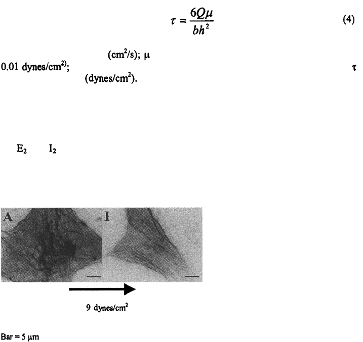
where Q is the flow rate is the viscosity of medium used in experiment (ca.
h is the channel height (0.022 cm); b is the slit width (2.5 cm); and is
the wall shear stress
7.2.2.
Examples of Cytoskeletal Importance During Fluid Flow
Figure 4. Human MSCs not exposed (A) or exposed (B), to
a fluid flow stimulus of
for 24 hr. Note the
general reorganization of the actin cytoskeleton of the cell
exposed to a fluid flow. Arrow indicates direction of flow.
