Salcido A. (ed.) Cellular Automata - Simplicity Behind Complexity
Подождите немного. Документ загружается.

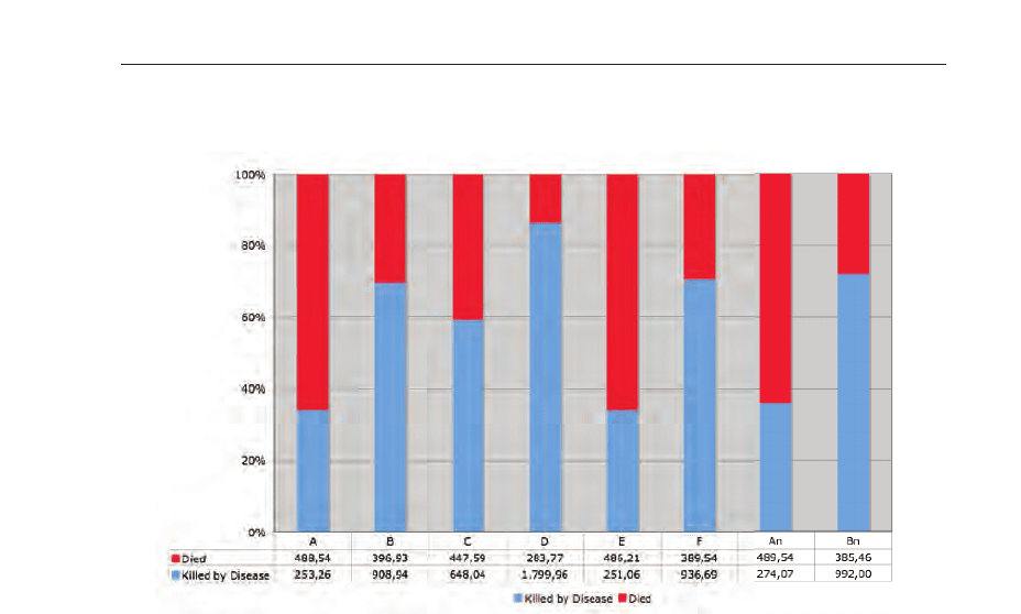
death and diseases fatal cases for the simulated scenarios A to F and An, and Bn. The
presented values are mean values per day.
Fig. 19. Percentage diagram with mean cases per day. The figure shows clearly, that in the
scenarios B, Bn, F, and especially D the death cases caused by the disease prevail.
When adding the natural births, it is possible to predict the population development during
the disease spread. Figure 20 shows, as expected, that the population decreases in each
scenario, but scenario D shows a dramatic decrease in the population.
The parameters used for the diseases life cycle were 3 days for the latent period, 10 days for
the infectious period, the incubation period was set to 3 days and the symptomatic one to 4
days. After 15 days, the individual get removed or recovers from the disease. Figure 21 shows
the ratio between the disease life cycle states. It is clearly visible that in simulation scenario
D the most infections occur and the disease is able to spread the most. Furthermore, when
comparing the scenarios A with An and B with Bn it can be figured out that the presence of
geological and demographic realities reduces the spreading in a natural way.
The pie chart presented in figure 22 shows the perceptual distribution of the fatal cases per
day.
The simulation showed that the geographic structure of the area is of importance as natural
barriers slow down the velocity of the spread. A slower velocity coupled with the change
of the natural behavior of the individuals helps to reduce cases of death. Because in this
simulation the temporary immunity was set to 100 days, the figures show a periodicity with
decreasing amplitude.
As a cellular automaton has a cellular space or cellular lattice, these models allow the
visualization of the automaton states at each time point. The regular lattice consists of several
individual cells, which interact using a neighborhood relation. In this simulation, a Moore
neighborhood instead of a von Neumann neighborhood or 2-radial neighborhood was taken
for the simulation. It has to be noted down that when simulating the scenarios using a
517
Biophysical Modeling using Cellular Automata
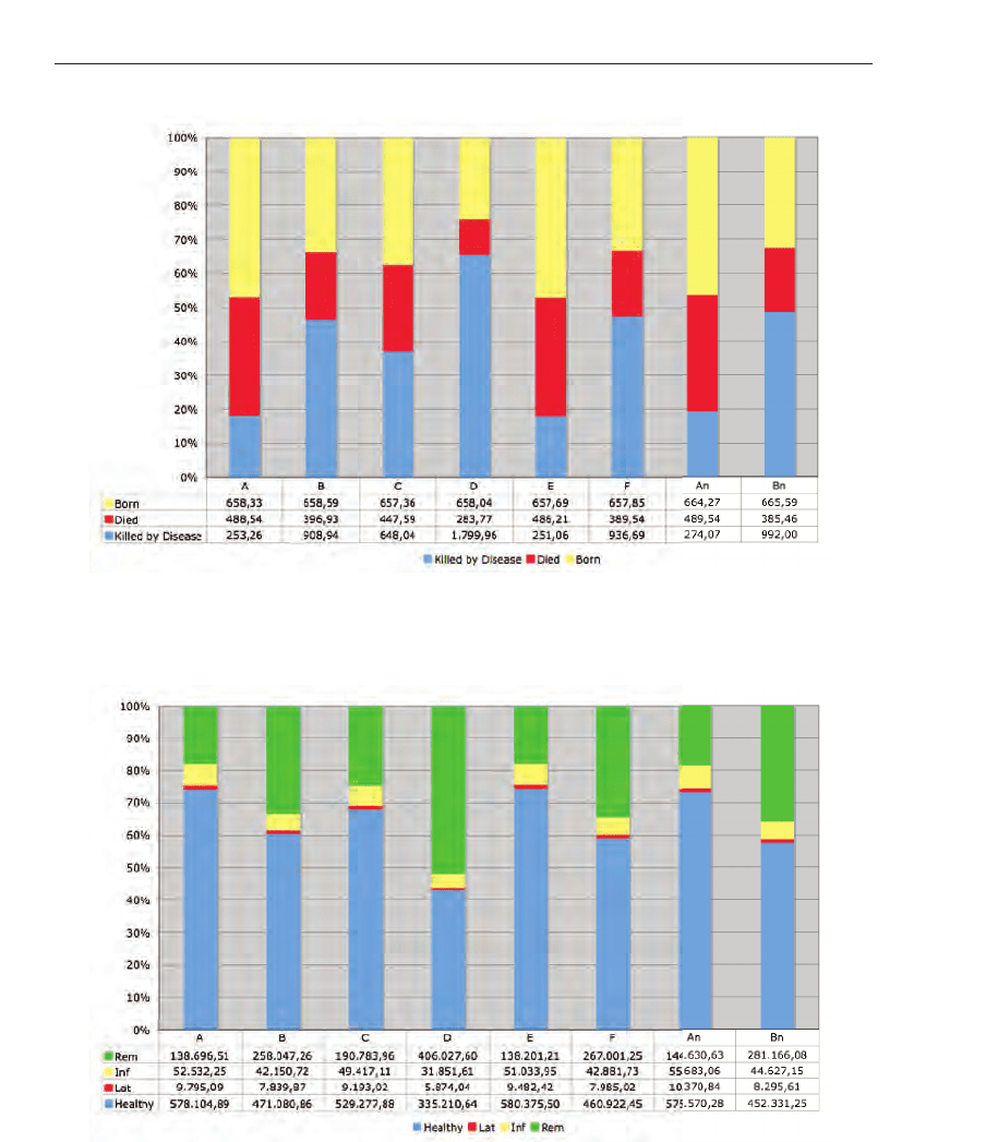
Fig. 20. Development of the population during the outbreak within the different scenarios.
Fig. 21. Tracking of the disease life cycle for the individuals for the different simulation
scenarios.
518
Cellular Automata - Simplicity Behind Complexity
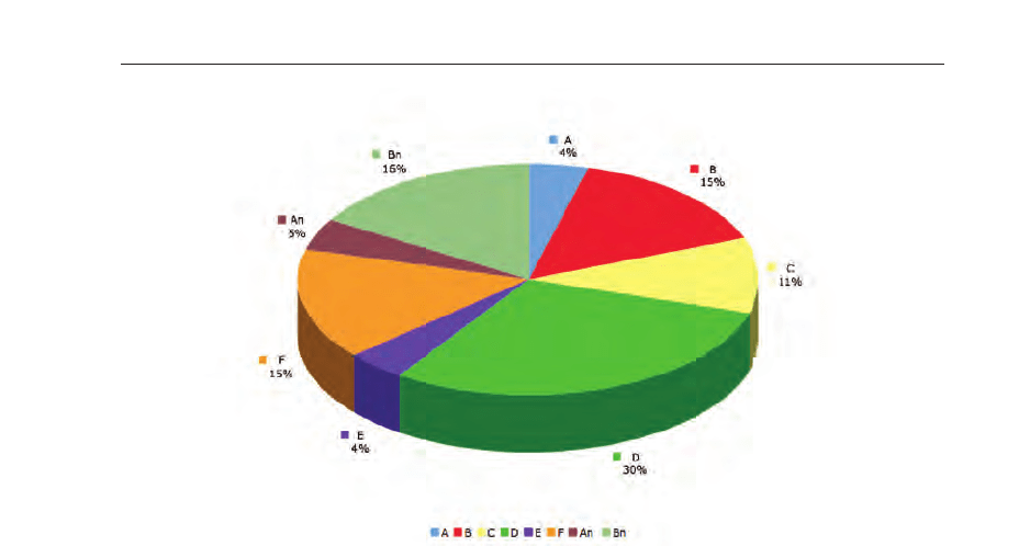
Fig. 22. Percential distribution of fatal cases per day computed by using mean value.
2-radial or von Neumann neighborhood the graphical representation is smoother and ringlike
compared to the presented ones, but the overall behavior is almost equal.
Figure 23 depicts the spatial results of the disease spread for scenario A. When analyzing
the spreading, one can see that the geographical properties and the different densities in the
valleys of Tyrol are responsible for slowing down the outbreak. However, the geographical
conditions are unable to stop the outbreak completely.
3.5.3.3 Computing time
The run time of the simulation for the state Tyrol simulation was μ = 51.87 [min] minutes
(σ
= 1.87 [ mi n ]) per scenario (simulation of 365 time steps per scenario) with four scenarios
started parallel on the same machine. The run time of the simulation for Austria was 5.4
[h]
hours per scenario. The simulation was performed using an Apple X-Serve 1.1 OS X Server
Version 10.4.10 with2x2GHzDual Core Intel Xeon processor and 2 GB of RAM installed.
The mean storage per scenario was about 10 MB (spatiotemporal snapshots per time step and
overall information). The maximum capacity of the framework is only limited by the available
memory. A simulation with 10 billion individuals, which is enough to simulate a spread of a
disease over the whole globe, would be possible, however, the simulation time would be high.
Since the insights, which wanted to be obtained on mechanisms and procedures of disease
spread, are based on extensive and complex simulations, it is of great importance to have
the possibility to run a large number of simulations or simulations on fine-grained models
in a short period without the worry of long simulation periods. It is vital to have the
opportunity to experiment with different parameters of the models, in terms of vaccination
strategies, behavioral patterns of individuals, model variations and so on. At the European
Grid Conference in Amsterdam a framework Wurz & Schuldt (n.d.) for seamless parallel
execution of various kinds of algorithms was published. The framework can be used to
schedule execution of software in parallel without the burden for the application developer
519
Biophysical Modeling using Cellular Automata
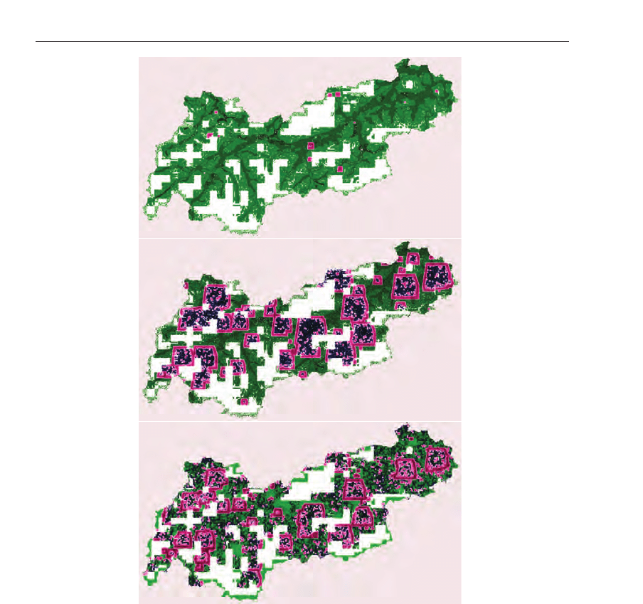
Fig. 23. The colors were used as following: green describes individuals in the state
susceptible (S), gentle-pink marks individuals as in state infective (I) and dark blue marks the
individuals as to be in state recovered (R). The first graphic depicts the simulation after 20
days. Although the simulation has started in the capital of Tyrol, in Innsbruck, after 20 days
there are some outbreaks in the west of the state and in the east, which results form the fact
that individuals are moving from cell to cell. Furthermore, unknown activities, which are
denoted as spontaneous infection, are responsible for these characteristics. The second image
shows a snapshot 80 days later at the time point 100 days after the infection started. It is
clearly visible that in the capital where the disease spread started most people are in state
recovered. Individual who did not die from the disease do have a temporary immunity. More
than 50% of the individuals are in state infected. The last image shows a snapshot at time
point 200 days after outbreak where individuals may get infected again. This represents the
second outbreak wave with smaller amplitude.
520
Cellular Automata - Simplicity Behind Complexity

to know details about the computational resources available, and that the framework,
acting as a middleware, allows for a dynamic adaptation of the scheduling process.The
framework is designed to cope with heterogeneous resources in a dynamic and rather instable
network, so that it can be used to utilize computation power available on the Internet or
the universities network to speed up simulations of the CA Framework or add additional
facets like visualization Ôon the flyÕ. The usage of grid computing in CA model simulation
lends itself to be perfect as CA models can be parallelized perfectly. In the first simulation, an
Apple Xgrid environment was used. Apple’s Xgrid technology enables to use ad hoc groups
of Macintosh system into low-cost supercomputer.
3.6 Summary
The framework enables the simulation of different communicable diseases by specifying the
disease parameters and demographic characteristics. Furthermore, the population can be
divided into subgroups, which enables to simulated different impacts of the disease on each
individual. The described scenarios demonstrated that CA and agent based models can be
used for simulating and visualizing the spread and the adherent impact of infectious diseases.
Furthermore, the simulation environment allows the access to any individual parameter at
any point in time of the simulation, which enables detailed statistical analysis. Although
the connections and the affiliated behavior between the individuals can be modeled using
different neighborhood relations, the behavior of any individual in these models depends on
functions using random numbers. For upgrading the behavior algorithms, social behavior
approaches should be used and the computation of the economic impact should be computed
for better creation of public health strategy plans for managing fatal diseases. The natural
manner Ð let us call it "Groundhog Day" - that most individuals do have a way of living
caused by their daily workflow, can not be modeled correctly, using such functions. To solve
this problem, virtual worlds could be helping. The well-known Second Life, where millions of
people do live an additional life and do also have a behavior, which is very similar compared
to their own, should be observed for simulations. One has to keep in mind, that building a
population model from census and demographic data statistically equal to one in the real
world would be very complex but also a deep impact to privacy, and therefore, afflicted with
many of problems.
Recapitulatory one can state, that the proposed CA framework is able to support public health
offices by providing them with information for creating plans to manage such situations and
to prevent serious long-term economic repercussions.
4. Imaging of the cardiac electrical function using cellular automaton approach
4.1 Introduction
Simulation of the electrocardiogram (ECG) and of the body surface potential (BSP) have
been a research topic during the last decades. Nowadays, because of the enormous computer
power available and because of extensive knowledge about cardiac electrophysiology from
the cellular to the tissue level, sophisticated three-dimensional approaches have been
developed. Although, the models became very attractive during the last years, there are still
extensive problems in making them useful for cardiovascular diagnosis and therapy. First,
the individual parameters like fibre architecture, conductivities and others are not available
to that extension needed. Second, today, the used cell membrane models have to consider
molecular function as well. Finally, the models should be validated based on human data from
the cellular to the organ level. This will imply further research necessary in the upcoming two
521
Biophysical Modeling using Cellular Automata

decades. The paper presented deals with an anisotropic ventricular and an isotropic atrial
model developed by our research group during the last 15 years. This in silico approach is
used in the electrocardiographic forward and inverse approach. Validation has been done
for the inverse formulation in 45 patients only. The whole-heart model presented uses the
bidomain source-field formulation. A detailed anatomical model of the atrium is considered
as well. The geometrical data is derived from individual magnetic resonance images. Fibre
architecture in the ventricle and anatomical features in the atrium, like Bachmann bundle or
others are considered based on literature data. The whole-heart model allows the calculation
of the de- and repolarization and of the three-dimensional potential pattern throughout the
entire heart muscle and volume conductor. Based on this simulated potential data, the 12-lead
standard ECG or different BSP maps can be visualized. Today, this in silico whole-heart
model environment is used for enhancing the understanding of the nature of the ECG in
the normal beat, for different arrhythmias, and for ischemia and infarction. A user-friendly
software environment allows interactive model generation, parameter adjustment, simulation
and visualization.
4.2 Methods: the forward problem
4.2.1 Volume conductor model
The volume conductor model (VCM) with the embedded cardiac source volume is the basis
for the electrocardiographic forward problem. The VCM consists of the compartments chest,
lungs, atrial and ventricular myocardium, and of the blood masses from an individual patient.
The morphological imaging data were acquired using a Magnetom Vision Plus 1.5 Tesla
scanner, Siemens Medical Solutions, Erlangen, Germany. For the lung and torso shape
extraction a T1 flash, non-contrasted axial data set during breath-hold (expiration, 10 mm
spacing) was used. The cardiac geometry (atrial and ventricular models) was acquired in
ECG-gated cine mode during breath-hold (expiration, oblique short-axis scans) with 4 and
6 mm spacing. The segmentation of the compartments was performed using a recently
developed VCM segmentation pipeline. The resulting labelsets were triangulated using
a standard marching cubes algorithm and optimized using Hammer B. (2001). In the
next VCM assembling step the tetrahedral mesh was created using the software package
Hypermesh (Altair Eng.), which allows to produce optimized tetrahedral meshes on the basis
of the high quality surface mesh. The whole heart VCM consists of 104,001 tetrahedrons and
18,170 nodes, respectively. The volume conductor (whole heart) model with its compartments
is depicted in Fig. 24.
4.2.2 Computation of cardiac activation sequences - the cellular automaton
The CA used in this study was developed Fuchsberger M. (1993); Killmann R. (1987; 1990);
Rosian M. (1991), modified and implemented in amiraDev 3.0™ Hayn D. (2002). Briefly, after
segmentation, triangulation and tetrahedral mesh generation different types of tissue were
assigned to the atria and ventricles with corresponding parameter setting for each tissue type.
Further, the CA needs the fiber structure assigned to each node of the tetrahedral model as
well as the refractory periods assigned to each tissue type.
In the models the types of tissue were the endocardia, epicardium and myocardium.
Furthermore, the sinus node, crista terminalis, Bachmann bundle, fossa ovalis, pectinate
muscles, coronary sinus, isthmus, the bundle of His, left and right bundle branch and the
Purkinje fibers were defined. Each type of tissue has its own set of parameters needed for
the computation of the activation times and the time dependent transmembrane potential
distribution. The fiber geometry was chosen such, that it fitted qualitatively well with the
522
Cellular Automata - Simplicity Behind Complexity
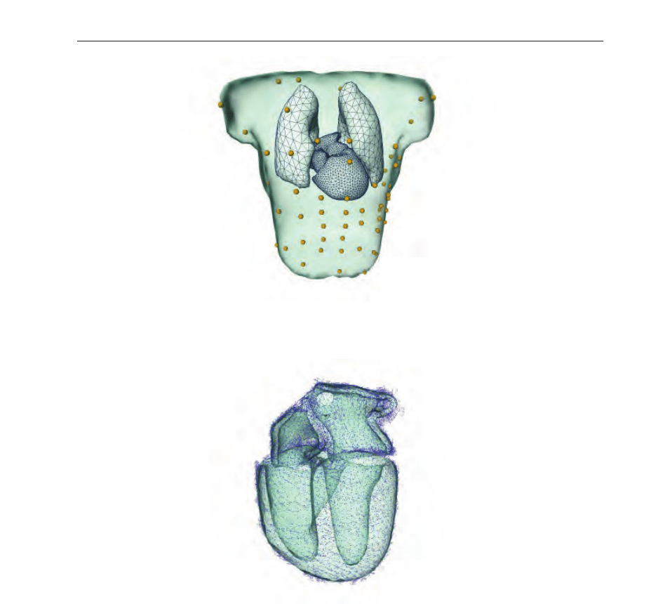
Fig. 24. VCM of an individual patient comprising chest surface, atria and ventricles with the
cavitary blood masses and both lungs displayed in an anterior-posterior view.
Fig. 25. Fiber orientation assigned to each node of the tetrahedral ventricular model for four
different views. The atria and ventricles are displayed in a transparent style.
findings described in literature Greenbaum R. A. et al. (1981); Mc Veigh E. et al. (2001);
Nielsen P. M. F. et al. (1991); Rijcken J. et al. (1999); Streeter Jr. D. D. et al. (1969). Figure 25
gives an impression of the ventricular fiber structure used in this study.
Another essential input for the CA are the effective refractory periods ERP defined for a heart
cycle length (CL) of 700 ms (termed ERP
700
). These can be adjusted for each tetrahedron
individually or for tetrahedrons belonging to one type of tissue. The effective refractory
period is defined as the time during which the cell cannot be excited by a stimulus of such
magnitude, which is twice as high as a stimulus (diastolic threshold) capable to excite a cell in
523
Biophysical Modeling using Cellular Automata

resting status. The ERP is computed during run time, as the values depend on each preceding
diastolic interval and on the current CL (detailed explanation can be found in Killmann R.
(1990); Wach P. et al. (1989)). It should be stated that the CL in this context is the time distance
between two proceeding excitation processes in one tetrahedron and does not necessarily
refer to a CL provoked by any pacemaker (e. g., sinus node, atrioventricular node or paced
rhythms). The run time ERP at the CL is calculated by
ERP
(CL)=ERP
700
+ A + B ·
CL −700
1000
− D ·
C
−CL
C
2
if CL < C and
ERP
(CL)=ERP
700
+ A + B ·
CL −700
1000
(10)
if CL
≥ C, respectively. Parameters A (offset value), B (slope of the function), C (a predefined
CL), and D (coefficient for the quadratical decrease) have to be defined for each type of tissue
individually. The values for these parameters were chosen for each tissue type according
to Killmann R. (1990). ERP
700
was set to 210 ms for the bundle of His, 290 ms for the
conjunction points, 280 ms for the left and right bundle branches, 320 ms for the Purkinje
fiber network and 260 ms for the ventricular myocardium. Parameter A is introduced for
enabling a simple change from physiological to pathological conditions for each tissue type
individually. The value for the slope parameter B was obtained by averaging data reported for
human hearts. Parameters C and D were set unequal zero for the ventricular epi-, endo- and
myocardium only as the relationship between CL and refractory periods shows up to have
a quadratically decreasing shape for these ventricular tissue types Killmann R. (1990) for the
case CL
< C.IfCL ≥ C, the ERP(CL) is assumed to increase linearly with the CL.
Computation of activation times
After selecting an arbitrary number of tetrahedrons assigned with an arbitrary time instant
for starting the excitation process the activation sequence is computed as follows. Every
tetrahedron of the cardiac model can take up three different states: excitable (e),refractory (R;
i. e., excited) or waiting (W; i. e., awaiting excitation). The last state has no physiological
meaning but was introduced due to algorithmic needs.
The simulation starts by selecting that tetrahedron with earliest excitation time and its status
is changed from e to R, i. e., the number of this tetrahedron is written into table R. The
’possible excitation times’ of the excited tetrahedron’s neighbors are calculated according to
the distance between the center of masses and conduction velocities for each connection and
stored in table W. In case no conduction between two neighboring tetrahedrons can occur the
conduction velocity is set to zero. Whether a conduction between the tissues the tetrahedra
are belonging to can or cannot occur is stored in an additional parameter set. In the next step
the two tables S and W are searched for the tetrahedron with the lowest excitation time. This
is the next tetrahedron to be excited and stored in table R and the corresponding table W is
cleared. If two or more tetrahedrons have the same starting or possible excitation time one
of them is arbitrarily chosen and the other one becomes/the other ones become the source
tetrahedron of the subsequent calculations. The propagation is then calculated as described
above, but now three cases may occur: The neighboring tetrahedron is
• in status e
→ the possible excitation time is stored in table W, i.e., the tetrahedrons’s status
is set to ’awaiting excitation’,
• already in waiting status and has been assigned a possible excitation time with
524
Cellular Automata - Simplicity Behind Complexity

– a lower value than the one calculated this time → no changes occur,
– a higher value than the one calculated this time
→ the possible excitation time is
changed to the lower value,
• in status R
→ no value is then stored for this point in table W.
Then the tables W and S are searched again and the sequence of instructions is performed for
the next source tetrahedron. The duration of status R – the period of time a tetrahedron cannot
be set into status W or e – is evaluated employing (10) each time a possible excitation is to be
assigned to that tetrahedron. The computation is finished, when no more starting tetrahedrons
in table S are left and W is empty or when the excitation time of the current source tetrahedron
is higher than the chosen upper limit for simulation duration predefined by the user.
Computation of transmembrane potentials
For modeling different shapes of ventricular and/or atrial action potentials the extended
Wohlfahrt formula described in Rosian M. (1991) is used:
ϕ
m
(t −τ)=α(t −τ) · β(t − τ) ·γ( t −τ)+K
10
[mV]. (11)
Parameter t is the current simulation time interval, τ represents the computed activation
time, α
(t − τ) [–] describes the shape of the depolarization, β(t − τ) [mV] the phase from
the beginning repolarization (enables modeling of a notch right after onset of depolarization)
to the plateau shape, and γ
(t − τ) [–] characterizes the repolarization process. The value
is 0 for all parameters of (11), when
(t − τ) < 0. Time t = 0 is considered as onset of
depolarization (time instant, when half of the depolarization amplitude is reached). With the
extended Wohlfahrt formula (11) the time dependent transmembrane potential shape for each
node of a cardiac tetrahedron can be calculated based on parameters specifically adjusted for
the types of cardiac tissue and based on the ’history’ of the preceding activation sequence
(relaxing phase, diastolic interval) and the computed activation times.
4.2.3 Extracellular potential computation
Based on the quasi static approximation of Maxwell’s equations for electromagnetic field
calculation and employing the bidomain theory Geselowitz D. B. & Miller 3rd W. T. (1983)
(therefore the subheading extracellular potential computation), the resulting differential
equations to be solved are
div
[
κ
b
gr a d
(
ϕ
)]
= −
div
[
˜σ
in
gr a d
(
ϕ
m
)]
(12)
for the cardiac region and
div
[
κ
c
gr a d
(
ϕ
)]
=
0 (13)
for all other compartments of the VCM. The tensor κ
b
is the bulk conductivity, i. e., the sum of
the electrical effective extracellular and effective intracellular conductivity ˜σ
in
. The potential ϕ
describes the extracellular, ϕ
m
the transmembrane potential. The tensor κ
c
holds the
electrical conductivities for all other compartments c. For the ventricular electrical anisotropic
conductivities, for the other compartments isotropic conductivities were assumed Bradley
et al. (2000).
Applying the FEM, considering the volume conductor model and the related boundary
conditions Seger M. et al. (2005) the equations (12) and (13) form together a system of algebraic
equations
Rφ
= Sφ
m
. (14)
525
Biophysical Modeling using Cellular Automata
The matrices R and S are the so-called stiffness matrices, the matrix φ describes the potentials
in all nodes of the tetrahedral mesh of the volume conductor, the matrix φ
m
contains the
transmembrane potentials in all source nodes of the ventricles. For forward simulation, the
matrix φ
m
is computed by the CA for discrete time steps. For inverting R on the left hand side
of equation (14), the matrix R has to be modified as this matrix is positive semi-definite due
to the Neuman’s boundary condition on the torso surface Fischer G. et al. (2002). Therefore, the
Wilson central terminal is used to define the reference potential on the torso surface Fischer G.
et al. (2002) to reveal a positive definite matrix
˜
R. The inversion of
˜
R is performed by a solver
based on the conjugated gradient method. This consequently leads to the desired potentials in
all nodes of the volume conductor model
φ
=
˜
R
−1
Sφ
m
. (15)
Apart from scaling of matrix
˜
R no additional preconditioning was performed.
4.3 Results
The environment for simulating the potential data and for visualizing the ECG or BSP maps is
based on amiraDev™ (TGS Europe Inc.), which was extended implementing the described
functionality using the plugin concept. Thus, a homogenous and user-friendly simulation
toolbox could be implemented.
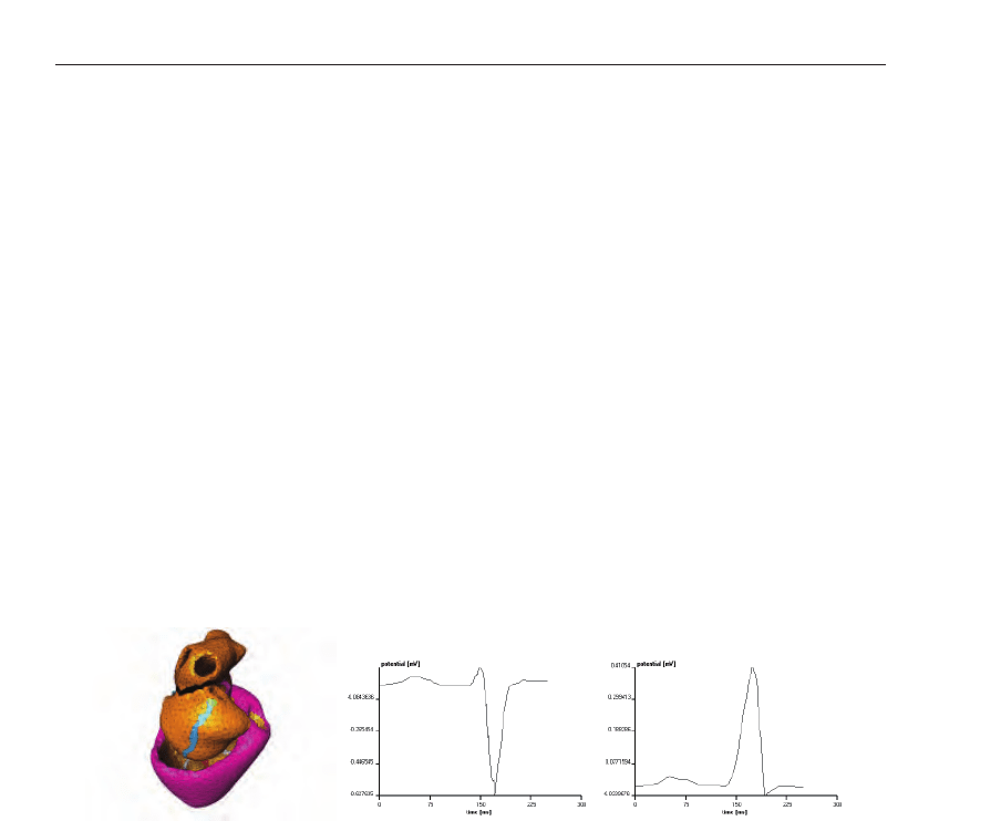
Figure 26 depicts a normal sinus rhythm, whereas in figure 27 an extra stimulus between the
right lower and upper pulmonary vein was simulated.
Fig. 26. ECG pattern simulation of a normal sinus rhythm. The gray boxes in the green area
(sinus node) mark the tetrahedron where the stimulation starts from. The second figure
shows the V3 and the third figure the V5 lead.
By varying the compartment specific parameters (intra- and extracellular conductivities,
transmembrane shape) and by specifying the starting point and time any simulation can be
performed and used for better understanding the ECG. Furthermore, the potential can be
visualized, which also contributes in a deeper understanding of electrical propagation in the
heart and the composition of the ECG patterns.
4.4 Discussion
We presented an in silico model environment for the simulation of cardiac de- and
repolarization and of the three-dimensional potential pattern throughout the entire volume
conductor. A cellular automaton and a bidomain-theory based source-field numerics are the
fundamental basics. Only a few simulation scenarios have been presented in this paper.
Limitations are given because of the relative course spatial discretization and because of not
considering heart muscle contraction. Hence, microscopic cardiac propagation effects can not
be simulated. Because we mostly had interest in investigating the macroscopic source-field
526
Cellular Automata - Simplicity Behind Complexity