Peterson D.R., Bronzino J.D. (Eds.) Biomechanics: Principles and Applications
Подождите немного. Документ загружается.


Arterial Macrocirculatory Hemodynamics 10-9
r
R(t)
1.0
0.60
0.44
0.5
0
–1.0
0.00
0.39
0.06
0.30
0.077
t=0.11
–0.4 –0.2 0 0.2 0.4 0.6 0.8 1.0
–0.5
w/w
m
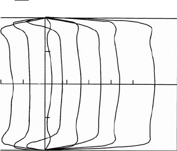
FIGURE 10.4 Velocity profiles obtained with a hot-film anemometer probe in the descending thoracic aorta of a
dog at normal arterial pressure and cardiac output. The velocity at t = time/(cardiac period) is plotted as a function
of radial position. Velocity w is normalized by the maximum velocity w
m
and radial position at each time by the
instantaneous vessel radius R(t). The aortic valve opens at t = 0. Peak velocity occurs 11% of the cardiac period after
aortic valve opening. (From Ling, S.C., Atabek, W.G., Letzing, W.G. et al. 1973. Circ. Res. 33: 198. With permission.)
for the increase in resistance of the large vessels, compromising the ability of the system to respond to
increases in demand during exercise. Eventually the circulation is completely dilated, and resting flow
begins to decrease. A blood clot may form at the site or lodge in a narrowed segment, causing an acute loss
of blood flow. The disease is particularly dangerous in the coronary and carotid arteries due to the critical
oxygen requirements of the heart and brain.
In addition to intimal thickening, the arterial wall properties also change with age. Most measurements
suggest that arterial elastic modulus increases with age (hardening of the arteries); however, in some cases
arteries become more compliant (inverse of elasticity) [Learoyd and Taylor, 1966]. Local weakening of the
wall may also occur, particularly in the descending aorta, giving rise to an aneurysm, which, if ruptures,
can cause sudden death.
Defining Terms
Aneurysm: A ballooning of a blood vessel wall caused by weakening of the elastic material in the wall.
Atherosclerosis: A disease of the blood vessels characterized by thickening of the vessel wall and eventual
occlusion of the vessel.
Collagen: A protein found in blood vessels that is much stiffer than elastin.
Elastin: A very elastic protein found in blood vessels.
Endothelial: The inner lining of blood vessels.
Impedance: A (generally) complex number expressing the ratio of pressure to flow.
Myogenic: A change in smooth-muscle tone due to stretch or relaxation, causing a blood vessel to resist
changes in diameter.
Newtonian: A fluid whose stress-rate-of-strain relationship is linear, following Newton’s law. The fluid
will have a viscosity whose value is independent of rate of strain.
Pulmonary: The circulation that delivers blood to the lungs for reoxygenation and carbon dioxide
removal.
Systemic: The circulation that supplies oxygenated blood to the tissues of the body.
Vasoconstrictor: A substance that causes an increase in smooth-muscle tone, thereby constricting blood
vessels.
9-10 Biomechanics
apposition of the valve leaflets can cause regurgitation, which is leaking of the blood being ejected back
into the atrium.
9.2.1 Mechanical Properties
Studies on the mechanical behavior of the mitral leaflet tissue have been conducted to determine the key
connective tissue components that influence the valve function. Histological studies have shown that the
tissue is composed of three layers that can be identified by differences in cellularity and collagen density.
Analysis of the leaflets under tensionindicated that the anterior leaflet wouldbe more capable of supporting
larger tensile loads than the posterior leaflet. The differences between the mechanical properties between
the two leaflets may require different material selection for repair or replacement of the individual leaflets
[Kunzelman et al., 1993a,b].
Studies have also been done on the strength of the chordae tendinae. The tension of chordae tendineae
in dogs was monitored throughout the cardiac cycle by Salisbury and co-workers [1963]. They found that
the tension only paralleled the left ventricular pressure tracings during isovolumic contraction, indicating
slackness at other times in the cycle. Investigation of the tensile properties of the chordae tendineae at
different strain rates by Lim and Bouchner [1975] found that the chordae had a non-linear stress–strain
relationship. They found that the size of the chordae had a more significant effect on the development of
the tension than did the strain rate. The smaller chordae with a cross-sectional area of 0.001 to 0.006 cm
2
had a modulus of 2 × 10
9
dyn/cm
2
, while larger chordae with a cross-sectional area of 0.006 to 0.03 cm
2
had a modulus of 1 ×10
9
dyn/cm
2
.
A theoretical study of the stresses sustained by the mitral valve was performed by Ghista and Rao
[1972]. This study determined that the stress level can reach as high as 2.2 × 10
6
dynes/cm
2
just prior to
the opening of the aortic valve, with the left ventricular pressure rising to 150 mmHg. A mathematical
model has also been created for the mechanics of the mitral valve. It incorporates the relationship between
chordae tendineae tension, left ventricular pressure, and mitral valve geometry [Arts et al., 1983]. This
study examined the force balance on a closed valve, and determined that the chordae tendinae force was
always more than half the force exerted on the mitral valve orifice by the transmitral pressure gradient.
During the past 10 years, computational models of mitral valve mechanics have been developed, with the
most advanced modeling being three-dimensional finite element models (FEM) of the complete mitral
apparatus. Kunzelman and co-workers [1993, 1998] have developed a model of the mitral complex that
includes the mitral leaflets, chordae tendinae, contracting annulus, and contracting papillary muscles.
From these studies, the maximum principal stresses found at peak loading (120 mmHg) were 5.7 × 10
6
dyn/cm
2
in the annular region, while the stresses in the anterior leaflet ranged from 2 × 10
6
to 4 × 10
6
dyn/cm
2
. This model has also been used to evaluate mitral valve disease, repair in chordal rupture, and
valvular annuloplasty.
9.2.2 Valve Dynamics
The valve leaflets, chordae tendineae, and papillary muscles all participate to ensure normal functioning
of the mitral valve. During isovolumic relaxation, the pressure in the left atrium exceeds that of the left
ventricle, and the mitral valve cusps open. Blood flows through the open valve from the left atrium to the
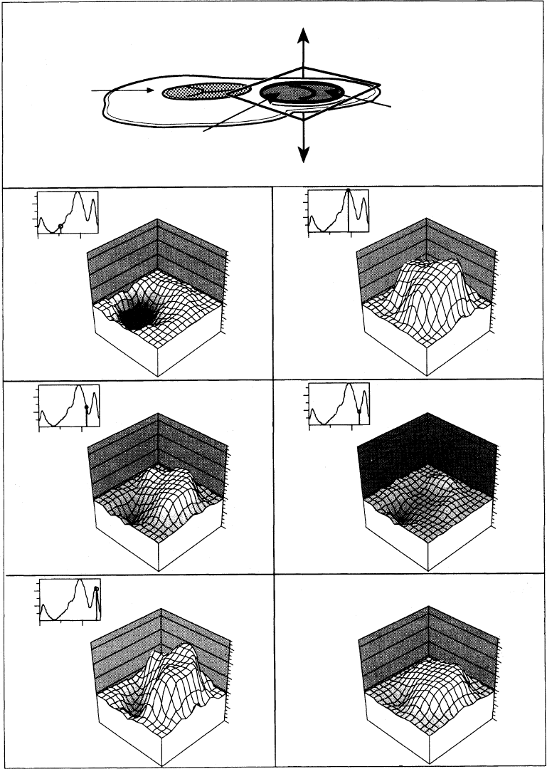
left ventricle during diastole. The velocity profiles at both the annulus and the mitral valve tips have been
shown to be skewed [Kim et al., 1994] and therefore are not flat as is commonly assumed. This skewing
of the inflow profile is shown in Figure 9.7. The initial filling is enhanced by the active relaxation of the
ventricle, maintaining a positive transmitral pressure. The mitral velocity flow curve shows a peak in the
flow curve, called the E-wave, which occurs during the early filling phase. Normal peak E-wave velocities
in healthy individuals range from 50 to 80 cm/sec [Samstad et al., 1989; Oh et al., 1997]. Following active
ventricular relaxation, the fluid begins to decelerateand the mitral valve undergoespartial closure. Then the
atrium contracts and the blood accelerates through the valve again to a secondary peak, termed the A-wave.
The atrium contraction plays an important role in additional filling of the ventricle during late diastole.

Heart Valve Dynamics 9-11
Apex of the heart
(a) (b)
(c) (d)
(e) (f)
L
Posterior
mitral leaflet
PR
Base of the heart
Anterior
mitral
leaflet
A
Aortic
valve
20.0
0.0
0.0
500.0
Velocity (cm/sec)
20.0
0.0
0.0
500.0
Velocity (cm/sec)
Velocity (cm/sec)
60
40
20
0
–20
–40
Velocity (cm/sec)
60
40
20
0
–20
–40
270 msec 470 msec
590 msec
mean
A
PR
L
A
PR
L
20.0
0.0
0.0
500.0
Velocity (cm/sec)
20.0
0.0
0.0
500.0
Velocity (cm/sec)
Velocity (cm/sec)
60
40
20
0
–20
–40
Velocity (cm/sec)
60
40
20
0
–20
–40
560 msec
A
Time (msec) Time (msec)
Time (msec) Time (msec)
PR
L
A
PR
L
20.0
0.0
0.0
500.0
Velocity (cm/sec)
Velocity (cm/sec)
60
40
20
0
–20
–40
Velocity (cm/sec)
60
40
20
0
–20
–40
670 msec
A
Time (msec)
PR
L
A
PR
L
FIGURE 9.7 Two-dimensional transmitral velocity profiles recorded at the level of the mitral annulus in a pig [Kim
et al., 1994]. (a) systole; (b) peak E-wave; (c) deceleration phase of early diastole; (d) mid-diastolic period (diastasis);
(e) peak A-wave; (f) time averaged diastolic cross-sectional mitral velocity profile. (Reprinted with permission from
the American College of Cardiology, J. Am. Coll. Cardiol. 24: 532–545.)

9-12 Biomechanics
In healthy individuals, velocities during the A-wave are typically lower than those of the E-wave, with a
normal E/A velocity ratio ranging from 1.5 to 1.7 [Oh et al., 1997]. Thus, normal diastolic filling of the left
ventricle shows two distinct peaks in the flow curve with no flow leaking back through the valve during
systole.
The tricuspid flow profile is similar to that of the mitral valve, although the velocities in the tricuspid
valve are lower because it has a larger valve orifice. In addition, the timing of the valve opening is slightly
different. Since the peak pressure in the right ventricle is less than that of the left ventricle, the time for
right ventricular pressure to fall below the right atrial pressure is less than the corresponding time period
for the left side of the heart. This leads to a shorter right ventricular isovolumic relaxation and thus an
earlier tricuspid opening. Tricuspid closure occurs after the mitral valve closes since the activation of the
left ventricle precedes that of the right ventricle [Weyman, 1994].
A primary focus in explaining the fluid mechanics of mitral valve function has been understanding the
closing mechanism of the valve. Bellhouse [1972] first suggested that the vortices generated by ventricular
filling were important for the partial closure of the mitral valve following early diastole. Their in vitro
experiments suggested that without the strong outflow tract vortices, the valve would remain open at
the onset of ventricular contraction, thus resulting in a significant amount of mitral regurgitation before
complete closure. Later in vitro experiments by Reul and Talukdar [1981] in a left ventricle model made
from silicone suggested that an adverse pressure differential in mid-diastole could explain both the flow
deceleration and the partial valve closure, even in the absence of a ventricular vortex. Thus, the studies
by Reul and Talukdar suggest that the vortices may provide additional closing effects at the initial stage;
however, the pressure forces are the dominant effect in valve closure. A more unified theory of valve closure
putforth byYellin andco-workers[1981]includesthe importanceofchordaltension, flowdeceleration,and
ventricularvortices,with chordaltensionbeinganecessary condition for the other two.Theiranimalstudies
indicated that competent valve closure can occur even in the absence of vortices and flow deceleration.
Recent studies using magnetic resonance imaging to visualize the three-dimensional flow field in the left
ventricle showed that in normal individuals a large anterior vortex is present at initial partial closure of the
valve, as well as following atrial contraction [Kim et al., 1995]. Studies conducted in our laboratory using
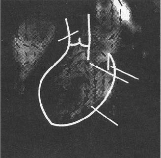
magnetic resonance imaging of healthy individuals clearly show the vortices in the left ventricle [Walker
et al., 1996], which may be an indication of normal diastolic function. An example of these vortices is
presented in Figure 9.8.
Another area of interest has been the motion of the mitral valve complex. The heart moves throughout
the cardiac cycle; similarly, the mitral apparatus moves and changes shape. Recent studies have been
LV
MV
AO
FIGURE 9.8 Magnetic resonance image of a healthy individual during diastole. An outline of the interior left ventricle
(LV) is indicated in white as are the mitral valve leaflets (MV) and the aorta (AO). Velocity vectors were obtained from
MRI phase velocity mapping and superimposed on the anatomical image.
Heart Valve Dynamics 9-13
conducted that examined the three-dimensional dynamics of the mitral annulus during the cardiac cycle
[Ormiston et al., 1981; Komoda et al., 1994; Pai et al., 1995; Glasson et al., 1996]. These studies have shown
that during systole the annular circumference decreases from the diastolic value due to the contraction of
the ventricle, and this reduction in area ranges from 10 to 25%. This result agrees with an animal study of
Tsakiris and co-workers [1971] that looked at the difference in the size, shape, and position of the mitral
annulus at different stages in the cardiac cycle. They noted an eccentric narrowing of the annulus during
both atrial and ventricular contractions that reduced the mitral valve area by 10 to 36% from its peak
diastolic area. This reduction in the annular area during systole is significant, resulting in a smaller orifice
area for the larger leaflet area to cover. Not only does the annulus change size, but it also translates during
the cardiac cycle. The movement of the annulus towards the atrium has been suggested to play a role in
ventricular filling, possibly increasing the efficiency of blood flow into the ventricle. During ventricular
contraction, there is a shortening of the left ventricular chamber along its longitudinal axis, and the mitral
and tricuspid annuli move towards the apex [Simonson and Schiller, 1989; Hammarstr
¨
om et al., 1991;
Alam and Rosenhamer, 1992; Pai et al., 1995].
The movement of the papillary muscles is also important in maintaining proper mitral valve function.
The papillary muscles play an important role in keeping the mitral valve in position during ventricular
contraction. Abnormal strain on the papillary muscles could cause the chordae to rupture, resulting in
mitral regurgitation. It is necessary for the papillary muscles to contract and shorten during systole to
prevent mitral prolapse; therefore, the distance between the apex of the heart to the mitral apparatus is
important. The distance from the papillary muscle tips to the annulus was measured in normal individuals
during systole and was found to remain constant [Sanfilippo et al., 1992]. In patients with mitral valve
prolapse, however, this distance decreased, corresponding to a superior displacement of the papillary
muscle towards the annulus.
The normal function of the mitral valve requires a balanced interplay between all of the components
of the mitral apparatus, as well as the interaction of the atrium and ventricle. Engineering studies into
mitral valve function have provided some insight into its mechanical properties and function. Further
fundamental and detailed studies are needed to aid surgeons in repairing the diseased mitral valve and
in understanding the changes in function due to mitral valve pathologies. In addition, these studies are
crucial for improving the design of prosthetic valves that more closely replicate native valve function.
References
Alam, M. and Rosenhamer, G. 1992. Atrioventricular plane displacement and left ventricular function.
J. Am. Soc. Echocardiogr. 5: 427–433.
Arts, T., Meerbaum, S., Reneman, R., and Corday, E. 1983. Stresses in the closed mitral valve: a model
study. J. Biomech. 16: 539–547.
Barlow, J.B. 1987. Perspectives on the Mitral Valve. F.A. Davis Company, Philadelphia, PA.
Bellhouse, B.J. and Bellhouse, F. 1969. Fluid mechanics of model normal and stenosed aortic valves. Circ.
Res. 25: 693–704.
Bellhouse, B.J. 1969. Velocity and pressure distributions in the aortic valve. J. Fluid. Mech. 37: 587–600.
Bellhouse, B.J. 1972. The fluid mechanics of a model mitral valve and left ventricle. Cardiovasc. Res.
6: 199–210.
Christie, G.W. 1990. Anatomy of aortic heart valve leaflets: the influence of glutaraldehyde fixation on
function. Eur. J. Cardio-Thorac. Surg. 6[Supp 1]: S25–S33.
David, H., Boughner, D.R., Vesely, I., and Gerosa, G. 1994. The pulmonary valve: is it mechanically suitable
for use as an aortic valve replacement? ASAIO J. 40: 206–212.
Davies, M.J. 1980. Pathology of Cardiac Valves. Butterworths, London.
Dunmore-Buyze, J., Boughner, D.R., Macris, N., and Vesely, I. 1995. A comparison of macroscopic lipid
content within porcine pulmonary and aortic valves: implications for bioprosthetic valves. J. Thorac.
Cardiovasc. Surg. 110: 1756–1761.
Emery, R.W. and Arom, K.V. 1991. The Aortic Valve. Hanley & Belfus, Philadelphia, PA.
9-14 Biomechanics
Glasson, J.R., Komeda, M., Daughters, G.T., Niczyporuk, M.A., Bolger, A.F., Ingels, N.B., and Miller, D.C.
1996. Three-dimensional regional dynamics of the normal mitral annulus during left ventricular
ejection. J. Thorac. Cardiovasc. Surg. 111: 574–585.
Gramiak, R. and Shah, P.M. 1970. Echocardiography of the normal and diseased aortic valve. Radiology
96: 1.
Ghista, D.N. and Rao, A.P. 1972. Structural mechanics of the mitral valve: stresses sustained by the valve;
non-traumatic determination of the stiffness of the in vivo valve. J. Biomech. 5: 295–307.
Hammarstr
¨
om, E., Wranne, B., Pinto, F.J., Puryear, J., and Popp, R.L. 1991. Tricuspict annular motion.
J. Am. Soc. Echocardiogr. 4: 131–139.
Hatle, L. and Angelsen, B. 1985. Doppler Ultrasound in Cardiology: Physical Principals and Clinical Appli-
cations. Lea and Febiger, Philadelphia, PA.
He,S., Fontaine, A.A., Schwammenthal, E., Yoganathan, A.P., and Levine, R.A. 1997. Integrated mechanism
for functional mitral regurgitation: leaflet elongation versus coapting force: in vitro study. Circulation
96: 1826–1834.
He, S., Lemmon, J.D., Weston, M.W., Jensen, M.O., Levine, R.A., and Yoganathan, A.P. 1999. Mitral
valve compensation for annular dilatation: in vitro study into the mechanisms of functional mitral
regurgitation with an adjustable annulus model. J. Heart Valve Dis. 8: 294–302.
Kilner, P.J., Yang, G.Z., Mohiaddin, R.H., Firmin, D.N., and Longmore, D.B. 1993. Helical and retrograde
secondary flow patterns in the aortic arch studied by three-directional magnetic resonance velocity
mapping. Circulation 88[part I]: 2235–2247.
Kim, W.Y., Bisgaard, T., Nielsen, S.L., Poulsen, J.K., Pederson, E.M., Hasenkam, J.M., and Yoganathan, A.P.
1994. Two-dimensional mitral flow velocity profiles in pig models using epicardial echo-Doppler-
cardiography. J. Am. Coll. Cardiol. 3: 673–683.
Kim, W.Y., Walker, P.G., Pederson, E.M., Poulsen, J.K., Houlind, K.C., and Oyre, S. 1995. Left ventricu-
lar blood flow patterns in normal subjects: a quantitative analysis of three-dimensional magnetic
resonance velocity mapping. 4: 422–438.
Komoda, T., Hetzer, R., Uyama, C., Siniawski, H., Maeta, H., Rosendahl, P., and Ozaki, K. 1994. Mitral
annular function assessed by 3D imaging for mitral valve surgery. J. Heart Valve Dis. 3: 483–490.
Kunzelman, K.S., Cochran, R.P., Murphee, S.S., Ring, W.S., Verrier, E.D., and Eberhart, R.C. 1993a. Dif-
ferential collagen distribution in the mitral valve and its influence on biomechanical behaviour. J.
Heart Valve Dis. 2: 236–244.
Kunzelman, K.S., Cochran, R.P., Chuong, C., Ring, W.S., Verner, E.D., and Eberhart, R.D. 1993b. Finite
element analysis of the mitral valve. J. Heart Valve Dis. 2: 326–340.
Kunzelman, K.S., Reimink, M.S., and Cochran, R.P. 1998. Flexible versus rigid ring annuloplasty for mitral
valve annular dilatation: a finite element model. J. Heart Valve Dis. 7: 108–116.
Kunzelman, K.S., Cochran, R.P., Verner, E.D., and Eberhart, R.D. 1994. Anatomic basis for mitral valve
modeling. J. Heart Valve Dis. 3: 491–496.
Leeson-Dietrich, J., Boughner, D., and Vesely, I. 1995. Porcine pulmonary and aortic valves: a comparison
of their tensile viscoelastic properties at physiological strain rates. J. Heart Valve Dis. 4: 88–94.
Levine, R.A., Trivizi, M.O., Harrigan, P., and Weyman, A.E. 1987. The relationship of mitral annular shape
to the diagnosis of mitral valve prolapse. Circulation 75: 756–767.
Lim, K.O. and Bouchner, D.P. 1975. Mechanical properties of human mitral valve chordae tendineae:
variation with size and strain rate. Can. J. Physiol. Pharmacol. 53: 330–339.
Lo, D. and Vesely, I. 1995. Biaxial strain analysis of the porcine aortic valve. Ann. Thorac. Surg. 60(Suppl II):
S374–S378.
Oh, J.K., Appleton, C.P., Hatle, L.K., Nishimura, R.A., Seward, J.B., and Tajik, A.J. 1997. The noninvasive
assessment of left ventricular diastolic function with two-dimensional and doppler echocardio-
graphy. J. Am. Soc. Echocardiogr. 10: 246–270.
Ormiston, J.A., Shah, P.M., Tei, C., and Wong, M. 1981. Size and motion of the mitral valve annulus in
man: a two-dimensional echocardiographic method and findings in normal subjects. Circulation 64:
113–120.
Heart Valve Dynamics 9-15
Pai, R.G., Tanimoto, M., Jintapakorn, W., Azevedo, J., Pandian, N.G., and Shah, P.M. 1995. Volume-
renderedthree-dimensionaldynamicanatomyofthemitral annulususing a transesophagealechocar-
diographic technique. J. Heart Valve Dis. 4: 623–627.
Paulsen, P.K. and Hasenkam, J.M. 1983. Three-dimensional visualization of velocity profiles in the ascend-
ing aorta in dogs, measured with a hot film anemometer. J. Biomech. 16: 201–210.
Raganathan, N., Lam, J.H.C., Wigle, E.D., and Silver, M.D. 1970. Morphology of the human mitral valve:
the valve leaflets. Circulation 41: 459–467.
Reul, H. and Talukdar, N. 1979. Heart valve mechanics. In Quantitative Cardiovascular Studies Clinical
and Research Applications of Engineering Principles. Hwang, N.H.C., Gross, D.R., and Patel, D.J., Eds.
University Park Press, Baltimore, MD, pp. 527–564.
Roberts, W.C. 1983. Morphologic features of the normal and abnormal mitral valve. Am. J. Cardiol. 51:
1005–1028.
Rossvoll, O., Samstad, S., Torp, H.G., Linker, D.T., Skjærpe, T., Angelsen, B.A.J., and Hatle, L. 1991. The ve-
locity distribution in the aortic annulus in normalsubjects: a quantitativeanalysisoftwo-dimensional
Doppler flow maps. J. Am. Soc. Echocardiogr. 4: 367–378.
Sahasakul, Y., Edwards, W.D., Naessens, J.M., and Tajik, A.J. 1988. Age-related changes in aortic and mitral
valve thickness: implications for two-dimensional echocardiography based on an autopsy study of
200 normal human hearts. Am. J. Cardiol. 62: 424–430.
Salisbury, P.F., Cross, C.E., and Rieben, P.A. 1963. Chordae tendinea tension. Am. J. Physiol. 25: 385–392.
Samstad, O., Gorp, H.G., Linker, D.T., Rossvoll, O., Skjaerpe, T., Johansen, E., Kristoffersen, K.,
Angelson, B.A.J., and Hatle, L. 1989. Cross-sectional early mitral flow velocity profiles from colour
Doppler. Br. Heart J. 62: 177–84.
Sanfilippo,A.J.,Harrigan, P., Popovic, A.D., Weyman, A.E., andLevine, R.A.1992. Papillary muscle traction
in mitral valve prolapse: quantitation by two-dimensional echocardiography. J. Am. Coll. Cardiol.
19: 564–571.
Scott, M.J. and Vesely, I. 1996. Morphology of porcine aortic valve cusp elastin. J. Heart Valve Dis.5:
464–471.
Silver, M.D., Lam, J.H.C., Raganathan, N., and Wigle, E.D. 1971. Morphology of the human tricuspid
valve. Circulation 43: 333–348.
Silver, M.A. and Roberts, W.C. 1985. Detailed anatomy of the normally functioning aortic valve in hearts
of normal and increased weight. Am. J. Cardiol. 55: 454–461.
Silverman, M.E. and Hurst, J.W. 1968. The mitral complex: interaction of the anatomy, physiology, and
pathology of the mitral annulus, mitral valve leaflets, chordae tendineae and papillary muscles. Am.
Heart J. 76: 399–418.
Simonson, J.S. and Schiller, N.B. 1989. Descent of the base of the left ventricle: an echocardiogiaphic index
of left ventricular function. J. Am. Soc. Echocardiogr. 2: 25–35.
Sloth, E., Houlind, K.C., Oyre, S., Kim, Y.K., Pedersen, E.M., Jørgensen, H.S., and Hasenkam, J.M. 1994.
Three-dimensional visualization of velocity profiles in the human main pulmonary artery using
magnetic resonance phase velocity mapping. Am.HeartJ.128: 1130–1138.
Sung, H.W. and Yoganathan, A.P. 1990. Axial flow velocity patterns in a normal human pulmonary artery
model: pulsatile in vitro studies. J. Biomech. 23: 210–214.
Thubrikar, M. 1990. The Aortic Valve. CRC Press, Boca Raton, FL.
Thubrikar, M., Heckman, J.L., and Nolan, S.P. 1993. High speed cine-radiographic study of aortic valve
leaflet motion. J. Heart Valve Dis. 2: 653–661.
Tsakiris, A.G., von Bernuth, G., Rastelli, G.C., Bourgeois, M.J., Titus, J.L., and Wood, E.H. 1971.
Size and motion of the mitral valve annulus in anesthetized intact dogs. J. Appl. Physiol. 30:
611–618.
Vesely, I. and Noseworthy, R. 1992. Micromechanics of the fibrosa and the ventricularis in aortic valve
leaflets. J. Biomech. 25: 101–113.
Vesely, I., Macris, N., Dunmore, P.J., and Boughner, D. 1994. The distribution and morphology of aortic
valve cusp lipids. J. Heart Valve Dis. 3: 451–456.
9-16 Biomechanics
Vesely, I. 1998. The role of elastin in aortic valve mechanics. J. Biomech. 31: 115–123.
Vollebergh, F.E.M.G. and Becker, A.E. 1977. Minor congenital variations of cusp size in tricuspid aortic
valves: possible link with isolated aortic stenosis. Br. Heart J. 39: 106–111.
Walker, P.G., Cranney, G.B., Grimes, R.Y., Delatore, J., Rectenwald, J., Pohost, G.M., and Yoganathan, A.P.
1996. Three-dimensional reconstruction of the flow in a human left heart by magnetic resonance
phase velocity encoding. Ann. Biomed. Eng. 24: 139–147.
Westaby, S., Karp, R.B., Blackstone, E.H., and Bishop, S.P. 1984. Adult human valve dimensions and their
surgical significance. Am. J. Cardiol. 53: 552–556.
Weyman, A.E. 1994. Principles and Practices of Echocardiography. Lea & Febiger, Philadelphia, PA.
Yellin, E.L., Peskin, C., Yoran, C., Koenigsberg, M., Matsumoto, M., Laniado, S., McQueen, D., Shore, D.,
and Frater, R.W.M. 1981. Mechanisms of mitral valve motion during diastole. Am. J. Physiol. 241:
H389–H400.
10-10 Biomechanics
Vasodilator: A substance that causes a decrease in smooth-muscle tone, thereby dilating blood vessels.
Viscoelastic: A substance that exhibits both elastic (solid) and viscous (liquid) characteristics.
References
Chandran, K.B. 1992. Cardiovascular Biomechanics. New York, New York University Press.
Friedman, M.H., Brinkman, A.M., Qin, J.J. et al. 1993. Relation between coronary artery geometry and
the distribution of early sudanophilic lesions. Atherosclerosis 98: 193.
Fung, Y.C. 1984. Biodynamics: Circulation. New York, Springer-Verlag.
Korteweg, D.J. 1878. Uber die Fortpflanzungsgeschwindigkeit des Schalles in elastischen. Rohren. Ann.
Phys. Chem. (NS) 5: 525.
Learoyd, B.M., and Taylor, M.G. 1966. Alterations with age in the viscoelastic properties of human arterial
walls. Circ. Res. 18: 278.
Liepsch, D., Thurston, G., and Lee, M. 1991. Studies of fluids simulating blood-like rheological properties
and applications in models of arterial branches. Biorheology 28: 39.
Ling, S.C., Atabek, W.G., Letzing, W.G. et al. 1973. Nonlinear analysis of aortic flow in living dogs. Circ.
Res. 33: 198.
Mills, C.J., Gabe, I.T., Gault, J.N. et al. 1970. Pressure–flow relationships and vascular impedance in man.
Cardiovasc. Res. 4: 405.
Milnor, W.R. 1989. Hemodynamics, 2nd ed., Baltimore, Williams and Wilkins.
Moens, A.I. 1878. Die Pulskurve,Leiden.
Nerem, R.M., and Seed, W.A. 1972. An in-vivo study of aortic flow disturbances. Cardiovasc. Res.6:1.
Womersley, J.R. 1957. The mathematical analysis of the arterial circulation in a state of oscillatory motion.
Wright Air Development Center Technical Report WADC-TR-56-614.
Young, T. 1808. Hydraulic investigations, subservient to an intended Croonian lecture on the motion of
the blood. Phil. Trans. R. Soc. Lond. 98: 164.
Further Information
A good introduction to cardiovascular biomechanics, including arterial hemodynamics, is provided by
K.B. Chandran in Cardiovascular Biomechanics. Y.C. Fung’s Biodynamics: Circulation is also an excellent
starting point, somewhat more mathematical than Chandran. Perhaps the most complete treatment of the
subject is in Hemodynamics by W.R. Milnor, from which much of this chapter was taken. Milnor’s book is
quite mathematical and may be difficult for a novice to follow.
Current work in arterial hemodynamics is reported in a number of engineering and physiological jour-
nals, including the Annals of Biomedical Engineering, Journal of Biomechanical Engineering, Circulation
Research, and The American Journal of Physiology, Heart and Circulatory Physiology. Symposia sponsored
by the American Society of Mechanical Engineers, Biomedical Engineering Society, American Heart As-
sociation, and the American Physiological Society contain reports of current research.
