Pecharsky V.K., Zavalij P.Y. Fundamentals of Powder Diffraction and Structural Characterization of Materials
Подождите немного. Документ загружается.

Experimental techniques 301
3.5.6
Effects of sample preparation on powder diffraction data
To summarize this section, a high quality specimen for powder
diffraction may be difficult to prepare and it is not as simple as it seems. The
task requires both experience and creativity. The adverse effects of an
improperly prepared sample can be illustrated by the following three figures,
shown in Rietveld format, where the experimental powder diffraction data
were collected in the Bragg-Brentano geometry from two different
specimens prepared from the same material, which was intentionally left in
the form of course powder.
In
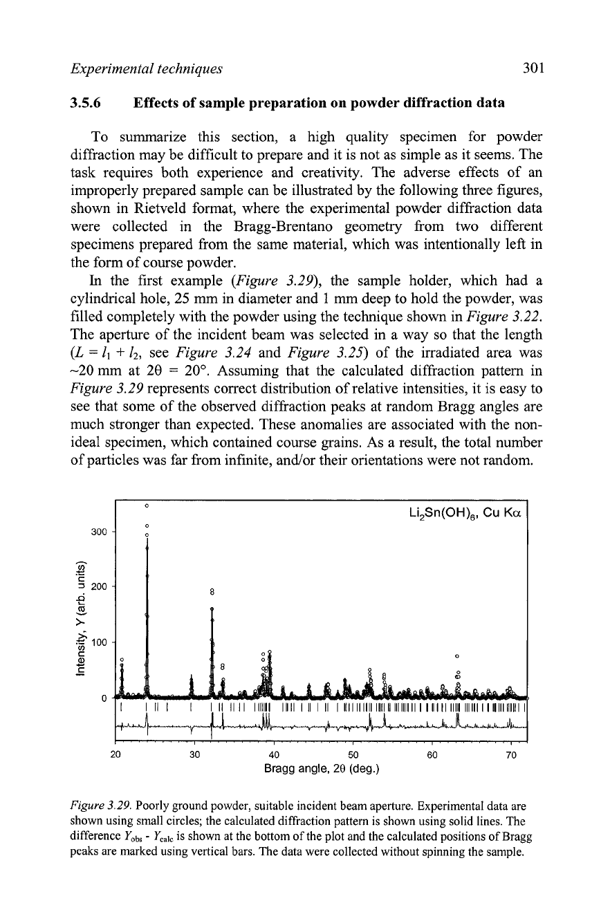
the first example (Figure 3.29), the sample holder, which had a
cylindrical hole, 25 mm in diameter and
1
mm deep to hold the powder, was
filled completely with the powder using the technique shown in Figure 3.22.
The aperture of the incident beam was selected in a way so that the length
(L
=
1,
+
12, see Figure 3.24 and Figure 3.25) of the irradiated area was
-20 mm at 28
=
20". Assuming that the calculated diffraction pattern in
Figure 3.29 represents correct distribution of relative intensities, it is easy to
see that some of the observed diffraction peaks at random Bragg angles are
much stronger than expected. These anomalies are associated with the non-
ideal specimen, which contained course grains. As a result, the total number
of particles was far from infinite, andlor their orientations were not random.
20
30
40
50 60
70
Bragg angle,
28
(deg.)
Figure 3.29.
Poorly ground powder, suitable incident beam aperture. Experimental data are
shown using small circles; the calculated diffraction pattern is shown using solid lines. The
difference
Yobs
-
Ycalc
is shown at the bottom of the plot and the calculated positions of Bragg
peaks are marked using vertical bars. The data were collected without spinning the sample.
3
02
Chapter
3
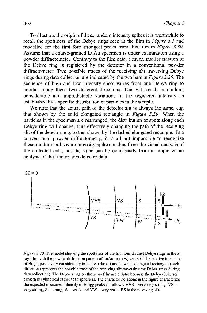
To illustrate the origin of these random intensity spikes it is worthwhile to
recall the spottiness of the
Debye rings seen in the film in
Figure
3.1
and
modelled for the first four strongest peaks from this film in
Figure
3.30.
Assume that a course-grained LuAu specimen is under examination using a
powder diffractometer. Contrary to the film data, a much smaller fraction of
the Debye ring is registered by the detector in a conventional powder
diffractometer. Two possible traces of the receiving slit traversing
Debye
rings during data collection are indicated by the two bars in
Figure
3.30. The
sequence of high and low intensity spots varies from one Debye ring to
another along these two different directions. This will result in random,
considerable and unpredictable variations in the registered intensity as
established by a specific distribution of particles in the sample.
We note that the actual path of the detector slit is always the same, e.g.
that shown by the solid elongated rectangle in
Figure
3.30. When the
particles in the specimen are rearranged, the distribution of spots along each
Debye ring will change, thus effectively changing the path of the receiving
slit of the detector,
e.g. to that shown by the dashed elongated rectangle.
In
a
conventional powder diffractometry, it is all but impossible to recognize
these random and severe intensity spikes or dips from the visual analysis of
the collected data, but the same can be done easily from a simple visual
analysis of the film or area detector data.
Figure
3.30.
The model showing the spottiness of the first four distinct Debye rings in the x-
ray film with the powder diffraction pattern of LuAu from
Figure
3.1.
The relative intensities
of Bragg peaks vary considerably in the two directions shown as elongated rectangles (each
direction represents the possible trace of the receiving slit traversing the
Debye rings during
data collection). The Debye rings on the x-ray film are elliptic because the Debye-Scherrer
camera is cylindrical rather than spherical. The character notations in the figure characterize
the expected measured intensity of Bragg peaks as follows: VVS
-
very very strong, VS
-
very strong, S
-
strong, W
-
weak and VW
-
very weak. RS is the receiving slit.
Experimental techniques 303
In
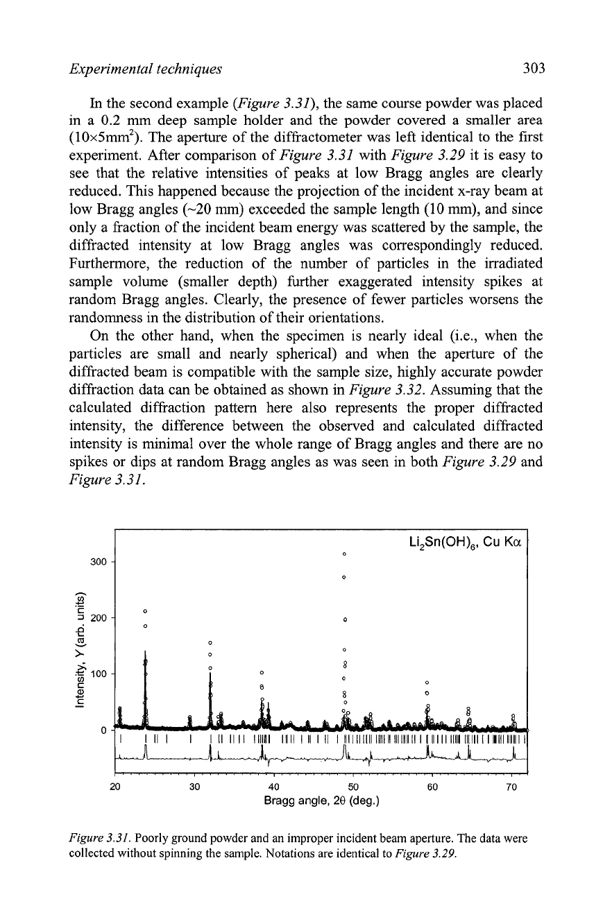
the second example (Figure 3.31), the same course powder was placed
in a 0.2 mm deep sample holder and the powder covered a smaller area
(10x5mm2). The aperture of the diffractometer was left identical to the first
experiment. After comparison of Figure 3.31 with Figure 3.29 it is easy to
see that the relative intensities of peaks at low Bragg angles are clearly
reduced. This happened because the projection of the incident x-ray beam at
low Bragg angles (-20 mm) exceeded the sample length (10 mm), and since
only a fraction of the incident beam energy was scattered by the sample, the
diffracted intensity at low Bragg angles was correspondingly reduced.
Furthermore, the reduction of the number of particles in the irradiated
sample volume (smaller depth) further exaggerated intensity spikes at
random Bragg angles. Clearly, the presence of fewer particles worsens the
randomness in the distribution of their orientations.
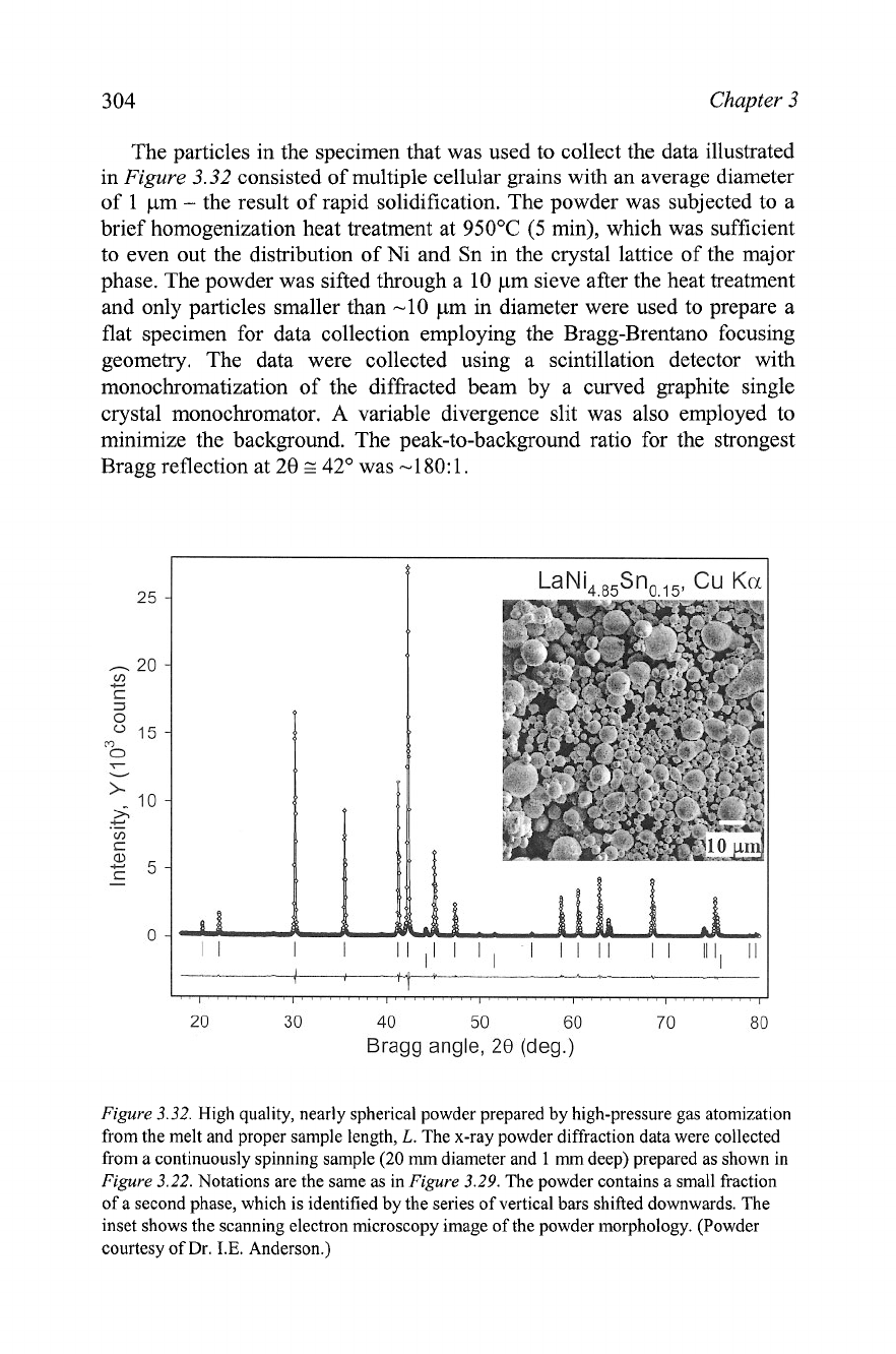
On the other hand, when the specimen is nearly ideal
(i.e., when the
particles are small and nearly spherical) and when the aperture of the
diffracted beam is compatible with the sample size, highly accurate powder
diffraction data can be obtained as shown in Figure 3.32. Assuming that the
calculated diffraction pattern here also represents the proper diffracted
intensity, the difference between the observed and calculated diffracted
intensity is minimal over the whole range of Bragg angles and there are no
spikes or dips at random Bragg angles as was seen in both Figure 3.29 and
Figure 3.31.
Figure
3.31.
Poorly ground powder and an improper incident beam aperture. The data were
collected without spinning the sample. Notations are identical to
Figure
3.29.
304
Chapter
3
The particles in the specimen that was used to collect the data illustrated
in
Figure
3.32 consisted of multiple cellular grains with an average diameter
of
1
pm
-
the result of rapid solidification. The powder was subjected to a
brief homogenization heat treatment at 950•‹C (5 min), which was sufficient
to even out the distribution of Ni and Sn in the crystal lattice of the major
phase. The powder was sifted through a 10 pm sieve after the heat treatment
and only particles smaller than -10 pm in diameter were used to prepare a
flat specimen for data collection employing the Bragg-Brentano focusing
geometry. The data were collected using a scintillation detector with
monochromatization of the diffracted beam by a curved graphite single
crystal monochromator. A variable divergence slit was also employed to
minimize the background. The peak-to-background ratio for the strongest
Bragg reflection at 28
z
42" was
-1
80: 1.
20 30 40 50 60 70 80
Bragg angle,
28
(deg.)
Figure
3.32.
High quality, nearly spherical powder prepared by high-pressure gas atomization
from the melt and proper sample length,
L.
The x-ray powder diffraction data were collected
from a continuously spinning sample
(20
rnrn
diameter and 1
mm
deep) prepared as shown in
Figure
3.22.
Notations are the same as in
Figure
3.29.
The powder contains a small fraction
of a second phase, which is identified by the series of vertical bars shifted downwards. The
inset shows the scanning electron microscopy image of the powder morphology. (Powder
courtesy of
Dr.
I.E.
Anderson.)
Experimental techniques
3.6
Data acquisition
When the sample for a powder diffraction experiment has been properly
prepared, the next step, i.e. acquiring experimental diffraction data is also
exceedingly important in obtaining reliable diffraction data. Needless to say,
many of the data acquisition variables can be adjusted to produce a better or
worse quality data set. These include instrumental parameters, i.e., the
wavelength (energy) of the x-rays, their monochromatization, apertures of
goniometer optics, power settings, and data collection parameters,
i.e.,
scanning mode, scan range, step in data collection, and counting time.
3.6.1
Wavelength selection
Usually the wavelength of the x-rays can be freely selected only when
using synchrotron radiation.
In
conventional laboratory conditions when the
wavelength of the x-rays needs to be changed, this normally means that the
x-ray tube has to be replaced, which should be followed by tube ageing and
realignment of the goniometer. These operations should be carried out by
trained personnel and they are rarely performed in practice to collect data
from a single sample. Therefore, the selection of the x-ray tube (and the
anode) type is usually done based on practical considerations,
i.e., based on
which type of materials are customarily studied, and what is the purpose of
the powder diffraction examination of the majority of samples.
The most typical wavelength selection in powder diffiactometry
is
with a
copper (Cu) anode, although other anode materials may be used. These
include chromium (Cr), iron (Fe), cobalt (Co) and molybdenum (Mo)
anodes. The corresponding characteristic wavelengths for these anode
materials are listed in
Table
2.1,
Chapter
2.
Long wavelengths (Cr, Fe, Co)
are preferred when the accuracy of lattice parameters is of greatest concern
since it is possible to measure intensity scattered at high Bragg angles.' Short
wavelength (Mo) can be used to examine a large volume of the reciprocal
lattice. However, since powder diffraction data are one-dimensional, the
resolution may be too low for complex crystal structures andlor materials
with low crystallinity when excessive Bragg peak overlap makes the
diffraction pattern extremely difficult to analyze and proces~.~
See Chapter 5, section 5.13 for details about various factors affecting precision of the unit
cell dimensions.
Short wavelength radiation may adversely affect resolution of the data collected using
goniometers with small radii (usually under -200 mm). When a large radius goniometer
(usually 250
mrn
or greater) is employed, the reduction of the wavelength from Cu- to Mo-
anode has little effect on pattern resolution because the widths of Bragg peaks decrease
with the decreasing wavelength. Regardless of the goniometer radius, the use of short
wavelengths requires a more precise alignment when compared to long wavelengths.
306
Chapter
3
Another important consideration in selecting x-ray wavelengths for a
powder diffraction experiment is whether or not the studied material
contains chemical elements with their K-absorption edges located just above
the used characteristic wavelength. For example, the K-absorption edge of
Co is -1.61
A. The strongest KallKa2 spectral lines of copper have
wavelengths -1.54 A. Hence, nearly all KallKa2 characteristic Cu radiation
will be absorbed by Co, and this type of x-ray tube is hardly suitable for x-
ray powder diffraction analysis of Co-based materials. The same is true for
Fe-based materials because the K-absorption edge of Fe is -1.74 A. On the
other hand, Co and Fe anodes are well-suited for collecting powder
diffraction data from Co- and Fe-containing substances using Co KallKa2
and Fe KallKa2 doublets.
Certain combinations of characteristic wavelengths and chemical
elements may cause considerable x-ray fluorescence (e.g. see
Figure
3.16).
This phenomenon is similar to the fluorescence in the visible spectrum and it
originates from the fact that electrons in an atom can be excited and removed
from their ground states by energy transfer from photons of sufficient
energy. Electrons from higher energy levels produce fluorescent x-rays when
they lower their energy by occupying the formed vacancies. Fluorescent
radiation is dissipated in all directions and it usually results in an increase of
the background. Chemical elements with strong true absorption will
generally produce strong x-ray fluorescent background.
3.6.2
Monochromatization
Either the diffracted or incident beam should be monochromatized during
data collection. When conventional x-ray sources are employed, additional
peaks due to the presence of weaker spectral lines with various energies in
the characteristic spectrum will "contaminate" the diffraction picture and
exaggerate peak overlap. The strongest unwanted wavelength for any anode
material is
KP1 (see
Table
2.1
in Chapter
2).
When a continuous
(synchrotron or neutron) spectrum is available, it would be impossible to see
discrete Bragg reflections as a function of Bragg angle without
monochromatization, as directly follows from Bragg's equation.
The simplest monochromatization tool that can be used in powder
diffractometry is a
P-filter (also see Chapter 2, section 2.3.2.1). Filter
materials have their K-absorption edges just above the wavelength of the
strongest
KP spectral line of the anode of the x-ray tube, and their
performance is based on how completely they absorb the characteristic
impurity wavelengths and how well they transmit the desired parts of the
characteristic x-ray spectrum. For example, to eliminate (absorb) nearly all
the
P-component of a copper anode
(h
=
1.39 A) but to transmit most of the
Experimental techniques 307
a-component (h
z
1.54
A)
the filter should be made from Ni (the K
absorption edge of Ni is -1.494. This results in nearly an eight-fold
difference in the linear absorption coefficients of Ni for
Kp and Ka parts of
the characteristic copper spectrum.
Various p-filters are most often used to monochromatize the diffracted
beam
(e.g. see Figure 3.6, left), but sometimes they are used to eliminate
Kp radiation from the incident beam in conventional x-ray sources. The
advantages of p-filters are in their simplicity and low cost. The
disadvantages include: (1) incomplete monochromatization because a small
fraction of Kp spectral line intensity always remains in the x-ray beam;
(2)
the intensity of the Ka spectral line is reduced by a factor of two or more,
and (3) the effectiveness of a p-filter is low for white x-rays above Ka and it
rapidly decreases below Kp. Therefore, p-filters are nearly helpless in
eliminating the background, especially when the latter is enhanced by x-ray
fluorescence (the filter itself fluoresces due to the true Kp absorption).
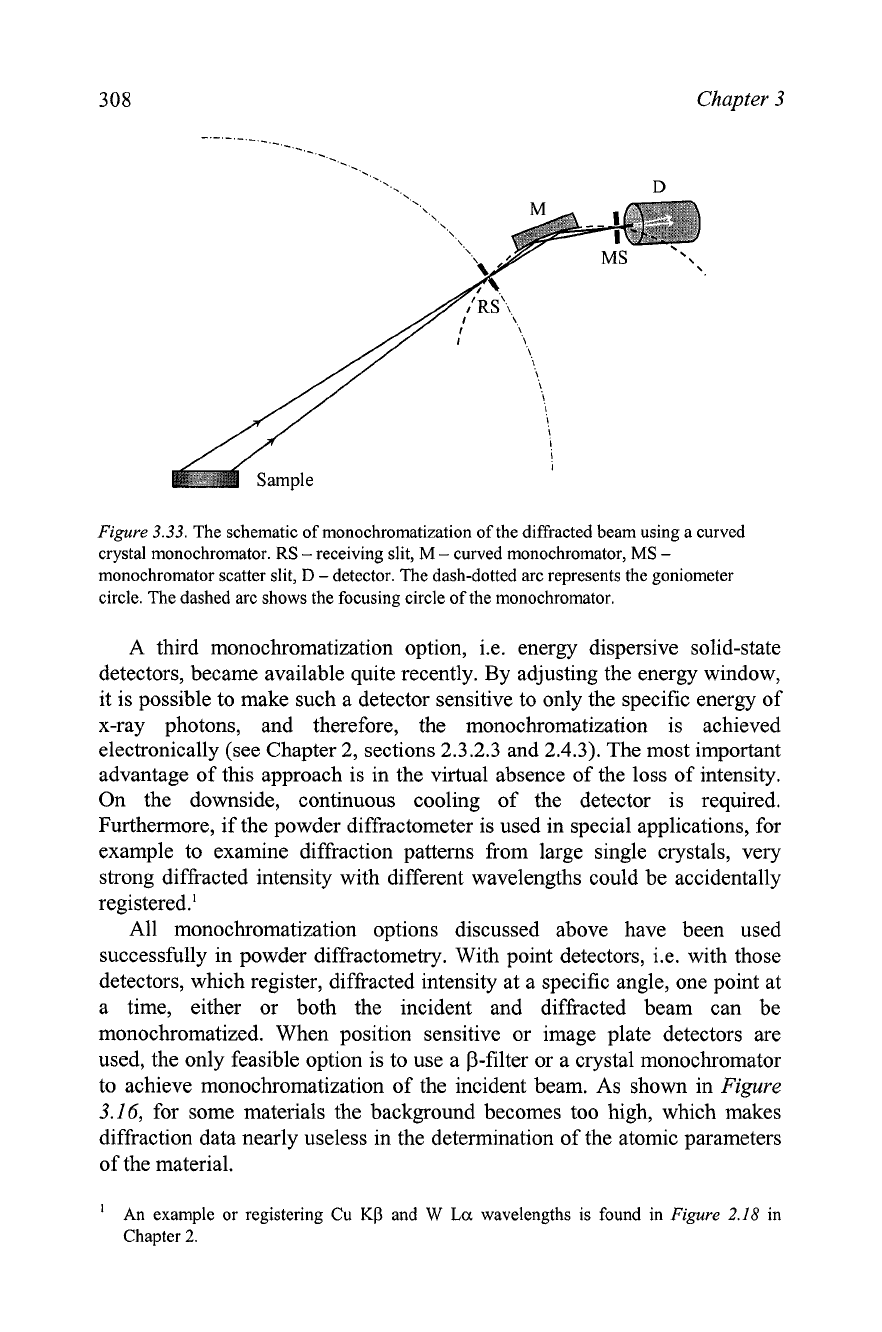
The most common monochromatization option used in modern powder
diffractometry is by means of crystal monochromators (see Figure 3.6, right
and Chapter 2, section 2.3.2.2). Monochromators transmit only specific,
narrowly selected wavelengths. As follows from the Bragg equation (nh
=
2dmsinOm), for a constant interplanar distance, dm, only one wavelength, h,
will be transmitted at a given monochromator angle, Om, assuming that n
=
1.
A great variety of crystal monochromators are used in practice, but the best
results are usually obtained using curved crystal monochromators, as shown
in Figure
3.33
since they achieve the most precise focusing of the x-ray
beam and therefore, lower the intensity losses.
A well aligned monochromator usually leaves only Ka, and Ka2
characteristic wavelengths and considerably reduces background when it is
used to monochromatize the diffracted beam.
In
fact, a diffracted beam
monochromator is very effective in nearly complete elimination of even
severe fluorescence (see Figure 3.16).
In
some instances, high quality curved
monochromators can be used to eliminate the Ka2 component in
combination with the relatively large focusing distance and narrow
monochromator slit. Spectral purity of x-rays is a definite advantage of this
monochromatization approach. Furthermore, if necessary, the
monochromator angle, Om, can be selected to eliminate the Kalla2 doublet
and to leave only the Kp part, which is truly monochromatic in light anode
materials, including the most commonly used Cu anode.
On a downside, high quality crystal monochromators are relatively
expensive, and they also reduce the intensity of characteristic x-rays by a
factor of two to three. It is worth noting that when the monochromator is
made from a low quality single crystal or when it is improperly aligned, the
resulting reduction of the transmitted intensity may be severe.
Chapter
3
Figure
3.33.
The schematic of monochromatization of the diffracted beam using a curved
crystal monochromator.
RS
-
receiving slit,
M
-
curved monochromator,
MS
-
monochromator scatter slit,
D
-detector. The dash-dotted arc represents the goniometer
circle. The dashed arc shows the focusing circle of the monochromator.
A third monochromatization option, i.e. energy dispersive solid-state
detectors, became available quite recently.
By
adjusting the energy window,
it is possible to make such a detector sensitive to only the specific energy of
x-ray photons, and therefore, the monochromatization is achieved
electronically (see Chapter 2, sections 2.3.2.3 and 2.4.3). The most important
advantage of this approach is in the virtual absence of the loss of intensity.
On the downside, continuous cooling of the detector is required.
Furthermore, if the powder diffractometer is used in special applications, for
example to examine diffraction patterns from large single crystals, very
strong diffracted intensity with different wavelengths could be accidentally
registered.'
All monochromatization options discussed above have been used
successfully in powder diffractometry. With point detectors,
i.e. with those
detectors, which register, diffracted intensity at a specific angle, one point at
a time, either or both the incident and diffracted beam can be
monochromatized. When position sensitive or image plate detectors are
used, the only feasible option is to use a
P-filter or a crystal monochromator
to achieve monochromatization of the incident beam. As shown in
Figure
3.16, for some materials the background becomes too high, which makes
diffraction data nearly useless in the determination of the atomic parameters
of the material.
'
An example or registering
Cu
KO
and
W
La
wavelengths is found in
Figure
2.18
in
Chapter
2.
Experimental techniques
309
3.6.3
Incident beam aperture
The aperture of the incident beam should be selected to match the
diffraction geometry and sample size. For the commonly used Bragg-
Brentano focusing geometry, the most important requirement is that at any
Bragg angle the length of the projection of the incident beam does not
exceed the length of the sample (see
Figure
3.24,
Figure
3.25 and Eq. 3.1).
This is achieved by a proper selection of the divergence slit (see
Figure
3.6).
When the slits are calibrated in degrees, the use of Eq. 3.1 to determine the
proper size of the divergence slit is straightforward.
In
the majority of
commercial diffractometers, however, slits are calibrated in mm and their
angular divergence can be estimated (also see Eq. 2.5 in Chapter 2) using the
following simple relationship, which assumes the infinitesimal size of the x-
ray tube focus.'
In
Eq. 3.5,
cp
is the angular divergence of the slit in degrees,
6
is the slit
opening in mm, and
r
is the distance from the x-ray tube focus to the
divergence slit in mm. It may be useful to check that the correct aperture of
the incident beam has been selected by mounting a fluorescent screen on the
sample holder at the lowest Bragg angle that will be examined during the
experiment. Alternatively, it is always possible to select a divergence slit
narrower than the acceptable maximum to ensure that the projection of the
incident beam fits within the sample length at the minimum Bragg angle.
Unnecessary reduction of the aperture of the incident beam, however, results
in the proportional reduction in the diffracted intensity, see
Figure
3.35,
below.
In
the transmission geometry the requirements are different. When a flat
transmission sample is used, the aperture of the incident beam is defined by
the largest Bragg angle of interest, since at
8
=
0 the sample is perpendicular
to the incident beam (and not parallel, as in the Bragg Brentano geometry).
Equation 3.1 then becomes as follows (where the notation are the same as in
Eq. 3.1)
As established in Chapter
2
(see
Figure
2.9), this is a valid approximation because the
typical projection of the I
mm
wide line focus of the x-ray tube, visible at a small take-off
angle (usually
5
to
6'),
results in the source size on the order of
0.1
mm.
3
10
Chapter
3
The opposite constraint on the relationships between the incident beam
size and sample size should be followed when a cylindrical transmission
sample is under examination: the projection of the beam should be large
enough to irradiate the whole sample at any position it may occupy during
spinning. This includes small precession around the goniometer axis. If this
requirement is not followed, the inhomogeneities in powder packing density
may cause sporadic changes in diffracted intensities.'
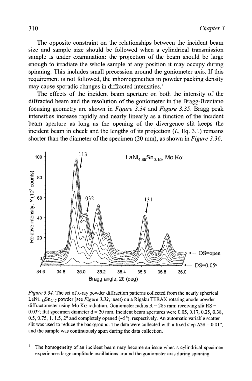
The effects of the incident beam aperture on both the intensity of the
diffracted beam and the resolution of the goniometer in the Bragg-Brentano
focusing geometry are shown in
Figure
3.34
and
Figure
3.35.
Bragg peak
intensities increase rapidly and nearly linearly as a function of the incident
beam aperture as long as the opening of the divergence slit keeps the
incident beam in check and the lengths of its projection
(L,
Eq.
3.1) remains
shorter than the diameter of the specimen (20 mm), as shown in
Figure
3.36.
34.6 34.8 35.0 35.2 35.4 35.6 35.8 36.0
Bragg angle,
20
(deg)
Figure
3.34.
The set of x-ray powder diffraction patterns collected from the nearly spherical
LaNi4,85Sno.ls powder (see
Figure
3.32,
inset) on a Rigaku TTRAX rotating anode powder
diffractometer using Mo
Ka
radiation. Goniometer radius
R
=
285
mm;
receiving slit RS
=
0.03"; flat specimen diameter d
=
20
mrn.
Incident beam apertures were 0.05, 0.17,0.25,0.38,
0.5,0.75, 1, 1.5,2" and completely opened (-5O), respectively. An automatic variable scatter
slit was used to reduce the background. The data were collected with a fixed step A20
=
O.OlO,
and the sample was continuously spun during the data collection.
The homogeneity of an incident beam may become an issue when a cylindrical specimen
experiences large amplitude oscillations around the goniometer axis during spinning.
