Olkkonen J. (ed.) Discrete Wavelet Transforms - Theory and Applications
Подождите немного. Документ загружается.


0
Shift-Invariant DWT for Medical Image
Classification
April Khademi, Sridhar Krishnan, Anastasios Venetsanopoulos
Department of Elec trical and Computer Eng ineering, University
of Toronto and Ryerson University
Canada
1. Introduction
The discrete wavelet transform (DWT) is g aining momentum as a feature extraction and/or
classification tool, because of its ability to localize structures with good resolution in a
computationally effective manner. The result is a unique and discrim inatory representation,
where important and inte resting structures (edges, details) are qu antified efficiently by few
coefficients . These coefficients m ay be used as features themselves , or features may be
computed from the wavelet domain that describe the anomalies in the data.
As a result of the potential that the DWT possesses for feature extraction and cl assification
applications, the current work focuses on its utility in a computer-aided diagnosis (CAD)
framework. CAD sy stems are computer-based methods that offer diagnosis supp ort to
physicians. The images are automatically analyzed and the p resence of pathology is identified
using quantitative measures (features) of dise ase.
With traditional radiology screening techniques, visually analyzing medical images is
labourious, time consuming, expensive (in terms of the radiologist’s time) and each individual
scan is prone to interpretation error (the error rate among radiologists is reported to hover
around 30% Lee (2007)). Additionally, visual analys is of radiographic images is subjective; one
rater may choose a particular lesion as a candidate, whi le another radiologist may find this
lesion insignificant. Consequently, some lesions are being missed or misinterpreted. To reduce
the error r ates, a secondar y opi nion may be obtained with a CAD system (automatically
reanalyze the images after the p hysician). Such methods are advantageous not only because
they are cost effective, but also because they are designed to objectively quantify p athology in
a robust, reliable and reproducible manner.
There has been a lot of research in CAD-system design for specific modalities or applications
with excellent results, i.e. see Sato et al. (2006) for CT, o r Guliato et al. (2007) for
mammography. Although these techniques may render good results for the particular
modality it was built for, the technique is not transferable and has little-to-no utility in other
CAD problems (cannot be applied to other images or d atabases) . Since CAD systems are being
employed widely, a framework that encompasses a variety of imaging modalities - not just a
single one - would be of value.
To this end, this work concerns the development of a generalized computer-aided diagnosis
system that is based on the DWT. It is considered generalized, since the same framework can
1
Shift-Invariant DWT for Medical Image
Classification
10

be applied to different images with no modifications. There are three image databases that
are used to test the generalized CAD system: small bowel, mam mogram and retinal images.
Although these images are very different from one another, a common attribute is noticed:
pathology is rough and heterogeneous, and healthy (normal) tissue is uniform. These images
are described in Section 2.
To quantify these differences between textures in normal and abno rmal images, a texture
analys is scheme based on human texture perception is proposed. To des cribe the
elemetary units of texture (which are needed for overall texture perception), important
features such as scale, f requency and orientation are used for texture discrimination. The
DWT is a perfect mechanism to highlight these space-localized features, since it offers
a high resolution, scale-invariant representation of nonstationary phenomena (such as
texture). Multiresolutional analysis, the wavel et transform, DWT with its properties and
implementations are discussed in Section 4.
Although the DWT has many beneficial qualities, the DWT is shift-variant. Therefore, any
texture metrics extracte d from the wavelet coefficients will also be shift-variant, reducing the
classification performance of our system. To combat this, a shift-invariant DWT (SIDWT)
is utilized to ensure that only translation invariant features are extracted (see Section 5).
To robustly quantify these texture elements (as described by the wavelet coefficients), a
multiscale texture analysis scheme is employed on the shift-invariant coefficients. At various
levels of decomposition, wavelet-domain graylevel cooccurrence matrices were implemented
in a variety of directions over all subbands to capture the orientation of such texture
elements. Texture features were extracted from each of the wavelet subbands to quantify the
randomness of the coefficients and they are classified using a linear classifier. The multiscale
texture analysis scheme and the classification technique are des cribed in S ection 6 and Section
7. Section 8 and Section 9 presents the results of the proposed generalized CAD framework for
all images and the concluding remarks, respectively. This work is a consolidation of several
research efforts Khadem i (2006) Khademi & Kr ishnan (2007) Khademi & Krishnan (2008).
2. Biomedical imagery
Three imaging modalities are utilized to test the classification system: mammography, retinal
and small bowel images. Each one of these image types are used to diagnose diseases from
a speci fic anatomical regio n. Although these images are qui te d ifferent from one another, the
current work develops a generalized framework for CAD that may be applied directly to each
of the images. The only apriori assumption is a very general one: the texture between normal
tissue and pathology is different.
The first modality, mammography, is an imaging technology which acquires an x-ray image
of the breast Ferreira & Borges (2001). They are currently the most effective method for early
detection of breast c ancers Cheng et al. (2006) Wei et al. (1995). A chall enging problem i n
human-based analysis of mammography is the discrimination between malignant and benign
masses. Incorrectly identifying the lesion type results in negative to positive biopsies ratios
as hi gh as 11:1 in some clini cs Rangayyan et al. (1997). Normal tissue masks the le sions and
breast parenchyma is much more prominent than the lesion itself Ferreira & Borges (2001).
To test the CAD system with mammog raphy images, a database is used where im ages contai n
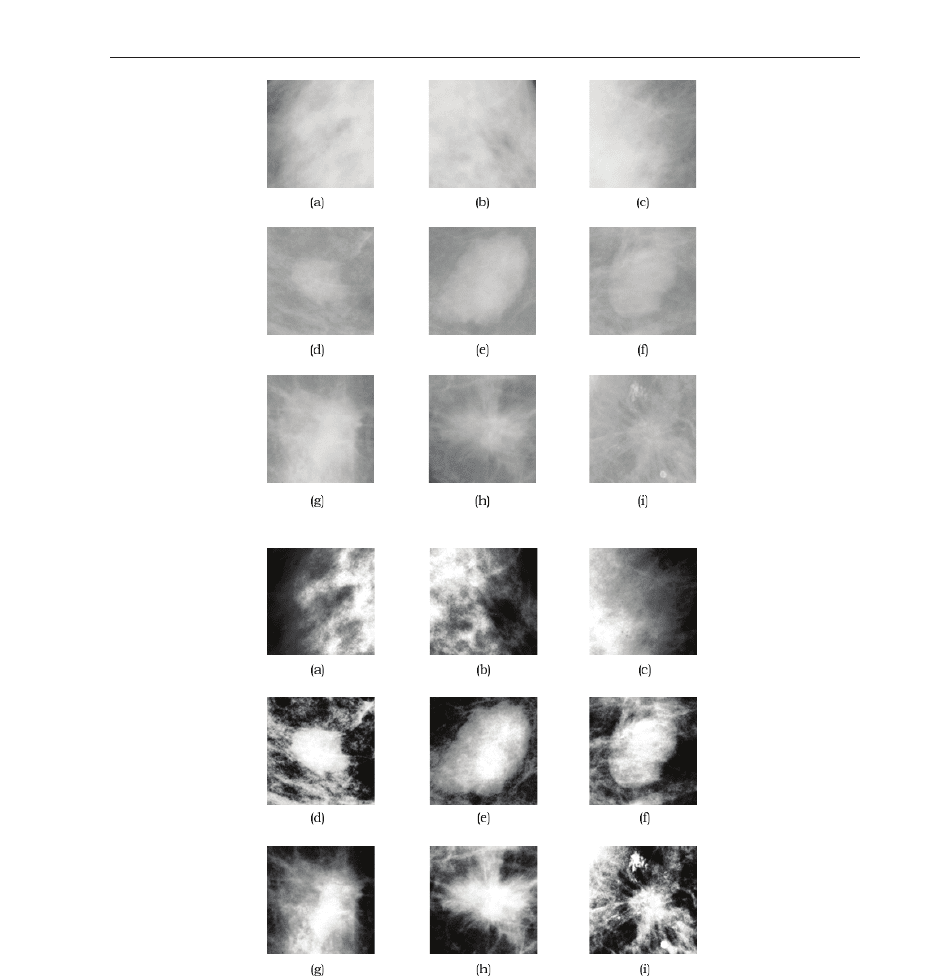
either a benign or malignant lesion(s). Examples of benign and malignant masses (along with
the contrast enhance d versi onS) are shown in Figure 1. Normal reg ions are also shown for
comparison.
180
Discrete Wavelet Transforms - Theory and Applications
be applied to different images with no modifications. T here are three image databases that
are used to test the generalized CAD system: small bowel, mam mogram and retinal images.
Although these images are very different from one another, a common attribute is noticed:
pathology is rough and heterogeneous, and healthy (normal) tissue is uniform. These images
are described in Section 2.
To quantify these differences between textures in normal and abno rmal images, a texture
analys is scheme based on human texture perception is proposed. To des cribe the
elemetary units of texture (which are needed for overall texture perception), important
features such as scale, f requency and orientation are used for texture discrimination. The
DWT is a perfect mechanism to highlight these space-localized features, since it offers
a high resolution, scale-invariant representation of nonstationary phenomena (such as
texture). Multiresolutional analysis, the wavel et transform, DWT with its properties and
implementations are discussed in Section 4.
Although the DWT has many beneficial qualities, the DWT is shift-variant. Therefore, any
texture metrics extracte d from the wavelet coefficients will also be shift-variant, reducing the
classification performance of our system. To combat this, a shift-invariant DWT (SIDWT)
is utilized to ensure that only translation invariant features are extracted (see Section 5).
To robustly quantify these texture elements (as described by the wavelet coefficients), a
multiscale texture analysis scheme is employed on the shift-invariant coefficients. At various
levels of decomposition, wavelet-domain graylevel cooccurrence matrices were implemented
in a variety of directions over all subbands to capture the orientation of such texture
elements. Texture features were extracted from each of the wavelet subbands to quantify the
randomness of the coefficients and they are classified using a linear classifier. The multiscale
texture analysis scheme and the classification technique are des cribed in S ection 6 and Section
7. Section 8 and Section 9 presents the results of the proposed generalized CAD framework for
all images and the concluding remarks, respectively. This work is a consolidation of several
research efforts Khadem i (2006) Khademi & Kr ishnan (2007) Khademi & Krishnan (2008).
2. Biomedical imagery
Three imaging modalities are utilized to test the classification system: mammography, retinal
and small bowel images. Each one of these image types are used to diagnose diseases from
a speci fic anatomical regio n. Although these images are qui te d ifferent from one another, the
current work develops a generalized framework for CAD that may be applied directly to each
of the images. The only apriori assumption is a very general one: the texture between normal
tissue and pathology is different.
The first modality, mammography, is an imaging technology which acquires an x-ray image
of the breast Ferreira & Borges (2001). They are currently the most effective method for early
detection of breast c ancers Cheng et al. (2006) Wei et al. (1995). A chall enging problem i n
human-based analysis of mammography is the discrimination between malignant and benign
masses. Incorrectly identifying the lesion type results in negative to positive biopsies ratios
as hi gh as 11:1 in some clini cs Rangayyan et al. (1997). Normal tissue masks the le sions and
breast parenchyma is much more prominent than the lesion itself Ferreira & Borges (2001).
To test the CAD system with mammog raphy images, a database is used where im ages contai n
either a benign or malignant lesion(s). Examples of benign and malignant masses (along with
the contrast enhance d versi onS) are shown in Figure 1. Normal reg ions are also shown for
comparison.
(a) Regular
(b) Enhanced
Fig. 1. Mammographic regions (128 × 128). (a)-(c) Normal regions , (d)-(f) circumscribed
benign masses, (g)-(i) spiculated malignant masses. The contrast enhanced versions of these
regions are also included to highlight the textural differences between lesions.
181
Shift-Invariant DWT for Medical Image Classification
The benign masses have a rounded appearance with a defined boundary, whil e the inside
of the mass is relatively uniform and radiolucent. This has also been noted by other others,
see Ferreira & Borges (2001) Rangayyan et al. (1997) Mudigonda et al. (2000). In contrast, the
malignant masses possess ill-defined boundaries, are of higher density (radiopaque) and have
an overall nonuniform appearance in comparison to the benign lesions. Furthermore, spicules
from malignant masses cause disturbances in the homogeneity of tissues in the s urrounding
breast parenchyma Rangayyan (2005). Since benign and malignant masses carry different
textural qualities, these textural differences will be exploited in the CAD syste m.
The second type of images are known as small bowel images. They are acquired by Given
Imaging Ltd.’s capsule endoscopy known as the PillCam
TM
SB video capsule. The PillCam
TM
is a tiny capsule (10mm × 27mm Kim et al. (2005)), which is ing ested f rom the mouth. As
natural peristalsis moves the capsule through the gastrointestinal tract it captures video and
wirelessly transmits it to a data recorder the patient is wearing around his or her waist Given
Imaging Ltd. (2006a). This video provides visualization of the 21 foot long small bowel, which
was originally seen as a “black box” to doctors Given Imaging Ltd. (2006b).
Video is recorded for approximately eight hours and then the capsule is excreted naturally
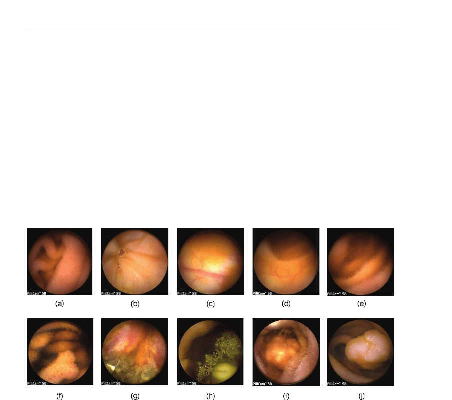
Fig. 2. Small bowel images captured by the PillCam
TM
SB, which exhibit textural
characteristics. (a) Healthy small bowel, (b) normal neocecal valve, (c) normal colonic
mucosa, (d) normal small bowel, (e) normal jejunum, (f) small bowel polyp, (g) small bowel
lymphoma, (h) GIST tumor, (i) polypoid mass, (j) small bowel polyp.
with a bowel movement Given Imaging Ltd. (2006a). Clinical results for the PillCam
TM
show
that it is a superior diagnostic method for diseases of the small intestine Given Imaging Ltd.
(2006c). The do w nfall of this technolo gy comes from the large amount of data which is
collected while the PillCam
TM
- the doctor has to watch and di agnose eight hours of footage!
This is quite a labourious task, which could cause the physicians to miss important clues due
to fatigue, boredom or due to the repetitive nature of the task. To combat missed pathologies,
the proposed CAD system could be used to double check the image data.
To test out the generalized CAD system, a small bowel image database is utilized that contains
both normal (healthy regions) and several abnormal images. As shown Figure 2(a)-(e), the
182
Discrete Wavelet Transforms - Theory and Applications

The benign masses have a rounded appearance with a defined boundary, whil e the inside
of the mass is relatively uniform and radiolucent. This has also been noted by other others,
see Ferreira & Borges (2001) Rangayyan et al. (1997) Mudigonda et al. (2000). In contrast, the
malignant masses possess ill-defined boundaries, are of higher density (radiopaque) and have
an overall nonuniform appearance in comparison to the benign lesions. Furthermore, spicules
from malignant masses cause disturbances in the homogeneity of tissues in the s urrounding
breast parenchyma Rangayyan (2005). Since benign and malignant masses carry different
textural qualities, these textural differences will be exploited in the CAD syste m.
The second type of images are known as small bowel images. They are acquired by Given
Imaging Ltd.’s capsule endoscopy known as the PillCam
TM
SB video capsule. The PillCam
TM
is a tiny capsule (10mm × 27mm Kim et al. (2005)), which is ing ested f rom the mouth. As
natural peristalsis moves the capsule through the gastrointestinal tract it captures video and
wirelessly transmits it to a data recorder the patient is wearing around his or her waist Given
Imaging Ltd. (2006a). This video provides visualization of the 21 foot long small bowel, which
was originally seen as a “black box” to doctors Given Imaging Ltd. (2006b).
Video is recorded for approximately eight hours and then the capsule is excreted naturally
Fig. 2. Small bowel images captured by the PillCam
TM
SB, which exhibit textural
characteristics. (a) Healthy small bowel, (b) normal neocecal valve, (c) normal colonic
mucosa, (d) normal small bowel, (e) normal jejunum, (f) small bowel polyp, (g) small bowel
lymphoma, (h) GIST tumor, (i) polypoid mass, (j) small bowel polyp.
with a bowel movement Given Imaging Ltd. (2006a). Clinical results for the PillCam
TM
show
that it is a superior diagnostic method for diseases of the small intestine Given Imaging Ltd.
(2006c). The do w nfall of this technolo gy comes from the large amount of data which is
collected while the PillCam
TM
- the doctor has to watch and di agnose eight hours of footage!
This is quite a labourious task, which could cause the physicians to miss important clues due
to fatigue, boredom or due to the repetitive nature of the task. To combat missed pathologies,
the proposed CAD system could be used to double check the image data.
To test out the generalized CAD system, a small bowel image database is utilized that contains
both normal (healthy regions) and several abnormal images. As shown Figure 2(a)-(e), the
normal small bowel images contain smooth, homogeneous texture elements with very little
disruption in uniformity except for folds and crevices.
Many typ es of pathologies are found in the small bowel image database ("abnormal" image
class), such as “Abnormal”: polyp, Kaposi’s sarcoma, carcinoma, etc. These diseases may
occur in various sizes, shapes, orientations and locations w ithin the gastrointestinal tract.
Abnormalities have some c ommon textural characteristics: the diseased region contains
many different textured areas simultaneously and these diseased areas are composed of
heterogeneous texture components. This may be seen in Figure 2(f)-(j).
The data for each patient is a series of 2D colour images. As the current chapter is focused
on grayscale p rocessing, the co lour images are converted to grayscale first. Additionally, each
image has been lossy JPEG compressed, so feature extraction is completed in the compressed
domain. Feature extraction in the compressed domain has become an imp ortant topic recently
Chiu et al. (2004) Xiong & Huang (2002) Chang (1995) Armstrong & Jiang (2001) Voulgaris &
Jiang (2001), since the prevalence of images stored in lossy formats far super sedes the number
of images stored in their raw format.
The last set of images are known as retinal images. Ophthalmologists use digi tal fundus
cameras to acquire and collect retinal images of the human eye Sinthanayothi n et al. (2003),
which includes the optic nerve, fovea, surrounding vessels and the retinal layer Goldbaum
(2002). Although screening with retinal imaging reduces the risk of serious eye impairment
(i.e. blindness caused by diabetic retinopathy by 50% Sinthanayothin et al. (2003)), it also
creates a large number of images which the doctors need to interpret Brandon & Hoover
(2003). This is exp ensive, time consuming and may be prone to human error. The current
automated system can be used to help the doctors with this diagnostic task by offering a
secondary opinion of the images.
The current database contains several normal (healthy) retinal images as well as several
images that contain a variety of patho logies. Exampl es of normal and abnormal retinal imag es
are shown i n Figure 3. He althy e yes are easily characteri zed by their overall homogeneous
appearance, as easily seen in Figure 3(a)-(c).
Eyes which contain disease do not possess uniform texture qualities. Three cases of abnormal
retinal images are shown in Figure 3(d)-(f). Diabetic retinopathy, which is characterized by
exudates or lesions (random whitish/yellow patches locatio ns Wang et al. (2000)) are shown
in Figure 3(a).
Another clinic al sign of diabetic retinopathy are microaneurysms and haemorrhages and
macular degeneration, which can cause blindness if it goes untreated. Macular degeneration
may be characterized by drusens, which appear as yellowish, cloudy blobs, which exhibit
no sp ecific size or shape Br andon & Hoover (2003). This is shown in Figure 3(e). These
pathologies disrupt the homogeneity of normal tissues. Other diseases include central retinal
vein and/or artery occlusion shown in Figure 3(f) (an oriented texture p attern which radi ates
from the optic nerve).
2.1 Texture for pathology discrimination
As shown in the previous subsection, pathological regions in the images have a heterogeneous
appearance and normal regions are uniform. Moreover, texture elements occur at a variety of
orientations, scales and locations. Thus the CAD system must be robust to all these variances,
but still remain modality- or database-independent (i.e. not tuned specifically for a modality).
Computing devices are becoming an integral part of our daily lives and in many times, these
183
Shift-Invariant DWT for Medical Image Classification
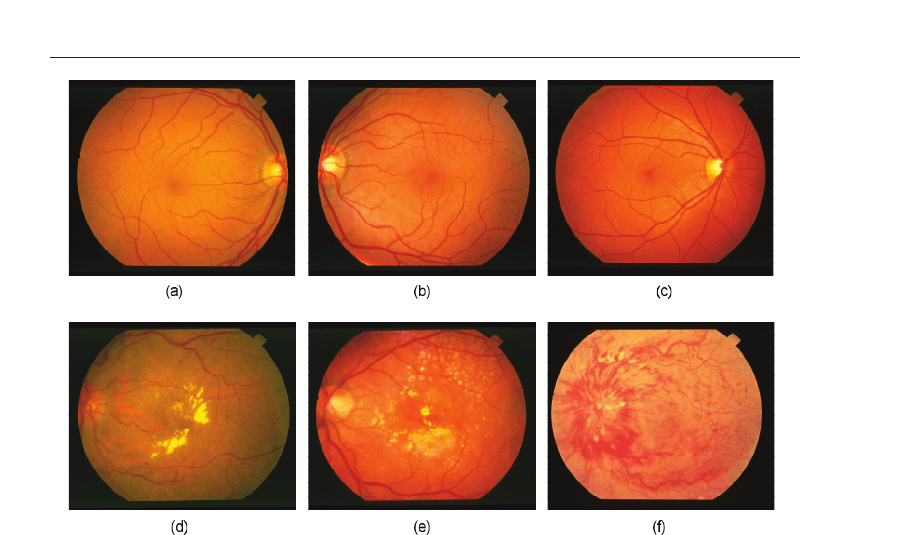
Fig. 3. Retinal images which exhibit textural characteristics. (a)-(c) Normal, homogeneous
retinal images, (d) background diabetic retinopathy (dense, homogeneous yellow clusters),
(e) macular degeneration (large, radiolucent drusens with heterogeneous texture properties),
(f) central retinal vein occlusion (oriented, radiating texture).
algorithms are designed to mimic human behaviour. In fact, this is the major motivatio n of
many CA D systems; to understand and analyze medical image content in the same fashion
as humans do. Since texture has been shown to be an important feature for discrimination in
medical images, understanding how humans perceive texture provides important clues into
how a computer vision system should be designed to discriminate pathology.
As shown, these images possess textural characteristics that differentiate between
pathological and healthy (no rmal) tissues. A common denominator is that the pathological
(cancerous) lesi ons seem to have heterogeneous, oriented texture characteristics, while the
normal images are relatively homog eneous. These differences are easily spotted by the human
observer and thus we want our system to also differentiate between these two texture types
(homogeneity and heterogeneity) for classification purposes.
To build a system that understands textural properties that is in line with human texture
perception, a human texture analysis model must first be examined. When a surface is viewed,
the human visual system can discriminate between textured regions quite easily. To describe
how the human visual s ystem can differentiate between textures, Julesz defined textons,
which are eleme ntary units of texture Julesz (1981). Textured regions can be decomposed
using these textons, which include elongated blobs, lines, terminators and more. It was found
that the freque ncy content, scale, orientation and period icity of these textons can provide
important clues on how humans differentiate between two or more textured areas Julesz
(1981). Therefore, to create a sys tem which mimics human unders tanding of texture for
pathology discrimination, it is necessary that the analysis system can detect the prop erties of
184
Discrete Wavelet Transforms - Theory and Applications

Fig. 3. Retinal images which exhibit textural characteristics. (a)-(c) Normal, homogeneous
retinal images, (d) background diabetic retinopathy (dense, homogeneous yellow clusters),
(e) macular degeneration (large, radiolucent drusens with heterogeneous texture properties),
(f) central retinal vein occlusion (oriented, radiating texture).
algorithms are designed to mimic human behaviour. In fact, this is the major motivatio n of
many CA D systems; to understand and analyze medical imag e content in the same fashion
as humans do. Since texture has been shown to be an important feature for discrimination in
medical images, understanding how humans perceive texture provides important clues into
how a computer vision system should be designed to discriminate pathology.
As shown, these images possess textural characteristics that differentiate between
pathological and healthy (no rmal) tissues. A common denominator is that the pathological
(cancerous) lesi ons seem to have heterogeneous, oriented texture characteristics, while the
normal images are relatively homog eneous. These differences are easily spotted by the human
observer and thus we want our system to also differentiate between these two texture types
(homogeneity and heterogeneity) for classification purposes.
To build a system that understands textural properties that is in line with human texture
perception, a human texture analysis model must first be examined. When a surface is viewed,
the human visual system can discriminate between textured regions quite easily. To describe
how the human visual s ystem can differentiate between textures, Julesz defined textons,
which are eleme ntary units of texture Julesz (1981). Textured regions can be decomposed
using these textons, which include elongated blobs, lines, terminators and more. It was found
that the freque ncy content, scale, orientation and period icity of these textons can provide
important clues on how humans differentiate between two or more textured areas Julesz
(1981). Therefore, to create a sys tem which mimics human unders tanding of texture for
pathology discrimination, it is necessary that the analysis system can detect the properties of
the fundamental units of texture (texture markers). In accordance to J ulesz’s model, textural
events will be detecte d based on their scale, frequency and orientation.
3. Feature invariance
To describe the textural characteristics of med ical images, a feature extraction scheme will
be used. The extracted features are fed into a classifier, which arrives at a decision related
to the diagnosis of the patient. Let
X⊂R
n
represent the signal space which contains all
biomedical images with the dimensions of n
= N × N. Since the images X can be expected
to have a very high dimensionality, using all these samples to arrive at a classification result
would be prohibitive Co ifman & Saito (1995). Furthermore, the original i mage sp ace
X is
also redundant, which means that all the image samples are not necessary for classification.
Therefore, to gain a more useful representation, a feature extraction operator f may map the
subspace
X into a feature space F
f : X→F, (1)
where
F⊂R
k
, k ≤ n and a particular sam ple in the feature space may be written as a feature
vector: F
= {F
1
, F
2
, F
2
, ···, F
k
}. If k < n, the feature space mapping would also result in a
dimensionality reduction.
Although it is important to choose features which provide the maximum discrimination
between textures, it is also important that these features are robust. A feature is robust if
it provides co nsistent results acros s the entire application domain Umbaugh et al. ( 1997). To
ensure robustness, the numerical descriptors should be rotation, scale and translation (RST)
invariant. In other words, if the image is rotated, scaled or translated, the extracted features
should be insensitive to these changes, or it should be a rotated, scaled or translated version
of the original features, but not modified Mallat (1998). This would be useful for cl assifying
unknown image samples since these test images will not have structures that have the same
orientation and size as the images in the training set Leung & Peterso n (1992). By ensuring
invariant features, it is possible to account for the natural variations and structures within the
retinal, mammographic and small bowel images.
As will be shown in the next section, such features are extracted from the wavelet domai n. If a
feature is extracted from a transform domain, it is also important to investigate the invariance
properties of the transform since any invariance in this do main also translates to an invariance
in the features. For instance, the 1-D Fourier spectrum is a well-known translation-invariant
transform since any translation in the time domain representation of the signal, does not
change the magnitude spectrum in the Fourier domai n
f
(t − τ
o
)⇔F(ω) · e
−jωτ
o
, (2)
for all real values of τ
o
. Similarly, scaling in time results in a easily definable reaction in the
frequency domain
f
(αt) ⇔
1
|α|
·
F
ω
α
, (3)
where α is an integer value.
Although the types of feature extraction algorithms that will be used have not yet been
discussed, prior to designing any feature extractor, it is important to understand the necessity
of robust features. The following sections will detail the analysis tool used to localize the
185
Shift-Invariant DWT for Medical Image Classification

texture events, as well as the feature extraction framework that is used to extract robust
features (in the RST-invariant sense).
4. Multiresolutional analysis
All signals and images may be cate gorized into one of two categories: 1) deterministic
or 2) non-deterministic (random). Deterministic signals allow for advanced prediction of
signal quantities, since the signal may be described by a mathematical function. In contrast,
instantaneous values of non-deterministic signals are unpredictable due to their random
nature and must be represented using a probabilistic model Ross (2003). This stochastic model
describes the inherent behaviour of the signal or image in question.
Random signals (both 1D and 2D) may be further classified into two groups: 1) stationary
or 2) nonstationary. A stationary signal (1D) is a signal which has a constant probability
distribution for all time instants. As a consequence, first order statistics such as the mean and
second order statisti cs such as variance mus t also remain constant. In contrast, a nonstationary
signal has a time-varying probability distribution which causes quantities computed from the
probability density function (PDF) to also be time-varying. For instance, the mean, variance
and autocorre lation function of a no nstationary signal would change with time. Since the
Fourier transform of the autocorrelation function is equal to the power spectral density (PSD)
of a signal (which is related to the spectral content), the PSD of a nonstationary si gnal is also
time- varying. Conseq uently, a nonstationary signal has time-varying spectral content.
The medical images (as with most natural images) are nonstationary since they have
spatially-varying frequency components. Texture is comprised of a variety of frequency
content (and may be found in any location in the image), and therefore texture is also a type of
nonstationary phenomena. Since textured regions provide important c lues that discriminate
between pathologies and/or healthy tissue, nonstationary analysis would add extra utility in
the sense that it would quantify or localize these textural elements. As discussed, the theory
of human texture perception is defined in terms of several features for texture discrimination:
the scale, frequency, orientation of textons. There fore, analyzing the scale, freque ncy and
orientation properties of textural elements by nonstationary image analysis is in accordance
to the human texture perception model.
The type of nonstationary image analysis tool that will be utilized is part of the
multiresolutional analysis family, and is known as the Discrete Wavelet Transform (DWT).
As will be discussed, wavele t transforms are optimal for texture localizatio n since the wavelet
basis have excellent joint space-frequency resolution Mallat (1998).
The section will begin by presenting the signal decomposition theory needed to understand
the fundamentals of the DWT. Following the introduction, the wavelet transform (with
desc riptions of the wavelet and scaling basis functions) are giv en, with emphasis given to
sig nal space definitions. The DWT is then defined using the filter-bank method which was
implemented by the lifting-approach for the 5/3 Le Gull wavelet.
4.1 Signal decomposition techniques
Signal decomposition techniques can b e used to transform the images into a representation
that highlights features of interest. As such decomposition techniques are used to define the
wavelet trans form and its variants, some brief background is given here.
A decomposition technique linearly expands a signal or image using a set of mathematical
functions. For a 1D signal, using a set of real-valued expansion coefficients a
k
, and a series
186
Discrete Wavelet Transforms - Theory and Applications

texture events, as well as the feature extraction framework that is used to extract robust
features (in the RST-invariant sense).
4. Multiresolutional analysis
All signals and images may be cate gorized into one of two categories: 1) deterministic
or 2) non-deterministic (random). Deterministic signals allow for advanced prediction of
signal quantities, since the signal may be described by a mathematical function. In contrast,
instantaneous values of non-deterministic signals are unpredictable due to their random
nature and must be represented using a probabilistic model Ross (2003). This stochastic model
describes the inherent behaviour of the signal or image in question.
Random signals (both 1D and 2D) may be further classified into two groups: 1) stationary
or 2) nonstationary. A stationary signal (1D) is a signal which has a constant probability
distribution for all time instants. As a consequence, first order statistics such as the mean and
second order statisti cs such as variance mus t also remain constant. In contrast, a nonstationary
signal has a time-varying probability distribution which causes quantities computed from the
probability density function (PDF) to also be time-varying. For instance, the mean, variance
and autocorre lation function of a no nstationary signal would change with time. Since the
Fourier transform of the autocorrelation function is equal to the power spectral density (PSD)
of a signal (which is related to the spectral content), the PSD of a nonstationary si gnal is also
time- varying. Conseq uently, a nonstationary signal has time-varying spectral content.
The medical images (as with most natural images) are nonstationary since they have
spatially-varying frequency components. Texture is comprised of a variety of frequency
content (and may be found in any location in the image), and therefore texture is also a type of
nonstationary phenomena. Since textured regions provide important c lues that discriminate
between pathologies and/or healthy tissue, nonstationary analysis would add extra utility in
the sense that it would quantify or localize these textural elements. As discussed, the theory
of human texture perception is defined in terms of several features for texture discrimination:
the scale, frequency, orientation of textons. There fore, analyzing the scale, freque ncy and
orientation properties of textural elements by nonstationary image analysis is in accordance
to the human texture perception model.
The type of nonstationary image analysis tool that will be utilized is part of the
multiresolutional analysis family, and is known as the Discrete Wavelet Transform (DWT).
As will be discussed, wavele t transforms are optimal for texture localizatio n since the wavelet
basis have excellent joint space-frequency resolution Mallat (1998).
The section will begin by presenting the signal decomposition theory needed to understand
the fundamentals of the DWT. Following the introduction, the wavelet transform (with
desc riptions of the wavelet and scaling basis functions) are giv en, with emphasis given to
sig nal space definitions. The DWT is then defined using the filter-bank method which was
implemented by the lifting-approach for the 5/3 Le Gull wavelet.
4.1 Signal decomposition techniques
Signal decomposition techniques can b e used to transform the images into a representation
that highlights features of interest. As such decomposition techniques are used to define the
wavelet trans form and its variants, some brief background is given here.
A decomposition technique linearly expands a signal or image using a set of mathematical
functions. For a 1D signal, using a set of real-valued expansion coefficients a
k
, and a series
of 1-D mathematical functions ψ
k
(t) known as an expansion set (ψ
k
(t)= ψ(t − k) for all
integer values of k), a signal f
(t) may be expressed as a weighted linear combination of these
functions
f
(t)=
∑
k
a
k
·ψ
k
(t) , k ∈ Z. (4)
If the members of the expansion set ψ
k
(t) are orthogonal t o one another:
�ψ
k
(t) , ψ
l
(t)� = 0, k �= l, (5)
then it is possible to find the expansion coefficients a
k
a
k
= �f (t), ψ
k
(t)�, (6)
where the inner product
�·� of two signals x(t) and y(t) is defined by
�x(t), y( t)� =
t
x
∗
(t) ·y(t)dt. (7)
The definition of an expansion set depends on various properties. For instance, if there is
a signal f
(t) which belongs to a subsp ace S ( f (t) ∈ S), then ψ
k
(t) will only be called an
expansion set for S if f
(t) can be expressed with linear combinations of ψ
k
(t) . The expansion
set forms a basis if the representation it provides is unique Burrus et al. (1998). Similarly, a basis
set may be defined first and then the space S spans all functions f
(t) which can be expressed
by f
(t)=
∑
k
a
k
·ψ
k
(t) .
For images, the basis functions may be dependant on both the horizontal and ver tical spatial
variables
(x, y ). This leads to 2D basis functions ψ
m,n
(x, y), where ψ
m,n
(x, y )=ψ(x − n, y −
m), for all (m, n) ∈ Z. Therefore, a 2D function (image) f (x, y), that belongs to the space of the
basis functions, may be rewritten as a linear expansion
f
(x, y)=
∑
m
∑
n
a
m,n
·ψ
m,n
(x, y), (8)
where a
m,n
are the 2-D expansion coefficients found by
a
m,n
= �ψ
m,n
(x, y ), f (x, y)�. (9)
Using decomposition techniques, a new representation is generated. In feature extraction
problems, we want this representation (i.e. coefficients a
k
or a
n,m
) to highlight the features we
are interested in. This requires us to choose basis functions that are tuned to the properties of
our image (i.e. nonstationary structure s). While choosing which basis set to use, one of the
main considerations is the functions’ s pace-frequency resolution.
Consider the basis function ψ
k
(t) which has an energy distribution that is concentrated near
the time instant k and is spread out over the interval Δt Mal lat (1998). This basis fu nction ψ
k
(t)
can identify time-localized features (at k) with a resolution of
1
Δt
. Similarly, in the frequency
domain, the Fourier representation Ψ
ξ
(ω) is concentrated in energy near the frequency ξ
and spre ad over the interval of Δω, which captures frequency-localized features (at ξ) with a
resolution of
1
Δω
.
Ideally, basis functions with infinitely small time and frequency would provide the best
representation since time-frequency structures would be represented with infinite resolution.
187
Shift-Invariant DWT for Medical Image Classification

However, this is not possible, because there is a direct trade off between time and
frequency resolution of basis functions as gov erned by the Heisenburg uncertainty principal
Burrus et al. (1998) Mallat (1998). The Heisenburg uncertainty principal states that resolution
of the time-frequency functions are lower bounded by
Δω
·Δt ≥ 1/2. (10)
Therefore, to capture nonstationary events with good space-frequency localization, we need
basis functions that aim to o perate near the theoretical lower bound. Many basis functions
offer solutions, but are not optimal for all applications. For example, the Short-Time Fourier
Transform (STFT) bases are not optimal because (1) they offer a fixed resolution for the
entire decomposition process (thus missing features that are comprised with different scales
and frequencies) , (2) do not offer an easy m ethod to access and manage the coefficients
and (3) creates a drastic increase in memory consumption and computational resources.
The following section will describe how the wavelet transform poses solutions to all these
problems.
4.2 Wavelet transforms
The wavelet transform offers solutions to all the problems associated with other basis
functions (such as the ST FT) Mallat (1989) Wang & Karayiannis (1998) Vetterli & Herley
(1992) Mallat (1998). It offers a multiresolutional representation (decomp oses the image using
various scale-frequency resolutions), which is achieved by dyadically changing the size of the
window. Space-frequency events are localized with good results since the changing window
function is tuned to events which have high frequency components in a small analysis
window (scale) or low frequency events with a large scale Burrus et al. (1998) . Therefore,
texture events could be efficiently represented using a set of multiresolutional basis functions.
Additionally, the discrete wavelet transform utilizes critical subsampling along rows and
columns and uses these subsampled subbands as the input to the next decomposition level.
For a 2-D image, this reduces the number of input samples by a factor of four for each level of
decomposition. This representation may be stored back on to the original image for minimum
memory usage and it also permits for an organized, computationally efficient manner to
access these subbands and extract meaningful features.
The wavelet transform utilizes both wavelet basis ψ
j,k
(t) and scaling basis φ
k
(t) functions.
The wavelet functions are used to localize the hi gh frequency content, whereas the scaling
function examines the low frequencies. The scale of the analysis window changes with each
decomposition level, thus achieving a multiresolutional representation. Starting with the
initial scale j
= 0, the wavelet transform of any function f (t) which belongs to L
2
(R) is found
by
f
(t)=
k=∞
∑
k=−∞
c(k) · φ
k
(t)+
j=∞
∑
j=0
k
=∞
∑
k=−∞
d(j, k) ·ψ
j,k
(t) , (11)
where c
(k) are the scaling or aver aging co efficients (low frequency material) defined by
c
(k)=c
0
(k)=�f (t), φ
k
(t)� =
f (t)φ
k
(t) dt, (12)
188
Discrete Wavelet Transforms - Theory and Applications