Ojima I. (ed.) Fluorine in Medicinal Chemistry and Chemical Biology
Подождите немного. Документ загружается.


502 Fluorine in Medicinal Chemistry and Chemical Biology
both higher spatial resolution ( ∼ 3 – 5 mm) and higher temporal resolution, which results
from this higher sensitivity. Because of the potential for MR spectroscopic techniques to
resolve metabolite resonances and assess the impact of multiple doses, this is a comple-
mentary modality to the PET kinetic information, even for fi rst - pass or single - dose
assessments.
Since the human in vivo concentration of exogenous fl uorinated drugs is often in the
micromolar range, spatial localization is often restricted to either the sensitive volume of
the detection coil [5, 9, 11, 27 – 33, 42, 45] , a single voxel in the organ of interest [5] , or
slabs of differing depth [5, 23, 56] . However, should the in vivo fl uorine concentration be
in the millimolar range, spatially localization using 2D or 3D chemical shift imaging (CSI)
techniques become feasible [5, 56] . If spatial localization is feasible, SNR can be traded
off either for increased spatial resolution or for decreased acquisition time to achieve better
temporally resolved spectra.
Surface RF coils (see Figure 19.1 ) are receiver coils (often made only of a loop of
copper wire or tubing) that are placed directly over the organ of interest and have a sensi-
tive depth of detection that is approximately half the coil diameter [5, 11, 15, 23] . If the
region of interest is superfi cial, this approach works well. However, if the organ is deep,
the subcutaneous fat and any other tissue within the coil ’ s sensitive volume may contribute
to the detected signal. All surface coils suffer from inhomogeneous receive fi elds. If used
to excite the nuclear spins, surface coils will also have spatially nonuniform excitation RF
fi elds. This means that the same fl ip angle cannot be obtained throughout the coil ’ s sensi-
tive volume. The impact of the inhomogeneities is magnifi ed if the same coil is used for
both excitation and detection. A larger (larger than the receive coil), more homogeneous
excitation surface coil is preferred to minimize the impact of the nonuniform limited
volume excitation RF fi elds [5, 56] .
Transmit – receive surface coils are generally used for MR spectroscopic studies in
organs other than the brain. If dual - frequency techniques such as NOE, polarization trans-
fer, or decoupling are to be utilized with surface coil excitation, or if relaxation time
measurements are to be performed, RF pulses that are insensitive to spatial and amplitude
(a) (b)
Figure 19.1 Surface coils can be as simple as a loop of copper wire or tubing. They are
placed directly over the organ of interest and have a sensitive depth of detection which is
approximately half the coil diameter. Examples of surface coils that may be used for either
transmit/receive or receive - only applications: (a) simple loop coil; (b) fi gure - eight coil.
Study of Metabolism of Fluorine-containing Drugs 503
inhomogeneity must be employed. Such RF pulses can have high power requirements and
may encounter specifi c absorption rate (SAR) limitations. Brain
19
F MR spectroscopy
generally utilizes a transmit – receive volume coil, which does not suffer nearly as much
from RF fi eld spatial inhomogeneities; however, SAR is particularly of concern with dual -
frequency sequences.
The inhomogeneity of the excitation RF fi elds from transmit – receive surface coils
can be used to advantage for selecting a sensitive volume slab based on approximate dis-
tance from the coil. Limited spatial selectivity can be achieved by adjusting the fl ip angle
to best select the depth of interest.
19.2.4 Metabolism, Binding, and Association
The signal intensity of an MR - visible fl uorinated compound is directly proportional to the
number of fl uorine spins present, similarly to other spin ½ nuclei. However, a number of
processes can restrict compound MR visibility and thereby impede quantifi cation from
spectra.
The appearance of fl uorine MR spectra can change if exchange, binding, or metabo-
lism is present [1, 9, 11, 14, 15, 19, 20, 23, 30 – 33, 42, 44, 59, 63, 68] . If the process is
slow compared with the Larmor resonance frequency difference between the two states
or species, then two distinct sets of resonances will be observed. However, if the process
is fast compared with the difference in Larmor frequency, the spectrum will have a single
resonance located at the weighted average of the chemical shifts of the two states or species
[14] .
Another common impact of in vivo processes is that the compound and its metabolic
products or states will undergo line broadening [1, 9, 11, 14, 15, 19, 20, 23, 30 – 33, 42,
44, 62, 63] . When one or more compounds have broad spectral lines, measurement of drug
metabolism in vivo may also be limited. Overlapping fl uorine resonances of the parent
drug and those of active and inactive metabolites [1, 11, 23, 41] at magnetic fi eld strengths
used for human MR spectroscopy may also occur, further confounding the results.
19.3 Applications of In Vivo Fluorine MR in Medicine and
Drug Development
Prior to using
19
F MR spectroscopy on a new compound in the clinic, the potential and
limitations should fi rst be explored in vitro and then in animals [19, 20] . If quantitative
fl uorine MR measures are desired, laboratory research may be needed prior to going into
the clinic with a new compound to reveal any potential diffi culties that might be encoun-
tered in vivo . Even with extensive pre - work, cross - species variation as well as selective
organ uptake may lead to different amounts of MR - visible signal [1, 31, 32, 41, 42, 44] .
Without the proper preliminary in vitro studies, spectroscopic signal changes measured in
vivo may be misinterpreted [12, 41, 44] .
Examples of spectral changes obtained from laboratory solutions, ex vivo specimens
and in vivo clinical measurements are shown in Figures 19.2 – 19.6 for tecastemizole
504 Fluorine in Medicinal Chemistry and Chemical Biology
(Soltara
™
), a monofl uorinated metabolite of the antihistamine astemizole (Hismanal)
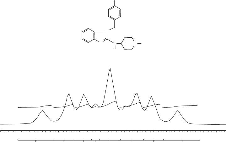
[11] . The spectrum of tecastemizole (see Figure 19.2 ) and its primary metabolite, 2 -
hydroxynorestemizole, are composed of a complex multiplet with several nonequivalent
1
H -
19
F couplings. At 7 T and in dimethyl sulfoxide (DMSO), the parent compound can be
distinguished from the metabolites; however, the four known metabolites are co - resonant
and cannot be differentiated. Since tecastemizole and its metabolites are all biologically
active, the lack of spectral resolution does not adversely impact biodistribution measure-
ments. Ideally, separable
19
F MR resonances would be observed for the parent compound
as well as its active and inactive metabolites. If they are not spectrally resolved, in vivo
measurement of fl uorinated drug metabolism by
19
F MR spectroscopy might have limited
utility.
F
(a)
(b)
N
N
N
H
H
–114.84 –114.86
1.00
1.57
1.40 3.02 1.40
0.64 2.24 0.98
–114.88 –114.90 –114.92 –114.94 –114.96 –114.98 ppm
N
Figure 19.2 (a) Chemical structure of tecastemizole. All known metabolites also have the
fl uorine atom in the para - position of the phenyl ring. (b) High - resolution, solution - state
19
F
spectrum without proton decoupling at 7 T of tecastemizole in DMSO.
( Source: Schneider E, Bolo NR, Frederick B et al. , Magnetic resonance spectroscopy for
measuring the biodistribution and in situ in vivo pharmacokinetics of fl uorinated compounds:
validation using an investigation of liver and heart disposition of tecastemizole, J. Clin. Pharm.
Ther. (2006) 31 , 261 – 273. Copyright (2006) John Wiley & Sons. Reprinted with
permission.)
Study of Metabolism of Fluorine-containing Drugs 505
Interactions between compounds and tissue constituents may also interfere with
detection by MR spectroscopy. In particular, compounds that interact with proteins and/or
other cellular components will have altered T
1
[20] and decreased T
2
[11] relaxation times.
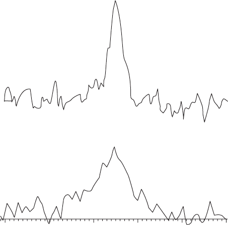
In a bovine serum albumin (BSA) solution, protein interactions caused the tecastemizole
19
F linewidth to increase (i.e., T
2
* and possibly T
2
decreased) and a 35% loss of total signal
(integrated area) was found in addition to a small frequency shift. After introduction to a
whole - blood solution (see Figure 19.3 ), protein binding also occurred, with 45% of the
integrated signal being lost immediately and 70% of the integrated fl uorine signal gone in
20 minutes. In a mixture of intact and disrupted human liver cells, an 85% loss of the
19
F
integrated signal area from tecastemizole occurred within one hour due to interactions with
the cellular components. Interactions between this compound with protein and cellular
constituents were strong enough to increase the correlation time, thus causing line broad-
ening and an overall loss of integrated signal intensity compared with that observed in
solution.
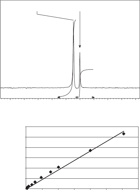
At 4 T, the fl uorine resonance frequency difference between tecastemizole and 2 -
hydroxynorestemizole in isotonic saline solution was found to be 6.7 Hz and a linear
response of the measured
19
F integrated signal (see Figure 19.4 ) was calibrated against
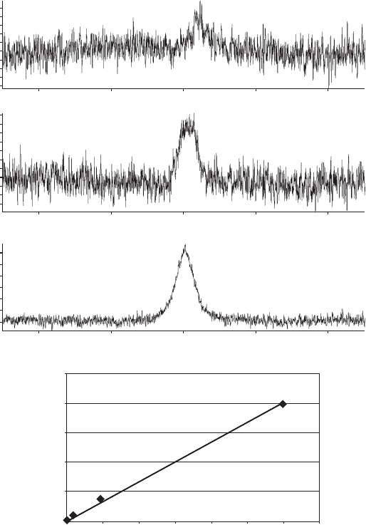
analytically validated concentrations (see Figure 19.4 b). When incorporated into either a
dog heart (see Figure 19.5 a) or liver, tecastemizole and its fl uorinated metabolites were
co - resonant in a single broad resonance (T
2
* = 1.4 ms; T
1
= 300 ms). A linear response of
–115.6 –115.8 –116.0
(b)
(a)
–116.2 –116.4 –116.6
Figure 19.3 Solution - state
19
F spectra at 7 T of 2.7 mM tecastemizole in a 50% whole blood
solution. (a) The integrated fl uorine signal is only 45% of that measured in DMSO because
of protein binding. (b) After 20 min, 70% of the integrated signal was lost.
506 Fluorine in Medicinal Chemistry and Chemical Biology
(b)
300
y = 0.9164x
R
2
= 0.9896
250
200
150
19
F MR measured
concentration (µM)
100
50
0
0 50 100 150 200 250 300 350
Analytical concentration (µM)
2500
(a)
2000 1500 1000
231.934h
0.000h
0.721
0.250
Hz
500 0 –500 –1000 –1500 –2000
Figure 19.4 Typical phantom calibration curve measurement. (a)
19
F MR spectrum of com-
pound and reference standard: (left) tecastemizole; (right) potassium fl uoride. (b) Calibration
curve of tecastemizole concentration measured analytically by LC - MS versus that measured
using
19
F MR.
integrated signal (area under the curve or AUC, see Figure 19.5 a - c) with tecastemizole
total fl uorine measured using liquid chromatography – mass spectrometry (LC - MS) was
found (see Figure 19.5 d) in the ex vivo specimen [11] . The calibration curve presented in
Figure 19.5 d, based on ex vivo spectroscopic measurements, provides a good indication
that the in vivo compound visibility should be linear with concentration and that sequestra-
tion of the compound is not expected over the dose ranges investigated. In vivo
19
F MR
spectra were also similar to those measured in the ex vivo specimens: the parent compound
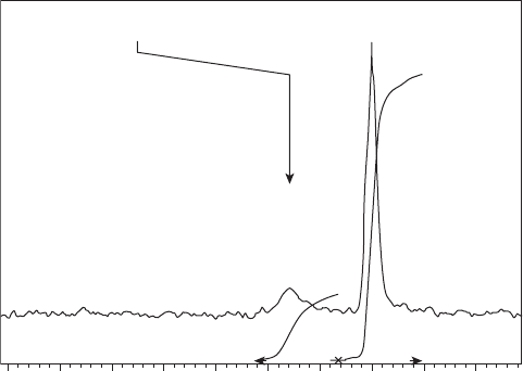
resonance was unresolvable from that of its metabolites (see Figure 19.6 ), and the broad
single resonance had similar relaxation times to that found ex vivo (see Table 19.3 ).
Study of Metabolism of Fluorine-containing Drugs 507
200
3.E+09
(d)
2.E+09
2.E+09
1.E+09
5.E+08
0.E+00
y = 2E + 06x
R
2
= 0.9966
0
19
F MR AUC
400 600 800
Analytical total fluorine (µmoles)
1000 1200 1400
(a)
1.50E5
1.25E5
Amplitude (–)Amplitude (–)
1.00E5
7.50E4
5.00E4
2.50E4
0
–2.50E4
–5.00E4
–7.50E4
1.25E5
1.00E5
7.50E4
5.00E4
2.50E4
0
–2.50E4
–5.00E4
–7.50E4
–1.00E5
–1.25E5
Amplitude (–)
1.00E5
2.00E5
3.00E5
4.00E5
5.00E5
0
–1.00E5
–2000 –1000 0 +1000 +2000
Frequency (Hz)
–2000 –1000 0 +1000 +2000
Frequency (Hz)
–2000 –1000 0 +1000 +2000
Frequency (Hz)
(b)
(c)
Figure 19.5 Typical ex vivo specimen calibration curve measurements.
19
F MR spectra of
ex vivo dog hearts at 4 T as a function of analytically measured tecastemizole concentration:
(a) 1.1 m M; (b) 359 m M; (c) 2149 m M. (D) Calibration curve of the ex vivo specimen
total fl uorine content measured analytically by LC - MS versus integrated AUC measured using
19
F MR.
19.4 Fluorine MR Spectroscopy in Cancer: Fluoropyrimidines
The impact of fl uorine MR spectroscopy as an in vivo analytical technique was demon-
strated by observations made with fl uorouracil (5 - FU) and has been the subject of excellent
review articles [3 – 5, 7 – 10, 46, 47, 59] . 5 - FU is an antimetabolite that has been used for
over 45 years in the treatment of several common cancers; however, signifi cant side - effects
508 Fluorine in Medicinal Chemistry and Chemical Biology
can occur, including cardiotoxicity and neurotoxicity. On the basis partly of evidence
obtained by in vivo
19
F MR spectroscopy, it was eventually hypothesized that the side -
effects resulted from transformation of FBAL, the primary catabolic metabolite, into fl uo-
roacetate, which has known cardiotoxicity and neurotoxicity [3 – 5, 7 – 10, 46] . To our
knowledge there have been no new clinical
19
F MR studies published on fl uoropyrimidine
drugs since the most recent review (2006) [4] . For an extensive discussion of the clinical
utilization of fl uoropyrimidine drugs (including 5 - FU), the metabolic pathway and modu-
lation by other medications, the reader is referred to these articles.
The metabolic pathways of 5 - FU and its prodrugs have been well characterized in
the liver as well as in liver tumors and metastases by in vivo
19
F MR spectroscopy. The
liver and extrahepatic spaces catabolize 5 - FU, which is subsequently excreted in the urine
[3 – 5, 7 – 10, 46, 59] . Active metabolites (fl uoronucleotides) are created by anabolism in
tumors. Even though clinical studies showed signifi cant individual subject variations [3,
5, 7 – 10, 26, 46, 47] , spectral characteristics such as resonance frequency, linewidth, relax-
ation time, and amplitude were not related to the therapeutic response. However, dynamic
processes, specifi cally the accumulation and retention of 5 - FU in the tumor, were indica-
tive of response. Patients showing tumor half - lives of free 5 - FU of 20 minutes or longer
observed in patients were characterized by Wolff et al. [47, 53] as “ trappers. ” While over
50% of the evaluated population were nontrappers, approximately 60% of the patients who
responded to therapy were trappers [3 – 5, 7 – 10] .
Use of in vivo
19
F MR spectroscopy to directly measure the pharmocokinetics of 5 - FU
in liver tumors and metastases enabled identifi cation of drug mechanism of action and
investigation of tumor pathophysiology. Similar studies were undertaken to evaluate the
ability of a range of medications to modulate the in vivo effectiveness of fl uorouracil and
its prodrugs [3 – 5, 7 – 10] . These studies have served as a model for other in vivo drug MR
3000 2500 2000 1500 1000 500 0 –500 –1000
791.016h
0.000h
0.191
0.912
Hz
Figure 19.6
19
F MR spectrum of tecastemizole in vivo at 4 T from the liver of a normal human
volunteer.
Study of Metabolism of Fluorine-containing Drugs 509
spectroscopic studies. In many respects, the analytic characterization of a potential
responder has evolved into the concept of personalized medicine.
In
19
F MR examinations of patients with liver tumors or metastases, the receive coil
sensitive volume typically encompassed the tumor, a signifi cant portion of normal tissue,
and possibly surrounding organs (such as the gallbladder, spleen, kidney, etc.). Initial
fi ndings of tumor trapping were based predominantly on large superfi cial liver tumors or
metastases that were evaluated by a surface coil placed in close proximity to the tumor.
However, since the sensitive region of the coil encompasses a considerable amount of
normal liver tissue, unequivocal proof of drug retention by the tumor was not possible.
This information required spatial localization and motivated technological improvements
to increase SNR and resolve the problems associated with chemical shift artifact [5, 24,
38, 56, 59] .
Because of the relatively low concentration of 5 - FU metabolites in vivo , it was chal-
lenging to perform volume - selective
19
F MR spectroscopy prior to technological advances.
An average 2 - fl uoro - β - alanine (FBAL) concentration of 0.92 ± 0.26 mmol/kg liver for the
fi rst 50 min post infusion was observed for doses varying from 750 mg to 2000 mg [43] ,
with a mean maximum concentration of 1.31 ± 0.33 mmol/kg liver, or, in a separate study,
1.0 ± 0.2 mM FBAL in the liver at 60 ± 10 min post infusion [56] .
To detect 5 - FU metabolism in the different liver regions, single - voxel acquisitions,
and 1D, 2D, or 3D chemical shift imaging (CSI) have been employed. CSI utilizes gradient
magnetic fi elds for spatial localization, identical to imaging techniques. Because the fi rst
gradient encoding (slab selection or 1D CSI) is based on frequency selectivity, the large
Larmor frequency differences between the 5 - FU and its metabolites results in chemical
shift artifacts along the slice select direction. This artifact means that the FBAL signal arises
from a spatially different slice from where the 5 - FU signal originates. At 1.5 T, the spatial
shift between 5 - FU and FBAL was 2.3 cm [5] . Methods to circumvent chemical shift arti-
facts include frequency - selective excitation RF pulses or frequency selective presaturation
RF pulses to isolate
19
F MR signal detection to only one chemical species [48 – 50, 56] .
In spite of the challenge presented by low in vivo concentrations and the chemical
shift artifact, spatially localized
19
F spectroscopy studies in the liver of patients receiving
5 - FU chemotherapy enabled advancement of the understanding of 5 - FU metabolism and
trapping. In addition, a catabolic resonance originating from the gallbladder was identifi ed
[57] , confi rming extrahepatic catabolism of 5 - FU. At 1.5 T, 2D CSI techniques with 8 × 8
localization voxel volumes of 6 cm × 6 cm × 4 cm (144 cm
3
) were acquired in 12.8 min
using a pulse repetition time (TR) of 60 ms and 12 800 excitations [50, 56] . 3D CSI local-
ization with voxel volumes of 4 cm × 4 cm × 4 cm (64 cm
3
) were acquired in 8.5 min using
a TR of 1 s and 512 excitations [50] or in 45 min using a TR of 260 ms and 10 240 excita-
tions [35] . Even smaller voxel volumes of 3 cm × 3 cm × 3 cm (27 cm
3
) were achieved in
45 min with double resonance (
1
H –
19
F) spectroscopy and an 8 × 8 × 8 resolution 3D CSI
acquisition [50] . However, the long acquisition times (45 min) for the 27 cm
3
3D CSI
acquisition precluded assessment of 5 - FU pharmacodynamics in vivo .
In vivo application of double resonance (
1
H –
19
F) spectroscopic techniques [50, 73,
74] , including a combination of NOE and proton decoupling, have proved to increase
SNR, which can be traded off for improved visibility or improved spatial or temporal
resolution of fl uorinated compounds. The double resonance decoupling, NOE, and
polarization transfer techniques used in vivo are similar to those used in solid state
510 Fluorine in Medicinal Chemistry and Chemical Biology
[71, 72] and animal spectroscopic evaluations. In vivo challenges include transmit coil
uniformity as well as high - power RF pulses that may limit duty cycle (TR) due to potential
heating (SAR) concerns [35, 36] . In addition, these techniques can only be used in vivo
when suffi cient signal (or suffi cient information) is present to correctly adjust the
parameters.
Klomp et al. [24] were able to double the SNR of their
19
F MR spectra at 1.5 T by
systematically reducing the noise factor of each component in the signal detection of the
MR system. With this increase, a 4 cm × 4 cm × 4 cm (64 cm
3
) 3D CSI acquisitions were
acquired in 4 min without polarization transfer or decoupling and these acquisitions were
subsequently used to evaluate fi ve patients receiving 5 - FU chemotherapy for treatment of
superfi cial and central liver metastases. The in vivo T
1
for 5 - FU in the liver following a
bolus injection was 380 ± 80ms, which was considerably lower than in earlier publications
(see Table 19.3 ).
A dramatic increase of in vivo
19
F MR SNR (a factor of 1.3 – 3) was then again
achieved by evaluating capecitabine metabolism in the liver at 3 T magnetic fi eld strength
compared with 1.5 T [38, 67] . At 3 T, the spectral resolution was also increased and allowed
differentiation of the FBAL – bile acid conjugate [57] from the primary FBAL resonance.
Due to increased SNR, which resulted both from use of optimized hardware and use of
3 T technology, Klomp et al. [68] were able to quantify the spatial distribution of
capecitabine and its prodrugs (including FBAL) in the liver of patients with advanced
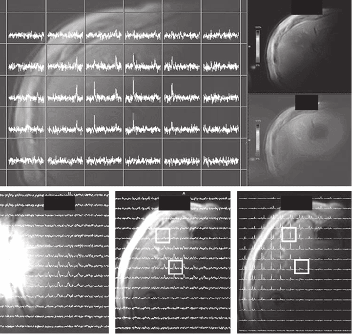
colorectal cancer using a 3D CSI acquisition with 10 × 10 × 10 resolution in a 9 min
acquisition (see Figure 19.7 ). This study found a nonhomogeneous spatial distribution of
capecitabine and FBAL, in agreement with Li et al. [56] . The order of magnitude of the
FBAL concentration [68] was also in agreement [56] ; however, the other capecitabine
metabolite concentrations differed signifi cantly. The fi nding of spatial heterogeneity of
compound distribution and metabolism must be accounted for when in vivo concentrations
are determined [56, 68] .
Kamm et al. [26] used the higher SNR in vivo
19
F MR confi guration at 1.5 T [24] to
examined the uptake and metabolism of 5 - FU modulated by trimetrexate in liver metas-
tases of colorectal cancer. All patients had one or more liver metastases of at least 2 cm
diameter within 8 cm of the skin surface. These criteria enabled inclusion of patients with
large superfi cial tumors, as well as patients with smaller metastases. Treatment response
was assessed during week 6 of the fi rst chemotherapy cycle using unlocalized
19
F MR
spectroscopy as well as before and after each 8 - week treatment cycle with either computed
tomography or ultrasound. The patients were divided into two groups, those with a larger
contribution of the
19
F signal from normal liver tissue (smaller tumors) and those with a
smaller contribution (larger tumors). In patients with larger tumors, a correlation was found
between increase in tumor size and the catabolite FBAL concentration, and the poorer
response to treatment was hypothesized to be correlated with higher degradation of 5 - FU
into catabolites. However, this relationship was not found when smaller tumors were
studied [26] . It is unclear whether these discrepant results were found because of the large
contribution of anabolic signal from the liver (e.g. partial volume effects due to lack of
spatial localization) or because of true differences in tumor drug metabolism. In addition
to examining patients with smaller liver metastases, the
19
F measurements were performed
after 5 weeks of chemotherapy and were timed to potentially detect only tumor cells
refractory to 5 - FU [26] .
Study of Metabolism of Fluorine-containing Drugs 511
In addition to 5 - FU, its fl uorinated prodrugs such as gemzar, fl oxuridine, capecitabine,
tegafur uracil, etc. have also been evaluated using
19
F MR spectroscopy, at least in the
laboratory or in animal models [3, 7 – 10] . Improved effi cacy of 5 - FU has been achieved
by using it in combination with other medications that either modulate its uptake
or/and increase its metabolism.
19
F MR has been used to measure the modulation of 5 - FU
(a) (b)
FBAL
cap
FBAL
cap
WaterFBALFBAL
(c)
(f)(e)(d)
Figure 19.7 Distribution of capecitabine and its metabolite FBAL in the liver of a patient
treated with oral capecitabine at 3 T. (a) Spatially localized
19
F MR CSI spectra overlaid on
the axial proton image of the liver acquired using the same surface coil. (b) and (c) Color
depiction of distribution of FBAL in the axial plane and capecitabine in the coronal plane,
respectively. (d) and (e) Distribution of FBAL in the coronal and axial planes, respectively,
depicted by CSI spectra. (f) Distribution of water signal in the axial plane. See color plate
19.7.
( Source: Klomp D., van Laarhoven H., Scheenen T., et al. Quantitative
19
F MR spectroscopy
at 3 T to detect heterogeneous capecitabine metabolism in human liver, NMR in Biomedicine
(2007) 20 , 485 – 492. Copyright (2007) John Wiley & Sons. Reprinted with permission.)
