Nielsen H. RNA: Methods and Protocols
Подождите немного. Документ загружается.

294 Mašek, Valášek, and Pospíšek
their 5 ´end prebound with a complex of three eIFs called eIF4F.
The 48S pre-initiation complex formed in this way then under-
goes specific conformational changes that enable this machinery
to star t scanning the 5
-untranslated region for the AUG start
codon in an optimal sequence context. Upon AUG recognition,
the GTP hydrolysis reaction is completed, most of the eIFs are
ejected, and a large 60S ribosomal subunit joins the 40S-mRNA-
Met-tRNA
i
Met
complex to produce the translation-competent
80S ribosome (monosome; for a general review on translation
initiation see (1)).
More than one 80S monosome can be translating an mRNA
at a time producing so called polysomes. The number of
polysomes on an mRNA reflects the initiation, elongation and
termination rates and is a measure of the translatability of the par-
ticular transcript under given conditions. Lower or higher than
average association of a particular mRNA with ribosomes indi-
cates its “strength” as well as a potential involvement of gene-
specific regulatory mechanisms.
Velocity sedimentation in sucrose gradients was introduced
more than 40 years ago for assessing translational fitness of the
cell (2). The polysome profile analysis has been routinely used to
monitor the translational status under various physiological con-
ditions (3–5), during stress and subsequent cell recovery (6–8)
(see Fig. 20.1), to reveal defects in ribosome biogenesis (9, 10),
to investigate functions of proteins involved in translation (11–
14), to deter mine the role of 5 ´UTR structures on translatability
of corresponding mRNAs (15), and for examination of miRNA
mediated translational repression (16, 17). The polysome profile
analysis is especially well established in yeast translation research.
However, the method can be easily modified for bacterial (18,
19), plant (20, 21) and mammalian cells (22, 23)aswellas
for the translation-competent cell-free systems (24). The general
use of polysome analysis can be further extended by collecting
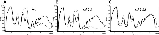
Fig. 20.1. An example of typical polysome profiles of three yeast strains subjected to an oxidative stress. One contains
the wild-type
RCK2
gene (a MAPKAP kinase operating downstream of HOG signaling pathway (7)). The other two con-
tain its mutant alleles. Cultures were harvested before (
thick lines
, no stress) or after the exposure to 0.8 mM t-butyl
hydroperoxide for 30 min (
thin lines
, 30 min with tBOOH). (a) Wild-type; (b)
rck2
Λ;(c)
rck2-kd
(a dominant negative
allele encoding catalytically inactive enzyme). In the wt and
rck2
Λ cells, the number of polysomes decrease upon stress
indicating an inhibition of general transla tion initiation. The polysome fraction is decreased to an even higher degree in
rck2
Λ
.
By contrast,
rck2-kd
fails to show polysome run-off due to a block in translation elongation. The charts were
generated by the Clarity software.
Polysome Analysis and RNA Purification from Sucrose Gradients 295
fractions from sucrose gradients followed by a variety of down-
stream applications including Western and Northern blotting,
qRT-PCR (25, 26), RNase protection assay (16), and microarray
analysis (27–30). High-throughput polysome fractionation using
deep 96-well plates has also been reported. However, it does not
seem to stand up to the high quality of resolution of the classical
setup (31).
The polysome pr ofile analysis has been described in the lit-
erature with a variety of modifications. The main concern has
to do with the choice of a stabilization reagent used to prevent
polysome run-off. The most widely used reagent is the antibiotic
cycloheximide that binds the 60S ribosomal subunit (32)andis
thought to block translation elongation by preventing release of
deacylated tRNA from the ribosome E site after translocation (33,
34), thus stalling the 80S ribosomes on mRNA in a polysomal
state. Usage of cycloheximide may be omitted in studies aimed
at examining defects in the elongation step as these usually pre-
vent polysome run-off that naturally occurs during the cell lysate
preparation in the absence of any stabilization agent (35).
Heparin, a highly sulfated glycosaminoglycan, is routinely
used to stabilize translational complexes pre-tr eated with cyclo-
heximide (36, 37) and to protect them against RNase activity
during preparation of cell extracts. However, inclusion of hep-
arin in extraction buffers seems to inhibit initiation of protein
synthesis (38, 39) and leads to artificial association of initiation
factors with pre-initiation complexes that do not reflect their nat-
ural state in the cell at the time of lysis (25). Hence, a new strat-
egy has recently been developed employing formaldehyde as a
cross-linking reagent to fix ribosomes on mRNAs in the living
yeast cells. This technique is believed to provide the best available
approximation of the native 43S/48S pre-initiation complexes
composition in vivo (40).
A decrease in the initiation rate results in the polysome run-
off with a concomitant increase in the amount of free 80S ribo-
somes seen as a monosomal peak in a polysome profile. The frac-
tion of vacant mRNA-free 80S ribosomes can be distinguished
from mRNA-bound monosomes on the basis of their different
sensitivity to high salt concentrations (41). The 80S couples dis-
sociate into individual subunits at 0.8 M KCl (41) or 0.7 M NaCl
(36) only if they are not associated with an mRNA.
When performing polysome analysis, a common task is cal-
culation of ratios of particular peak areas in order to deter-
mine what proportion of the translational machinery is actively
engaged in translation. Consensually, only polyribosomes are con-
sidered to be actively translating ribosomes because the mono-
somal peak contains an unknown proportion of mRNA-free 80S
couples. Therefore the translational rate is usually expressed as the
polysome-to-monosome (P/M) ratio, which, in theory, decreases
296 Mašek, Valášek, and Pospíšek
with translation initiation defects but increases with defects in
elongation. The P/M ratio determination may not have a true
predicative value in those cases where a particular mutant causes
accumulation of free ribosomal subunits as a consequence of
either a defect in ribosome biogenesis or reduced ability of
mRNAs to be translated or as a result of inhibition or slowing
down initiation complex assembly (see Fig. 20.2a).
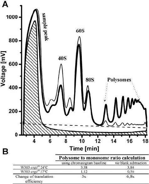
Fig. 20.2. Polysome profile normalization strategies and the P/M ratio calculations. a
Polysome profiles of the W303 strain carrying a temperature-sensitive allele of an essen-
tial
CEG1
gene coding for a guanylyl transferase subunit of the yeast capping enzyme
(45). The culture was grown at a semipermissive temperature of 24
◦
C(
black thick line
)
and then shifted to a non-permissive 37
◦
C for additional 12 h (
dark grey thin line
). A
sample peaks indicate a volume and a total absorbance of yeast cell lysates deprived
of ribosomes or PEB buffer loaded on sucrose gradients (
blank
). Following peaks cor-
respond to 40S and 60S ribosomal subunits, to the 80S monosome and to polysomes.
Identities of the two unmarked peaks, which typically appear in the
ceg1
ts
polysome
profiles, are unknown. The
dashed grey line
depicts the actual chromatogram baseline
calculated by Clarity software after overlaying both chromatograms and transposing the
curves to the same position. This baseline connects the lowest points of the curves.
Shaded area
corresponds to a blank tube containing only PEB. Raw data were exported
into a tab-delimited-text format and displayed with the help of OriginPro
R
software.
b Polysome-to-monosome area ratios were determined either using the chromatogram
baselines or by subtraction of the blank area from the polysome profile areas. The inclu-
sion of a blank tube in this experiment permitted more accurate determination of P/M
ratios.
Polysome Analysis and RNA Purification from Sucrose Gradients 297
Calculation of peak areas represents another pitfall (see
Fig. 20.2). In the majority of published experiments, the peak
areas are subtracted either from the baseline corr e sponding to
detector zero or from the baseline extrapolated by the application
of chromatography software, which usually connects the lowest
points of curves. These methods of peak ar ea determination might
not be as accurate as often believed. Discrepancies may be caused
by extraction buffers containing TritonX-100 and/or other com-
pounds exhibiting a substantial absorbance at 254 nm. The ar ea
of the first peak detected in a polysome profile (the sample peak)
thus mostly reflects the amount of TritonX-100 in the sample
loaded on the sucrose gradient and, indirectly, also corresponds to
the sample volume. If the sample peaks do not differ substantially
and if the equal sample volumes were loaded, it is recommended
to overlay and compare polysome profile chromatograms based
on the first sample peak. Blank tubes containing only extraction
buffer can be used to circumvent many difficulties and provide
us with a more realistic baseline reflecting absorbance of extrac-
tion buffer across the polysome profile and allowing for a more
exact determination of the P/M ratio (see Fig. 20.2). As for the
data acquisition followed by their post-analysis modifications as
well as for the peak ar e a calculations, we take an advantage of
ISCO gradient analyzer connected with the data-acquisition PC
card in combination with the Clarity chromatography software
(DataApex Company; www.dataapex.com). This software allows
not only smooth on-line data acquisition but also many logistic
operations such as baseline shifting, profiles zooming in/out and
peak editing, combining and dividing. The Clarity software also
supports graphical editing of profile curves including their over-
laying (see Fig. 20.1) as well as saving and exporting raw or edited
data in various formats (see Fig. 20.2). The subtraction of blank
area from polysome profiles can be carried out if the same artifi-
cial detector zero line is inserted at the beginning of all readings.
Such artificial baselines ensure easy comparison and recalculation
of the measured data between samples in addition to the blank
sample subtraction. The calculation of profile areas can be further
achieved after raw data export to the suitable spread-sheet calcu-
lator (e.g., OriginPro
R
) without inserting artificial detector zero
line (see Fig. 20.2). Substantial differences in the net profile areas
after blank subtraction in a single experiment usually indicate that
unequal lysate concentrations were loaded on gradients.
Unexpected discrepancies in the polysome profile analysis
may also be caused by degradation of RNA during a crude
cell extract preparation and subsequent procedures. It is recom-
mended to check the RNA quality in lysates electrophoretically
prior to the analysis. We have recently introduced a simpler and
less hazardous TAE/formamide agarose gel electrophor esis that
is particularly suitable for RNA separation in crude cell extracts
298 Mašek, Valášek, and Pospíšek
containing large amounts of proteins, DNA and other contami-
nating molecules. We have also demonstrated that this technique
can be successfully used for analysis of unpurified or partially puri-
fied sucrose gradient fractions as well as for high quality resolu-
tion of purified polysomal RNA that is perfectly suitable for the
subsequent Northern blot analysis (42)(see Fig. 20.3).
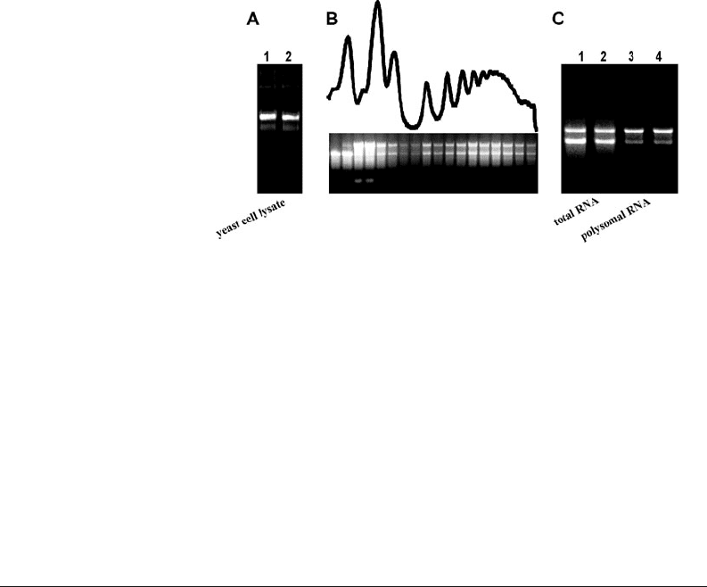
Fig. 20.3. Application of TAE-formamide agarose gel electrophoresis at various steps of
the yeast polysome profile analysis. a Quality assessment of a crude yeast cell extract.
Formamide was added to a yeast cell lysate to a final concentration of 60% (v/v). Loading
dye was supplemented with 1% SDS (Section 3.5 , Option 1). b Electrophoresis of yeast
polysome profile fractions. The profile corresponds to the lysate shown in (a; line 1)
that has been loaded onto a 7–50% sucrose gradient and centrifuged in a SW41 rotor
for 3 h at 35,000 RPM at 4
◦
C. 0.5-mL fractions were collected starting from a layer
where the small ribosomal subunits sediment. RNA was coarsely purified according
to the protocol described in Section 3.5, Option 2. c Comparison of whole-cell RNA
(lines 1, 2) purified by the acid-phenol method directly from the yeast and RNA samples
purified from polysome fractions (lines 3, 4) by the protocol presented at Section 3.5,
Option 3. All samples originated from the same yeast culture.
2. Materials
2.1. Yeast Culture
and Preparation
of Cell Lysate
1. SC medium: 2% (w/v) glucose, 0.65% (w/v) yeast nitrogen
base, 50 mg/L of each auxotrophic supplement
2. YPD medium: 2% (w/v) glucose, 1% (w/v) yeast extract, 2%
(w/v) bactopeptone
3. RNAse-free deionized water (see Notes 1 and 2)
4. Cycloheximide stock 10 mg/mL
5. Polysome extraction buffer (PEB): 20 mM Tris-HCl, pH
7.4, 140 mM KCl, 5 mM MgCl
2
, 0.5 mM DTT, 1% (v/w)
TritonX-100, 0.1 mg/mL cycloheximide, 0.2 mg/mL hep-
arin (ammonium salt)

Polysome Analysis and RNA Purification from Sucrose Gradients 299
6. Glass beads, acid washed (0.45–0.55 mm in diameter, see
Note 3)
2.2. Gradient
Preparation
1. Gradient solution 1: 20 mM Tris-HCl, pH 7.4, 140 mM
KCl, 5 mM MgCl
2
, 0.5 mM DTT, 0.1 mg/mL cyclohex-
imide, 0.2 mg/mL heparin, 7% (w/v) sucrose (see Note 4)
2. Gradient solution 2: 20 mM Tris-HCl, pH 7.4, 140 mM
KCl, 5 mM MgCl
2
, 0.5 mM DTT, 0.1 mg/mL cyclohex-
imide, 0.2 mg/mL heparin, 50% (v/w) sucrose
3. Solution 3: 20 mM Tris-HCl, pH 7.4, 140 mM KCl, 5 mM
MgCl
2
, 0.1 mg/mL cycloheximide, 60% (w/v) sucrose (see
Note 5)
2.3. RNA Isolation
from Polysomal
Profiles
1. GuITC: 6 M guanidium thiocyanate, 0.25 M sodium acetate
2. 96 and 75% ethanol
3. RNAase-free deionized water
4. Acid phenol, p H 4.0–5.2
5. Chloroform: Chloroform:Isoamylalcohol (24:1)
6. 6 M LiCl
7. 3 M sodium acetate, pH 5.2
2.4. RNA
Electrophoresis
1. 50× TAE: to prepare 1 L, add 242 g Tris, 100 mL of 0.5 M
EDTA, pH 8.0, and 57.1 mL of glacial acetic acid
2. Agarose
3. Deionized formamide
4. Ethidium bromide (1 mg/mL)
5. 10× Loading Dye: 50 mM Tris-HCl, pH 7.6, 0.25% (w/v)
Bromophenol Blue, 60% (v/v) glycerol
3. Methods
3.1. Yeast Culture
and Preparation
of Cell Lysate
1. Inoculate a yeast strain of interest into 40 mL of a suit-
able medium and grow it in 250-mL Ehrlenmayer flask
at the desired temperature for 12–24 h to early stationary
phase. Use either rich YPD medium or defined SC minimal
medium depending on the type of experiment; an incuba-
tion temperature of 28
◦
C works well for most yeast strains
(see Note 6 on culture conditions and Note 7 for a proce-
dure for making extracts from mammalian cells).
2. Inoculate approximately 40 μL of the stationary culture to
75 mL of fresh medium in a 250-mL Ehrlenmayer flask
300 Mašek, Valášek, and Pospíšek
and incubate the cells for 12–16 h with vigorous shak-
ing until the cultures reach mid-exponential growth phase
(OD
660
=0.4–0.6).
3. At the time of harvest, add 750 μL of cycloheximide from
the stock solution and chill cells by adding one spoon of
crushed ice (approx. 20 g of ice, 25% of total culture vol-
ume), gently shake several times and keep on ice for 5 min.
All subsequent steps have to be carried out on ice and with
pre-chilled tubes and centrifuges.
4. Transfer the cells into two 50-mL Falcon tubes and cen-
trifuge them for 5 min at 3,000×g at 4
◦
C. Resuspend the
pellets by adding 3 mL of ice-cold PEB and pool the aliquots
in one tube (see Note 8). Centrifuge once again for 5 min at
3,000×g.
5. Repeat the washing step by adding 6 mL of ice-cold PEB.
6. Resuspend the cells in 700 μL of ice-cold PEB and trans-
fer the resulting cell suspension into a pre-chilled 1.5-mL
Eppendorf tube containing 450 μL of pre-chilled glass
beads.
7. Break the cells by vigorous agitation with 30 oscillations/s in
a bead-beater for 3 min (e.g., MM301, Retsch). Tube hold-
ers should be pre-chilled at –20
◦
C for at least 1 h. Appropri-
ate conditions for efficient cell lysis can vary with different
equipment and should be set empirically (see Note 9).
8. Clear cell lysates by centrifugation at 8,000×g for 5 min.
9. Immediately proceed to loading of the cell lysates onto
sucrose gradients. If necessary, lysates can be stored at –70
◦
C
for no longer than several days or shipped on dry ice at this
stage.
3.2. Gradient
Preparation and
Centrifugation (for
SW41 Beckman
Rotors)
1. Measure the concentration of nucleic acids in the lysate spec-
trophotometrically and optionally check the RNA integrity
(see Section 3.5, Option 1). About 10–15 OD
260
units
should be loaded on the gradient, optimally at a volume less
than 400 μL, but not exceeding 800 μL(see Note 10 for
up-scaling).
2. Linear sucrose gradients can be prepared in several ways (see
Note 11). We usually prefer making gradients with the use
of a commercial gradient maker (Hoefer SG-50, Fig. 20.4).
For preparation of one 7–50% sucrose gradient, fill cham-
ber A with 6.3 mL of gradient solution 1 (7% sucrose) and
chamber B, which is closer to the outlet, with 6.3 mL of
gradient solution 2 (50% sucrose). The combined volumes
make up for the maximal capacity (12 mL) of SW41 cen-
trifugation tubes (Beckman, Ultra-ClearTM tube) and the
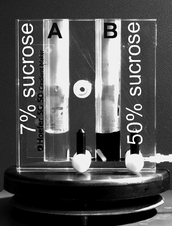
Polysome Analysis and RNA Purification from Sucrose Gradients 301
Fig. 20.4. Use of a gradient maker for preparation of linear sucrose gradients. C hamber
A and B are filled with the same volume of 7 and 50% sucrose solutions, respectively.
After connecting the chambers, gentle continuous mixing in chamber B generates a
concentration gradient which flows into a centrifugation tube. More detailed instructions
are described in Section 3.2, Step 2.
dead volume of the Hoefer SG-50 gradient maker that is
around 800 μL. After filling both chambers, add a stir bar
into chamber B, open the tap connecting the chambers and
force out any bubbles from the connecting tube, if neces-
sary. Open the outlet tap and suck the solution towards the
end of the connected elastic tubing by a pipette. Then turn
on the magnetic stirrer, adjust the position of the gradient
maker, and apply an appropriate speed of swirling to provide
gentle but complete mixing in chamber B. Place the end of
the elastic tubing at the bottom of a centrifugation tube and
start pouring the gradient by slow continuous movement of
302 Mašek, Valášek, and Pospíšek
the tubing towards the top. A slow pouring is important for
preparation of a high-quality undisturbed gradient and can
be achieved by proper adjustment of the distance between
the tube outlet and the level of the sucrose solutions in the
gradient maker.
3. Carefully load the lysate on the top of the gradient. Bal-
ance pairs of tubes to be centrifuged carefully with PEB (see
Note 12).
4. Put the tubes into a pre-cooled SW41 rotor according the
Beckman instructions (see Note 13). Centrifugation condi-
tions are summarized in Table 20.1.
Table 20.1
Gradient ranges and centrifugation conditions (using a Beckman SW41 rotor) for
visualization of different translational complexes
Translational
complexes to
be resolved
Sucrose
concentration
range (%)
Time of
centrifuga-
tion Speed (rpm) Citation
Eukaryotic 40S, 60S
subunits, 80S
ribosome and
polysomes
4.5–45
7–50
15–50
2.5
3
2.5
39,000
35,000
40,000
(25)
(7)
(5)
Bacterial 30S, 50S
subunits, 70S
ribosome and
polysomes
5–40
10–40
2.5
2.5
35,000
35,000
(19)
(46)
40S–80S 15–40
5–40
4.5
2
39,000
27,000
(36)
(9)
40S–60S 7.5–30
5–30
5
8
41,000
27,000
(25)
(9)
3.3. Data Acquisition
and Normalization
1. Place the centrifugation tube onto a Tube Pier cer of the UA-
6 UV/Vis detector (ISCO, Inc.) and carefully mount it in
a holder. Start pushing up 60% sucrose (Solution 3) from
the bottom of tube by switching on the peristaltic pump and
adjusting flow rate to 2.4 mL/min. Monitor absorbance at
254 nm continuously.
2. Absorbance profiles can be recorded either by chart recorder
which is an integral part of the ISCO instrument or by an
external data-acquisition module equipped with an appro-
priate software. We recommend Clarity from DataApex
(www.dataapex.com).
Polysome Analysis and RNA Purification from Sucrose Gradients 303
3. If various ribosomal complexes are subjected to further anal-
ysis of their RNA and/or protein content, collect fractions
corresponding either to the desired peaks or to fixed volumes
(see Note 14).
3.4. RNA Isolation
from Polysome
Profiles (for SW41 or
SW28 Profiles Split
into Two Fractions)
1. To prevent degradation of samples by RNases, mix them
with an equal volume of GuITC and vortex well immedi-
ately upon collecting the fractions. Add an equal volume of
96% ethanol to precipitate nucleic acids from the samples
and incubate them overnight at –20
◦
C, which is usually suf-
ficient for quantitative precipitation. Optionally, RNA can
be analyzed electrophoretically at this step (see Section 3.5,
Option 2).
2. Transfer samples into 28-mL centrifuge tubes and spin
down precipitated nucleic acids for 20 min at 25,000×g
at 4
◦
C. For SW28 fractions, pool aliquots in one
28-mL centrifuge tube by repeating the centrifugation
step.
3. Wash the pellets with 5 mL of 75% ethanol. Decrease the
volume of 75% ethanol for easier transfer of pellets into
1.5-mL Eppendorf tubes. At this step, the isolation pro-
cedure can be interrupted and samples can be stored at
–70
◦
C.
4. Centrifuge the samples at 21,000×g for 15 min at room
temperature. Aspirate the ethanol and apply a second short
spin followed by r emoval of residual ethanol and air-drying
of the pellet to completely get rid of ethanol (beware that
over-drying can result in difficulties in resuspension of the
pellet).
5. Dissolve pellets by adding 400 μL of DEPC-treated water.
Add 400 μL of acidic phenol and vortex for 5 min. Incu-
bate samples for 1 min at room temperatur e, then add 400
μL of chloroform and repeat vortexing for 5 min. Cen-
trifuge samples for 20 min at 21,700×g at 4
◦
C(orsee Note
15).
6. Transfer the aqueous phase containing the RNA into a new
1.5-mL Eppendorf tube.
7. Adjust volumes to 750 μL by RNase-free water, then add
250 μL of 6 M LiCl (final concentration 1.5 M), vor-
tex, and leave overnight at –20
◦
C. Centrifuge samples for
20 min at 25,000×g at 4
◦
C(see Note 16).
8. Completely remove the supernatants and wash the pellets
with 1 mL of 75% ethanol. Centrifuge samples for 15 min
at 21,700×g at 4
◦
C.
9. Repeat Step 8.