Marshall L. Stoller, Maxwell V. Meng-Urinary Stone Disease
Подождите немного. Документ загружается.


Chapter 31 / Indications and Outcomes of PCNL 609
609
From: Current Clinical Urology, Urinary Stone Disease:
A Practical Guide to Medical and Surgical Management
Edited by: M. L. Stoller and M. V. Meng © Humana Press Inc., Totowa, NJ
31
Percutaneous Nephrolithotomy
Indications and Outcomes
Paul K. Pietrow, MD
CONTENTS
HISTORY
INDICATIONS
CONTRAINDICATIONS
OUTCOMES
COMPLICATIONS
REFERENCES
Key Words: Nephrolithiasis; kidney; percutaneous access; nephrolitho-
tomy.
HISTORY
The management of renal calculus disease underwent drastic changes in the early
1980s with the arrival of percutaneous surgery and shockwave lithotripsy within sev-
eral years of each other. Previously, patients were managed with an array of open
procedures, including pyelolithomy, ureterolithotomy and anatrophic nephrolitho-
tomy. The opportunity to effectively manage renal calculi in a percutaneous manner
has drastically reduced patient morbidity when compared to an open, flank approach.
Fernstrom and Johansson were the first to describe a percutaneous approach to the
renal collecting system for the management of calculi (1). Much of the early pioneer-
ing efforts were performed at the University of Minnesota and were made possible by
the arrival of improved equipment and of an effective ultrasonic device that could be
used to destroy and remove stones of varying compositions (2). Although the avail-
ability of improved access devices, nephroscopes and lithotrites have made this pro-
cedure more facile and safe, the basic principles and techniques have not changed
dramatically over the past 20 yr.

610 Pietrow
INDICATIONS
Although significantly less morbid than open surgery, percutaneous nephrolithotomy
(PCNL) is still the most invasive approach to urinary lithiasis when compared to ureter-
oscopy or extracorporeal shockwave lithotripsy (SWL). Consequently, this technique is
generally reserved for specific needs (Table 1). Bulky stones greater than 2 cm are
frequently best treated with a percutaneous approach to minimize repeat treatments and
trauma to the kidney. Complete stone clearance can also avoid the risk of steinstrasse if
the ureter is unable to accommodate a large bolus of stone debris. These concerns are
especially true of staghorn calculi or complex stones that occupy multiple calyces. Some
stones are simply too dense or organic in nature to respond well to SWL and may be best
served with an initial percutaneous approach. Calcium oxalate monohydrate calculi are
noted to be the densest stones and often experience incomplete fragmentation (3,4).
Cystine stones are also resistant to shockwave energy, likely owing to their organic
origin and their tight crystalline formation. Additionally, residual cystine fragments can
easily act as “seed calculi” leaving the patient with multiple smaller stones rather than
their initial large calculus. In light of these difficulties, many authors advocate a percu-
taneous approach for cystine stones even less than 2 cm in order to achieve complete
stone clearance (5,6).
Some stones smaller than 2 cm may be best treated with a percutaneous approach if
they reside within difficult anatomic locations. Calyceal divertula have a tight neck that
will not allow the easy passage of stone debris. If the diverticulum is inaccessible from
a retrograde ureteroscopic approach, then percutaneous access can clear any stone
material and allow either destruction of the urothelial lining or creation of a wider
infundibular orifice (or both) (7,8). Owing to obvious anatomic constraints, this tech-
nique is best reserved for posterior calyces.
Stones within a lower pole calyx may have a difficult time clearing out of this region
into the pelvis and down the ureter following SWL. A well-controlled large randomized
trial has demonstrated that percutaneous nephrolithotomy is superior to SWL for lower
pole calculi (9). The difference was particularly dramatic for stones larger than 1 cm.
Table 1
Indications for Percutaneous Nephrolithotomy
Strong
• Calculi >2–3cm
• Staghorn calculi
• Complex calculi (multiple calyces)
Possible
• >1 cm calcium oxalate monohydrate calculi
• >1 cm cystine calculi
• Calyceal diverticular calculi
• >1 cm lower pole calculi
• >1 cm renal calculi in patients with urinary diversions
• >1 cm renal transplant calculi
• Calculi associated with UPJ obstruction
• Large proximal ureteral calculi

Chapter 31 / Indications and Outcomes of PCNL 611
Several authors have identified anatomic features of the lower pole collecting system
that might allow for the easy clearance of fragments after SWL. These include the
presence of an obtuse infundibular/renal pelvis angle (>70°), a short infundibular length
(<3 cm) and a wide infundibular neck (>5 mm) (10).
Patients with urinary diversions and moderate-to-large calculi may be best served
with PCNL if retrograde access is too difficult or if the presence of infected stones and
colonized urine preclude the use of shockwave lithotripsy. These patients fare well if
they are covered with broad spectrum antibiotics and pre-placement of the percutaneous
access 1–2 d before their procedure (11).
Urinary lithiasis within a kidney transplant may require a percutaneous antegrade
approach if the stone or the patient are not amenable to SWL. In addition, transplant
ureters can be difficult to access in a retrograde fashion owing to angulation and narrow-
ing at the uretero-vesical anastomosis.
The presence of co-existing pathology may also require a percutaneous approach.
Patients with large calculi and a ureteroplevic junction obstruction (UPJO) may be best
served by performing a PCNL and an antegrade endopyelotomy in the same sitting. Care
must be taken to discern the difference between a primary UPJO causing stone or a stone
causing a secondary UPJO.
CONTRAINDICATIONS
Several conditions can preclude the use of a percutaneous route to remove renal
calculi (Table 2). An uncontrolled coagulopathy (either pathophysiologic or pharmaco-
logic) puts the patient at too great of a risk for severe hemorrhage. Because each kidney
receives 5–10% of the total cardiac output with each beat of the heart, uncontrolled
hemorrhage can be rapid and require transfusion, selective arterial embolization or even
nephrectomy. Significant amounts of irrigating fluids are absorbed during a PCNL
owing to extravasation into the retroperitoneal space and the opening of venous sinuses
within the renal parenchyma. The presence of an active urinary tract infection, there-
fore, places the patient at a greater risk of bacteremia of even septicemia with vascular
collapse.
Renal anatomic abnormalities may make it difficult to adequately and safely access
the collecting system. This includes renal ectopia (e.g., pelvic kidney) or fusion anoma-
lies (e.g., horseshoe kidney). Although these anomalies are not absolute contraindications
for the performance of a PCNL, good spatial orientation, excellent preoperative imaging
and a very healthy respect for surrounding viscera and vasculature are crucial (12).
Patients with a solitary kidney should be approached with care. Any surgical interven-
tion runs the risk of permanent injury to the sole functioning renal unit.
Table 2
Contraindications for Percutaneous Nephrolithotomy
Absolute Relative
• Uncontrolled coagulopathy • Ectopic kidney
• Active urinary tract infection • Fusion anomalies
• Severe dysmorphism
• Morbid obesity
612 Pietrow
OUTCOMES
The technical success of percutaneous nephrolithotomy has made this technique the
standard of care for many calculi and scenarios as outlined in the Indications section. A
careful assessment of results, however, reveals that the expected outcomes and risks vary
with the specific application.
Staghorn Calculi
Staghorn calculi (either partial or complete) represent a unique challenge for the
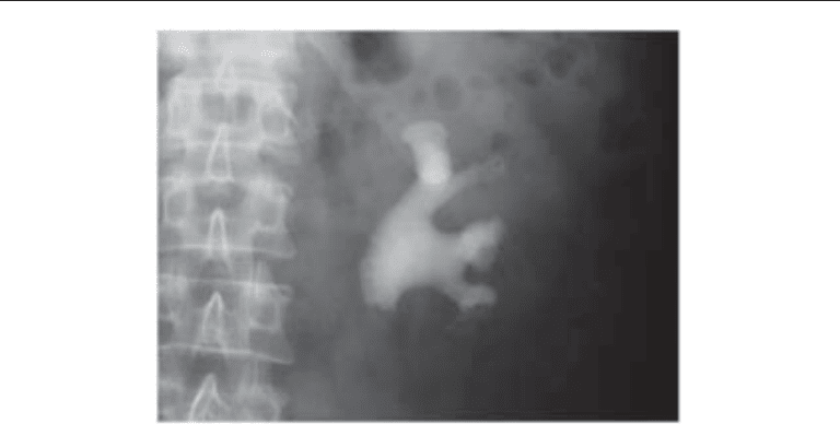
endourologist (Fig. 1). The sheer bulk of the stone burden and the complex branched
anatomy of the collecting system require careful planning regarding access and plurality
of access tracts. Additionally, these calculi are typically associated with an infectious
etiology. Antibiotics are never able to completely penetrate the interstices of a complex
stone, running the risk of a release of bacteria and endotoxin during stone ablation (13).
Despite these warnings, however, PCNL remains the standard of care for staghorn cal-
culi, replacing anatrophic nephrolithotomy and other open approaches.
Results from many authors have demonstrated the superiority of PCNL over SWL
alone. In 1994, the evidence was compelling enough to prompt the AUA Guidelines
Committee to recommend PCNL as first line therapy, often combined with SWL to
ablate calculi in difficult to reach calyces (14).
Second-look PCNL may also be added to the end of this regimen to “clean-up” any
residual fragments left after the shockwave lithotripsy. This technique has been dubbed
“sandwich therapy.” Stone-free rates tend to range from 70 to 100% with this approach,
with acceptable complication rates. Randomized trials have been performed comparing
a combination of PCNL with SWL vs SWL alone. One particularly noteworthy analysis
from Meretyk et al. demonstrated a stone-free rate of 74% with combination therapy vs
22% with SWL alone (15). Nearly half of all shockwave lithotripsy patients had septic
episodes whereas the combination patients had an 8% rate. Finally, 26% of the
monotherapy patients required ancillary procedures over 6 mo vs 4% of the combination
patients over one additional month.
Fig. 1. Large, branched staghorn calculus completely filling the collecting system of a left
kidney.

Chapter 31 / Indications and Outcomes of PCNL 613
Chandhoke has demonstrated that initial combination therapy is more cost effective
than shockwave monotherapy (16). This is especially notable when the stone surface
area exceeds 500 mm
2
when measured in its greatest dimensions. This implies a calculus
at least as large as 2.2 2.2 cm, clearly within the parameters of a typical staghorn
calculus.
More recently, some centers have moved away from a sandwich technique, relying on
aggressive PCNL at the first sitting. This may require the use of a supracostal access or
a multi-tract access at the first procedure (17–19). These same approaches may remove
so much stone burden that there is no longer a need for the “meat” portion of the sandwich
(SWL) or the second-look PCNL.
Others have moved away from the removal of all residual fragments, electing to treat
small remnants with aggressive medical therapy (20). This therapy should be directed
by the information gained from a complete metabolic evaluation and/or stone analysis.
Anatomic abnormalities (UPJ obstruction, caliceal diverticulum) or genetic predisposi-
tion (cystinuria) that played a role in the formation of the original calculus should be
addressed if possible. Residual fragments should be small and expected to have a rea-
sonable chance of spontaneous passage. The presence of infected fragments is generally
frowned on as these can easily act as “seed” calculi and reinfect the patient’s urinary
tract.
Simultaneous Bilateral Percutaneous Nephrolithotomy
Several authors from various institutions and nations have described the performance
of bilateral PCNL in one operative setting to avoid the costs and risks of multiple anes-
thetics (21–24). All authors provide the caveat that the decision to proceed on to the
second side is determined by the clinical result and the relative ease of performance of
the first side. Significant hemorrhage, prolonged stone extraction time, multiple access
tracts and instability of vital signs all warrant the cessation of surgery after completion
of the initial side. The most symptomatic side is usually approached first. Alternatively,
the renal unit most at risk of injury owing to obstruction or bulky stone burden may be
treated initially. Two of the series compared bilateral PCNL patients to either unilateral
PCNL or staged PCNL (22,23). Both demonstrated increased transfusion requirements
for the bilateral patients but noted that this was more closely related to number of
nephrostomy tracts employed and the relative stone burden addressed. That is, smaller
stone burdens addressed with one nephrostomy tract on each side appeared to lose as
much blood as an equal stone burden on a unilateral patient treated via two nephrostomy
tracts.
These same authors report that they are able to proceed on to bilateral PCNL in the
majority of patients selected. Both Dushinski and Maheshwari reported that 94–96% of
patients were able to be treated with a bilateral approach when surgically planned (21,
24). However, none of the authors are able to identify how many patients were screened
for a bilateral approach and deemed too risky for consideration of such an undertaking.
Judicious patient screening, careful technique on the initial side, and sound intraopera-
tive judgment are all crucial for the safe performance of a bilateral, synchronous neph-
rolithotomy.
Supracostal Access
As the kidney lies within the retroperitoneum, the lower pole is displaced ventrally
and laterally by the body of the psoas muscle. As a result, the upper pole of the kidney
614 Pietrow
represents the most superficial access point during a posterior percutaneous approach.
Entry into the lower pole can be hampered by the presence of adipose tissue and/or large
buttocks. In addition, entry into the collection system sometimes requires unimpeded
endoscopic access to the ureteropelvic junction or proximal ureter. For these reasons,
some authors have advocated the use of an upper pole access during PCNL. Depending
on the height of the kidney in the retroperitoneum, this may require that the tract gains
entry above the 12th or even the 11th rib. This approach has been termed “supracostal”
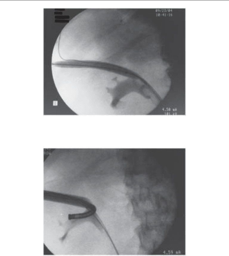
(Fig. 2). Although not without risk, this approach offers excellent visualization of the
upper pole, the lower pole and the UPJ. The use of flexible nephroscopy also allows
reasonable access into many (if not all) of the lateral calyces (Fig. 3).
Fig. 3. A flexible cystoscope is introduced through the upper pole access and directed into an
upper, lateral calyx to confirm complete stone clearance.
Fig. 2. Dilating balloon traversing percutaneous tract via puncture above the 12th rib. Calculi are
visible as filling defects within the renal pelvis.

Chapter 31 / Indications and Outcomes of PCNL 615
Indeed, Wong and Leveillee have demonstrated that a primary upper pole access is
effective for a series of staghorn and complex calculi that were all at least >5 cm in size
(17). By employing flexible endoscopes, a Holmium laser, nitinol baskets and second-
look PCNL, the authors were able to able to render 95% of the patients stone-free. This
did require an average of 1.6 procedures per patient. Complications were reasonable.
They report a 2% transfusion rate, a 3% incidence of pneumothorax and 12% of the
patients experiencing a postoperative febrile episode.
Gupta et al. reported a series of 62 patients who underwent supracostal access for
PCNL. All tracts were above the 12th but below the 11th ribs (25). With this approach,
90% of the patients were either stone-free or reduced to clinically insignificant frag-
ments. The staghorn patients fared well with an 84% stone-free rate. Chest complica-
tions developed in 5% of the patients, all of which were managed with chest tube
decompression. The authors conclude that fear of chest complications should not pre-
clude the use of supracostal access if it is needed for adequate access. As expected, they
also advocate the acquisition of an upright chest radiograph at the end of every
supracostal PCNL.
Munver et al. have also outlined an extensive series of patients, reporting on the
complication rates of 98 supracostal access tracts (26). Seventy-two of these tracts
were above the 12th rib, whereas 26 were above the 11th. Not surprisingly, the higher
the access tract the greater the rate of complications. Seven patients (7%) had intra-
thoracic problems, all managed with chest tube drainage. Of note, the authors also
compared the supracostal patients to a contemporary series of subcostal PCNL patients
and noted only one intrathoracic event out of 202 subcostal tracts (0.5%).
Multiple Access Percutaneous Nephrolithotomy
Clustered or staghorn calculi may be too complex to completely reach with the rigid
nephroscope through one single access tract. As noted previously, some remnant stones
can be reached with a flexible nephroscope through one access (especially if it is through
the upper pole). Alternately, some have advocated the placement of additional
nephrostomy tracts to reach isolated calyces and bulky remnants (27). Indeed, this tactic
has been employed since the early development of the percutaneous technique (28).
Most authors report increases in transfusion rates as the number of tracts increase.
Tubeless PCNL/Reduced Drainage PCNL
Although percutaneous nephrolithotomy is clearly effective, there is no doubt that it
creates significant discomfort. Many endourologists have tried various measures to
reduce postoperative pain without sacrificing efficacy or safety. One approach involves
the placement of a small drainage catheter at the end of the procedure. It is presumed that
the reduced diameter of the nephrostomy tube can reduce pain after the operative pro-
cedure. Pietrow and colleagues randomized patients to standard 22 Fr catheter drainage
vs 10 Fr drainage utilizing a small locking loop catheter (29). Patients demonstrated
significantly less pain in the immediate perioperative period if they had the smaller tube,
although this advantage waned quickly and was no different at the 14 d postoperative
follow-up. Importantly, there was no difference in complications or in hematocrit change
between the two groups. Moreover, many of these patients were treated through a
supracostal approach, which is generally considered more painful.
Others have also studied this modification. A recent investigation by Liatkos et al.
randomized patients to receive either a standard 24 Fr Malecot re-entry catheter or an

616 Pietrow
18 Fr catheter with a tail-less stent following PCNL (30). This study found that those
patients with the smaller tube and the tail-less stent had less pain than the standard cohort.
Of note, these authors did not assess the patients’ perception of pain until their 2-wk
postoperative visit. Although the patients appeared more comfortable, it is hard to dis-
tinguish whether this difference is caused by the smaller drainage catheter or to the
presence of a tailless stent.
Bellman has promoted a tubeless technique in selected patients. In this approach, the
full PCNL is performed in the usual fashion (31,32). If the patient meets strict criteria,
an internal ureteral stent is placed, the nephrostomy access tract is removed and the
skin is closed. Patients must not require a second-look procedure and must therefore
be rendered stone-free during their operation. Significant intraoperative hemorrhage,
large stone volume, prolonged procedure times and a risk of infection have all been
used as exclusion criteria for a tubeless procedure. Using these parameters, Bellman
and colleagues have demonstrated decreased postoperative pain and decreased analge-
sic requirements from those treated in a standard fashion. They do not report any
increase in complications nor in transfusion requirement. This technique has been
duplicated by others with similar results, confirming its safety and efficacy.
The ability to apply this technique beyond these strict guidelines has been proposed
and several have explored methods to close the tract to limit postprocedural hemorrhage.
Noller and colleagues have employed fibrin glue to seal the access tract at the end of the
procedure and have reported satisfactory results with small changes in hematocrit and
no need for transfusion (33).
Clayman and colleagues have used a novel hemostatic agent (Flo-Seal Matrix, Bax-
ter Healthcare Corp., Deerfield, IL) to fill the access tract before removal of the final
safety wire (34). A ureteral occlusion balloon is placed at the edge of the calyx to keep
the material next to the parenchyma and out of the collecting system. Both of these
approaches may allow a wider application of the tubeless technique. All authors repeat
the caveat that the procedure must be uncomplicated and that there must be no need for
re-entry into the collecting system.
In an interesting prospective, randomized study, Desai et al. compared standard large
bore drainage to small catheter drainage and a tubeless technique (35). The patients met
reasonable, but strict exclusion criteria that prevented an increased risk of complication
from any of the methods. The authors found that patients in the tubeless group had the
least postoperative analgesic requirement, followed by the small-bore catheter group.
Additionally, these patients had the shortest duration of urine leak through the
nephrostomy tract. As in earlier studies, there was no difference in transfusion require-
ments among the three groups.
Intracorporeal Lithotriptors
The performance of a successful percutaneous nephrolithotomy is dependent on the
ability to gain safe access into the collecting system and on the ability to fragment and
remove calculi once the stone has been encountered. The development of an effective
handheld ultrasonic device has been crucial to the efficacy of this procedure. After its
introduction in the early 1980s, it quickly became the instrument of choice owing to its
ability to fragment calculi of all compositions. The hollow channel and continuous suction
allow debris to be removed at the same time, thereby keeping the operative field free of
fragments and clot. Although the ultrasonic device can address all stones, it can be slow
and tedious when applied to very hard calculi such as calcium oxalate monohydrate.

Chapter 31 / Indications and Outcomes of PCNL 617
Pneumatic devices have also been employed. These instruments use compressed
nitrogen gas to drive a piston in the instrument, which subsequently fragments the calculi
into increasingly smaller pieces. These pieces must be extracted with a grasper or with
an ultrasonic device once the stone has been ablated. Although quite effective for stones
of all densities, the time required to remove all the fragments can also be significant and
tedious.
More recently, a combination device has been introduced that places a thin pneumatic
probe within the central channel of an ultrasonic probe. Early studies from several
investigators are promising and demonstrate faster operative times with no increase in
complications. Pietrow et al. compared two cohorts of patients, one treated with the
combination device and the second with an ultrasonic device alone (36).
The combination instrument was able to treat all stones regardless of composition.
Operations were quicker with the new instrument, with all gains coming from reduced
fragmentation time. Hofman et al. have reported similar results, noting a decrease in
disintegration time of 30–50% when applied to an in vitro model. The device was equally
effective in a clinical setting, reducing operative times through the rapid disintegration
of the target calculus (37).
COMPLICATIONS
The list of potential complications from PCNL is long and varied. The percutaneous
access must traverse multiple tissue layers and planes, including skin, subcutaneous fat,
muscle, fascia, perirenal fat, and parenchyma. In addition, difficulties can arise from the
location of the access tract, trauma to the renal parenchyma, injury to the collecting
system, hemorrhage, fluid shifts or even the physiologic stress of surgery. Rarely, other
structures may be abnormally displaced and are at risk of inadvertent damage. An incom-
plete list includes: colon, small bowel, spleen, liver, gallbladder, and the great vessels.
Transfusion rates generally run in the low single digits, but have been reported as high
as 50% in some older series. Hemorrhage can arise from the parenchyma of the kidney,
from branches of the renal vasculature or from the torn edges of the urothelium. As
mentioned in previous sections, increasing the number of access tracts will increase the
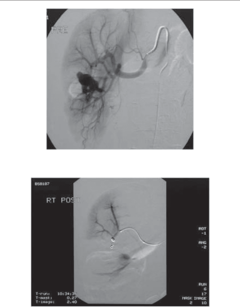
need for transfusion. A rare but impressive source of postoperative hemorrhage is from
the formation of an arteriovenous fistula along the percutaneous tract (Fig. 4). These
patients can present with massive hematuria and usually require embolization (Fig. 5)
or even nephrectomy.
Urinary tract infection and sepsis are always a risk during this procedure owing to
colonization of the urine and sequestration of bacteria within the calculi. Careful atten-
tion to preoperative urine cultures and the liberal use of perioperative antibiotics can
minimize the risk of overwhelming infections. Patients with known or suspected struvite
calculi should be treated with extra care. Rubenstein et al. have demonstrated that even
patients with neurogenic bladders and known urine colonization can be safely treated
with this technique (11). These authors have advocated the placement of the nephrostomy
access one day before the scheduled procedure to allow time for observation and the
early treatment of signs of systemic infection.
Colonic perforation has been reported from multiple authors and sites (38–40). This
particular complication is thought to be caused by the presence of a retrorenal colon or
caused by the placement of the access tract in too lateral a location. Very thin patients
may be at higher risk, as are females and left-sided procedures. Surgeons may not notice
the problem until after the case has been completed, because the nephrostomy access
618 Pietrow
Fig. 4. Angiogram demonstrating a large pseudoaneurysm in the right kidney. This patient
presented 2 wk after uneventful PCNL with massive gross hematuria.
Fig. 5. Same patient after selective embolization of the pseudoaneurysm.
sheath will likely tamponade the defect and “bypass” the injury. Patients may develop
significant fevers or abdominal pain in the postoperative period. Feculent material may
be visible in the urine, the urine leak may cause watery diarrhea or stool may exude from
the nephrostomy tract. Most authors recommend the placement of a ureteral catheter or
stent to attempt to divert the urine down towards the bladder and away from the
nephrocolonic fistula. In addition, the nephrostomy should be pulled back into the colon