Jan Lindhe. Clinical Periodontology
Подождите немного. Документ загружается.


REHABILITATION BY MEANS OF IMPLANTS: CASE REPORTS • 987
Table 42-5. Periodontal findings and prognosis at completion of active therapy
Clinical periodontal examination
Prognosis
Pocket depth
Furcation
involvement
questionable
maintain
Tooth
m
b d
114 3 2 2 3
x
13 3 2 2 2
x
12 2 1 2 2
x
11 2 2 2 1
x
21 2 1 2 1
x
22 2 1 2 1
x
23 2 2 2 2
x
124 3 2 3 2
x
47 4 3 4 2 b
x
46 4 2 3 2
I
x
45 3 2 4 2
x
44 3 2 3 1
x
43 3 2 2 1
x
42 2 1 3 1
x
41 2 2 2 1
x
31
2
1 3 1
x
32 2 2 3 1
x
33 3 2
3
1
x
34 3
1 3
2
x
35 3 2 4 3
x
36 4 2 3 2
I
x
37 3 3 3 3
I
x
Concluding remarks
This case demonstrates the sequence of therapy when
dental implants are to be placed in patients with peri
odontal disease. Successful completion of cause-re-
lated therapy including professional as well as per-
sonal plaque removal represents a
conditio
sine qua non
for implantation with predictable success. Further-
more, in situations where minimal amounts of bone
are available for implant placement, the concept of
shortened dental arches will limit therapy to standard
dental procedures. Finally, proper attention to per-
sonal and professional plaque removal will allow the
maintenance of a successful treatment outcome.
PATIENT 3
IMPLANTS USED TO RESTORE
FUNCTION IN THE MAXILLA
Initial examination
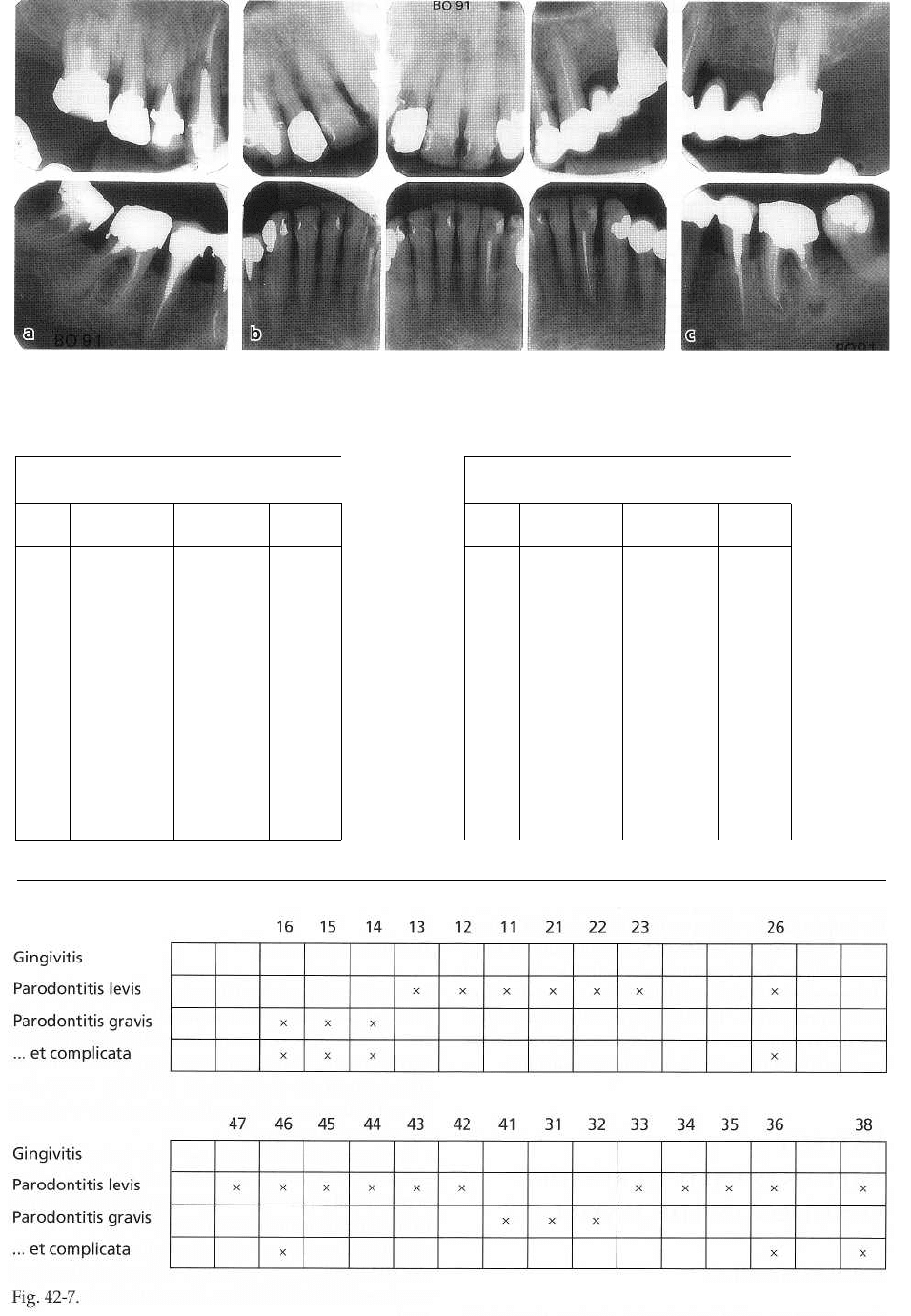
The condition of the dentition of this 49-year-old
woman is illustrated in the radiographs of Fig. 42-6
and the periodontal status (pocket depth, furcation
involvement, tooth mobility and diagnoses) from the
clinical examination in Fig. 42-7. The analysis of the
data discloses a case of moderately advanced peri-
odontal disease. The overall plaque score was 3
5
0
/0
and
the corresponding bleeding on probing (BoP) score
was 60%.
Treatment planning
The combined periodontal, endodontal and caries le
-
sions in both tooth 16 and the root fractures in teeth 15
and 14 called for extraction of these three teeth. In the
maxilla the following teeth were judged as maintain-
able: teeth 13, 12, 11, 21, 22, 23. Tooth 26, which dis
-
played both periodontal and endodontal lesions, was
regarded as questionable.
In the mandibular dentition both first molars (36
and 46) were furcation involved and displayed signs
of periapical pathology. In the mandible the following
teeth could be maintained: 47, 45... 35 and 37. From a
rehabilitation point of view, the mandible offered no
marked problem. The maxillary restoration, however,
was more difficult. Different alternatives were consid
-
ered.
988 • CHAPTER
42
Fig. 42-6a-c.
Case: B.O. female, 49 years
PLI: 35%
BoP: 60%
Periodontal charting
Tooth
Pocket depth
Furcation
involvement
Tooth
mobility
M
B
D
L
18"
1.7
'
16 6 7 6
11
m,b,d:lll
3
15 6 6 1
14 5 5 8 2
13 6 4 6 4 1
12 5 4 6 4 1
11 4 5 4 1
21 5 5 5 1
22 6 5 5
23 6 5 5
24
2
.
5
26 6 4 8 5
m: II, b: I
21
Tooth
Pocket depth
M
B
D
L
Furcation
involvement
Tooth
mobility
48
47 6 4 6 4
46 4 5 4 4
b,
I:
II
45 6 6
44 5 6 4
43 4 4
42 5 5 1
41 5 4 5 1
31 5 4 1
32 4 4 6 4 1
33 4 4
34 5 6 4
35 6 6 4
36 4 4
III
37
38 5 5 4
Diagnosis
REHABILITATION
BY
MEANS OF IMPLANTS: CASE REPORTS • 989
Fig. 42-8a-d.
Alternative 1
A removable partial denture anchored with attach-
ments or clasps to the canines/incisiors. This solution
was not readily acceptable to the patient.
Alternative
2
A fixed, cross-arch bridge extending from tooth 13 (
with cantilever in position 14) to tooth 26 (palatal
root).
Alternative 3
One implant-supported bridge distal of tooth 13, and
one tooth-supported fixed bridge extending from 23
to 26 (provided that at least one root in the molar tooth
could be maintained following furcation therapy and
root resection).
Treatment
Initial therapy
The cause-related therapy included oral hygiene in-
struction and meticulous scaling and root planing.
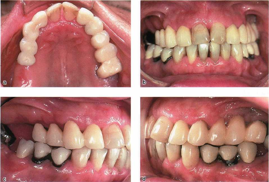
Teeth 16, 15 and 14 were extracted and a temporary
restoration placed on 13 and 12 with cantilevers on
positions 15 and 14 (Fig. 42-8). The cantilever in posi
-
tion 15 was in infraocclusion. The existing restoration
22...26 was removed and replaced with a temporary
reconstruction in acrylic. Teeth 26, 36 and 46 were
exposed to endodontic therapy.
Corrective therapy
A re-examinmation performed 3 months after the end
of the initial therapy phase disclosed that the patient's
self-performed plaque control was excellent. Several
deep pockets with positive BoP still existed. Flap sur
-
gery was performed in both the maxilla and mandible
to get access for proper scaling and root planing.
During surgery tooth 26 was subjected to root resec
-
tion and the remaining roots were subsequently fitted
with a post.
Implant therapy
The edentulous region of the right maxilla was anes
-
thetized. An incision, extending from region 13 to
region 16, was made at the top of the crest and full
thickness flaps were raised. Three fixture sites were
prepared in positions 14, 15 and 16. Fixtures of the
Astra Tech Implants
®
Dental System were installed;
diameter = 3.5 mm; length = 13 mm (position 14), 11
mm (position 15), and 11 mm (position 15). Abutment
connection was 6 months later in a second stage pro
-
cedure.
Restorative therapy
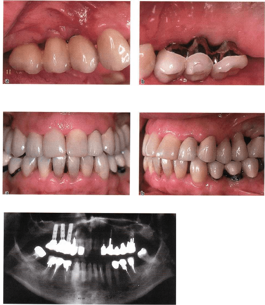
Two bridges — porcelain fused to gold — were fabri-
cated; one for the implant section (position 14... 16)
(
Fig. 42-9) and one for the tooth section (22... 26) (Fig.
42-10). In addition, single crowns were produced for
teeth 13 and 12 (Fig. 42-11).
990 • CHAPTER
42
Fig. 42-9a,b.
Fig. 42-10a,b.
Fig. 42-11.
Maintenance therapy
the implant and the tooth segments — will allow the
The supportive care included recall appointments
patient to exercise proper plaque control (Fig. 42-12).
with the dental hygienist every 3 months for plaque
control evaluation and, if indicated, professional de-
bridement at tooth sites exhibiting positive BoP val-
ues.
Concluding remarks
This case illustrates how implants can be used to
restore function in a periodontally compromised pa-
tient, and how the design of the restoration — both in
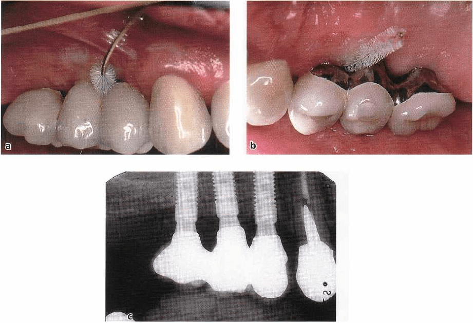
REHABILITATION BY MEANS OF IMPLANTS: CASE REPORTS • 991
Fig. 42-12a-c.
PATIENT 4
IMPLANTS USED IN A
CROSS-ARCH BRIDGE
RESTORATION
Initial examination
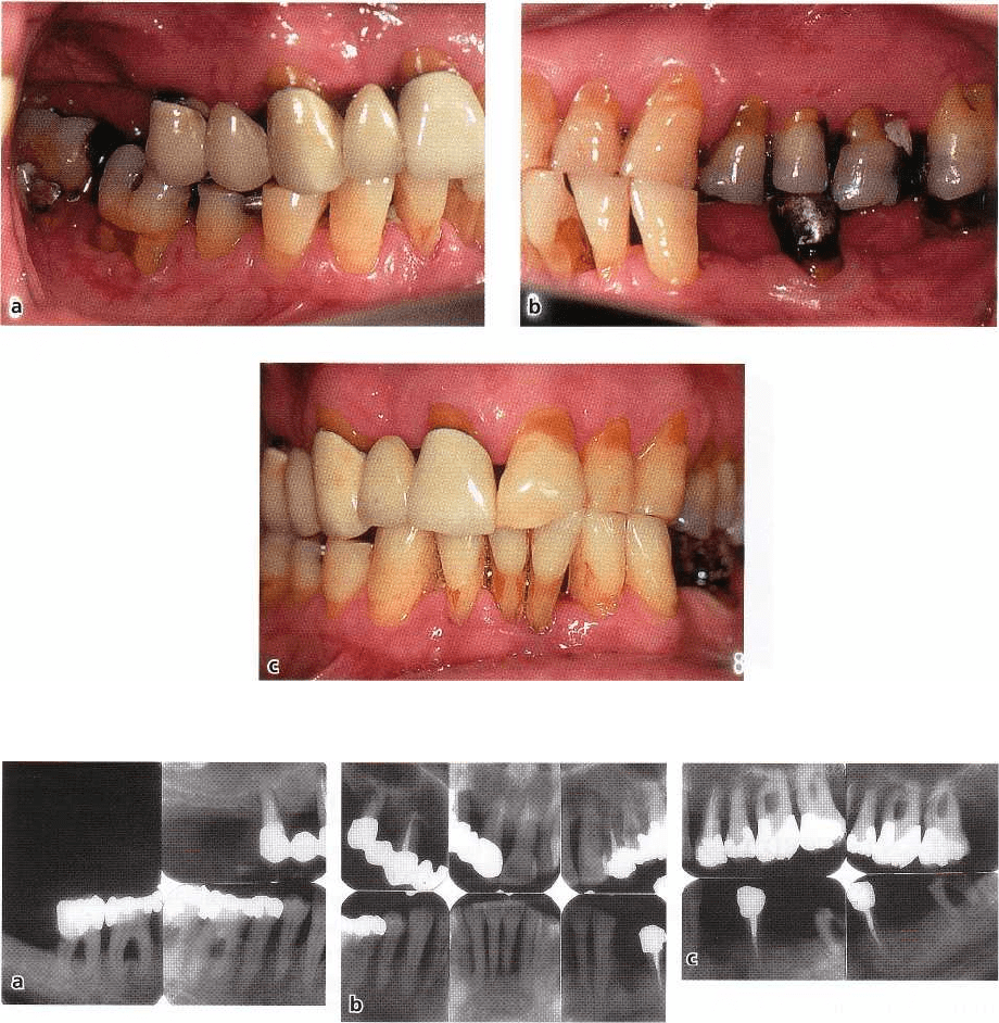
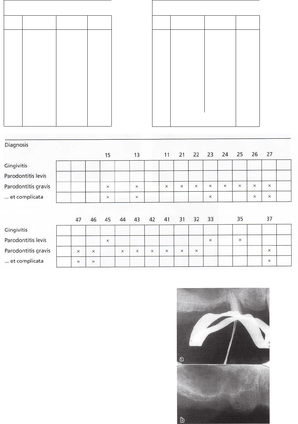
The clinical and radiographic characteristics of this
58-year-old man are illustrated in Figs. 42-13 and 42-14
and the periodontal conditions (pocket depth, furca-
tion involvement, tooth mobility and diagnoses) from
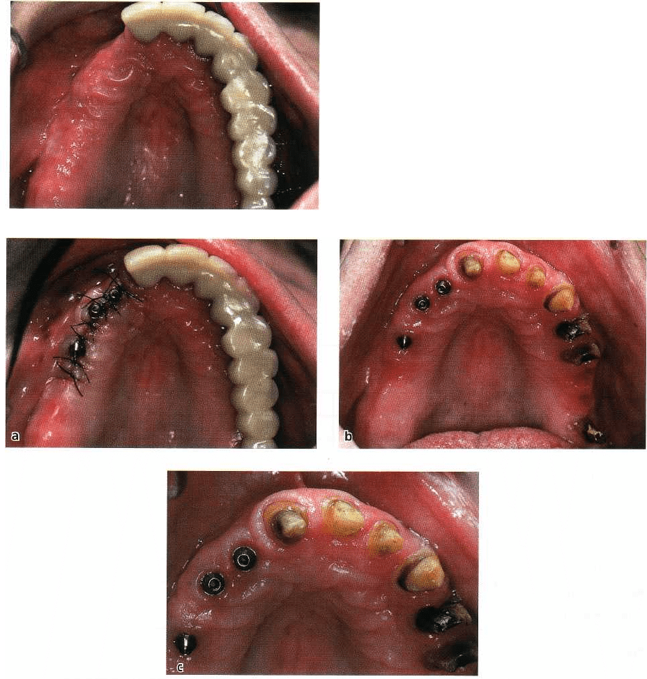
the initial examination in Fig. 42-15. The analysis of
the data disclosed a case of severely advanced peri-
odontal disease. At several teeth the destruction of the
periodontal tissues had reached a level close to their
apices. Tooth 15 exhibited a wide apico-marginal coun
munication, all remaining molars exhibited furcation
involvement of degree III. The overall plaque score
was 92% and the corresponding bleeding on probing
-
BoP - score was 86%.
Trea Intent planning
Tooth 15 required immediate endodontic therapy, al
-
though the prognosis was very questionable. The root
canal was debrided, widened and a calcium hydrox-
ide filling was placed. A clinical and radiographic
examination 8 weeks later disclosed that no proper
healing had occurred, the apico-marginal communi-
cation persisted and tooth 15 was extracted (Fig. 42
-
16). The severely involved mandibular molars (47, 46
and 37) as well as teeth 31 and 41 could not be main
tained and were scheduled for extraction. Teeth 13, 26
and 27 were regarded as questionable from a treat-
ment point of view. The remaining teeth in the maxilla
and mandible could be maintained following peri-
odontal therapy.
The prosthetic rehabilitation of the case was consid
ered difficult but different options could be
examined.
Alternative 1
A fixed, cross-arch bridge extending from tooth 13 (
with cantilever in position 14) to tooth 26 (27) could
have been considered, had the prognosis for tooth 13
been good. This particular tooth, however, was root
filled, exhibited advanced marginal bone loss and was
not regarded as a proper final abutment for a cross
arch splint.
Alternative
2
A removable partial denture anchored with attach-
ments to a fixed bridge extending from tooth 11 to 25.
This would allow the extraction of the two question-
able maxillary molars (26, 27).
992 • CHAPTER 42
Fig. 42-13a-c.
Fig. 42-14a-c.
Alternative 3
One implant-supported bridge distal of tooth 11, and
one tooth-supported fixed bridge extending from 11
to 25 (alternatively to 26/27, provided that at least one
root in this region could be maintained following
furcation therapy and root resection).
Treatment
Initial therapy
The cause-related therapy included oral hygiene in-
struction and meticulous scaling and root planing.
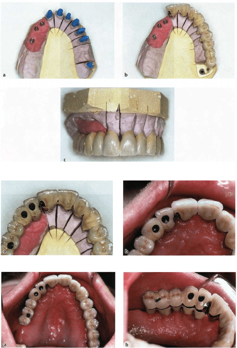
Teeth 15, 13, 47, 46, 31, 41 and 37 were extracted. A
temporary fixed bridge extending from 11 to 27 was
inserted (Fig. 42-17) and a removable partial denture
was produced to restore function in the right maxilla.
A temporary fixed bridge for the mandible extending
from 35 to 45 was also inserted. Teeth 26 and 27 were
exposed to endodontic therapy.
Corrective therapy
A re-examination performed 3 months after the end of
the initial therapy phase disclosed that the patient's
self-performed plaque control was excellent. Al-
though a marked pocket depth reduction had oc-
curred, several deep pockets still existed. Flap surgery
was performed in both the maxilla and mandible to
get access for proper scaling and root planing. During
surgery it was observed that all roots of 26 had to be
removed, while the palatal root of 27 could be main-
tained. This root was subsequently fitted with a
post.
Implant therapy
The edentulous region of the right maxilla was anes
-
thetized. An incision, extending from region 11 to
REHABILITATION BY MEANS OF IMPLANTS: CASE REPORTS • 993
Case: L.F. male, 58 years
PLI: 92%
BoP: 86%
Periodontal charting
Tooth
Pocket depth
M
B
D
L
Furcation
involvement
Tooth
mobility
18"
1/7
w
15 6 8 6
14
-
13 8 7 7
12
11 8 8 7
21 6 6 6
22 6 8 6
23 10 8 8
24 7 5 8
25 6 7 6
26 8 8 8 8 m,b,d:Ill
21 9 8
10 4
m,b,d:Ill
2
,
8
Tooth
Pocket depth
M
B
D
L
Furcation
involvement
Tooth
mobility
48
47 6 6 6
III
46 5 6 8 8
III
45 6 6 5
44 8 6 5
43 8 6 6
42 6 8 4 1
41 6 6 4 3
31 6 6 6 3
32 6 4 6 5 1
33 5 4
3-4
35 4 4
36
37 not probed
38
Fig. 42-15.
was made at the top of the crest and full thickness flaps
were raised. Three fixture sites were prepared in posi-
tion 12, 13 and 15. Fixtures of the Astra Tech Implant
s
®
Dental System were installed; diameter = 3.5 mm;
length =13 mm (position
1205
mm (position 13), and
9 mm (position 15). Abutment connection was made
6
months later in a second stage procedure (Fig. 42-18).
Restorative therapy
A cross-arch bridge made of porcelain fused to gold
was fabricated for the mandible. This bridge extended
from 35 to 45 and had one cantilever unit distal to both
35 and 45.
In the maxilla, two bridges - porcelain fused to gold
-
were fabricated; one for the implant section and one
for
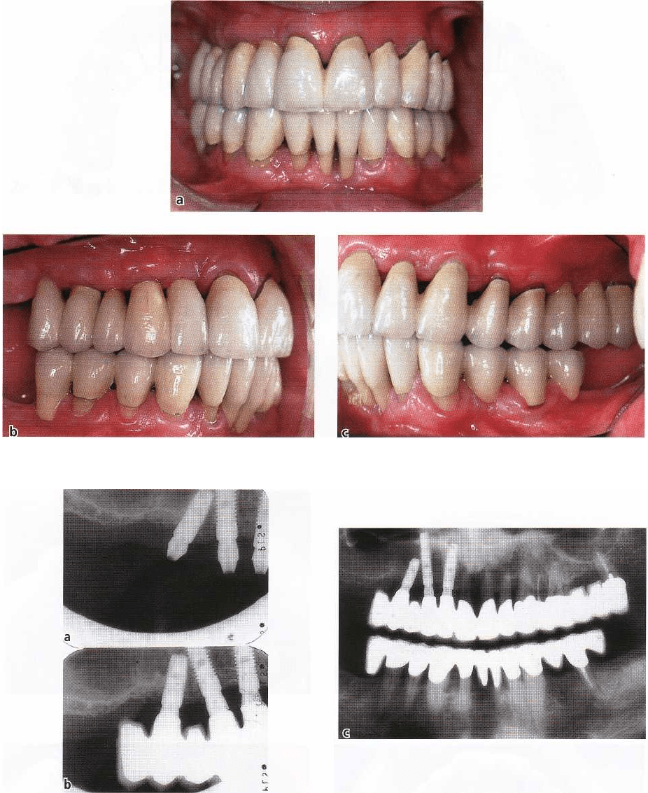
the tooth section (Fig. 42-19). The two bridges were
connected with an attachment that allowed movement
in apicocoronal direction but prevented horizon
tal
deflection (Fig. 42-20). The restoration at the try-in
appointment is illustrated in Fig. 42-21. The outcome
Fig. 42-16a,b.
994 • CHAPTER 42
Fig. 42-17.
Fig. 42-18a-c.
of treatment is illustrated in clinical photographs and
radiographs in Figs. 42-22 and 42-23.
Maintenance therapy
The supportive care included recall appointment with
the dental hygienist every 3 months for plaque control
evaluation and, if indicated, professional debride-
ment at tooth sites exhibiting positive BoP values.
Concluding remarks
This case illustrates how implants can be (1) used to
restore function in a periodontally compromised pa-
tient, (2) combined with a conventional bridge with an
attachment that allows apicocoronal movement of the
tooth-carrying segment but prevents horizontal de-
flection.
REHABILITATION BY MEANS OF IMPLANTS: CASE REPORTS • 995
Fig. 42-19a-c.
Fig. 42-20a,b.
Fig. 42-21a,b.
996 • CHAPTER 42
Fig. 42-22a-c.
Fig. 42-23a-c.
