Glusker J.P., Trueblood K.N. Crystal Structure Analysis: A Primer
Подождите немного. Документ загружается.

Unit cell outline
111 face (dull)
Dull face of cystal
Shiny face of crystal
B
A
SS
Zn
Zn
SS
Zn
Zn
SS
Zn
Zn
−h,−k,−l
h,k,l
(111) polarity sense
(a)
(b)
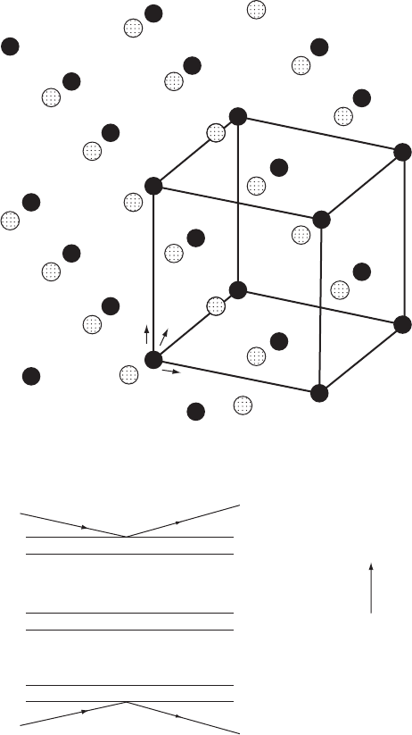
Fig. 10.5 Polarity sense of zinc blende.
(a) The structure of zinc blende, showing the arrangement of zinc and sulfur atoms.
Two views are shown, one down an axis and the other to show the planes of atoms
in the [111] direction. Zinc blende is often called sphalerite. Zn black, S stippled.
(b) Reflections from the two faces of zinc blende (dull and shiny) will have different
relative path differences for the zinc and the sulfur atoms (compare with Fig-
ure 10.4). If the radiation is near the absorption edge of zinc, the two types of
reflections will have different intensities, allowing one to determine (as did Coster,
Knol, and Prins in 1930) that the dull face has zinc atoms on the surface and the
shiny face has sulfur atoms on the surface.
Anomalous scattering and absolute configuration 159
O(6)
HO
H
COOH
COOH
COOH
COOH
H
H
COO
–
COOH
COOH
COOH
C
C
C
COOH
H
H
HO
O(4)
O(1)
O(7)
O(2)
O(3)
–
OOC
COO
–
Newman
Fischer
CH
2
COOH
OH
OH
OH
OH
H
H
H
H
H
OH
HHO
H
O(5)
(a)
(b)
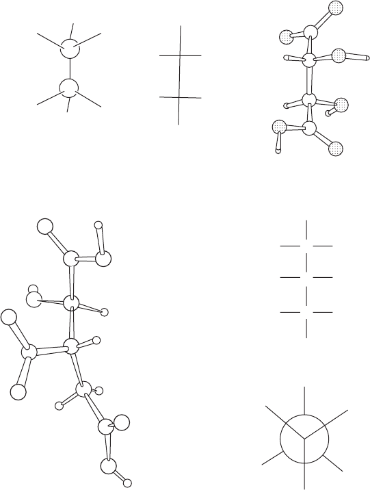
Fig. 10.6 Absolute configurations of biological molecules.
(a) Absolute configuration of (+)-tartaric acid (dextrorotatory tartaric acid). Note that
in the actual structure (right) the chain of four carbon atoms has effectively a planar
zigzag arrangement. In the formula on the left, and by convention in all “Fischer
formulas,” vertical carbon chains are represented as planar but with successive
bonds always directed into the page. Thus, in the formulas in the center and left
here, the lower half of the molecule has been rotated 180
◦
relative to the upper
half as compared with the actual structure. This affects the conformation but
not the absolute configuration of the molecule. The conformation of tartaric acid
illustrated on the left is a possible one for this molecule, but it is of higher energy
(because bonds are eclipsed) than that shown on the right, the conformation
observed in the crystals studied by Bijvoet et al. (1951). Still other conformers may
exist in solution or in other crystals.
(b) Absolute configuration of the potassium salt of (+)-isocitric acid (isolated
from the plant Bryophyllum calycinum). Fischer and Newman formulas are
shown. The correct designation of this enantiomer is 1R:2S-1-hydroxy-1,2,3-
propanetricarboxylate. The torsion angles are shown in Figure 12.5.
From Acta Crystallographica B24 (1968), p. 585, Figure 4.
160 Anomalous scattering and absolute configuration
fortunately that which was found was the one arbitrarily chosen from
the two possibilities half a century earlier by Fischer (Fischer, 1890,
1894), so the current organic chemistry textbooks did not have to be
changed. The absolute configurations of many other molecules have
been determined either by X-ray crystallographic methods or by chem-
ical correlation with those compounds for which the absolute configu-
ration had already been established (see Figure 10.6b). Values of anom-
alous scattering factors, especially those near the absorption edge, have
been measured in detail with synchrotron radiation (see, for example,
Templeton et al., 1980).
But how can absolute configuration be represented? The R/S sys-
tem of doing this involved assigning a priority number to the atoms
around an asymmetric (carbon) atom so that atoms with greater atomic
number have the higher priority (Cahn et al., 1966). If two atoms have
the same priority, their substituents are considered until differentia-
tion of priorities can be established (otherwise, of course, the central
atom is not asymmetric). Then the structure is viewed with the atom
of lowest priority directly behind the central (carbon) atom and the
other substituents are examined. Then if the order of the substituents
going from highest to lowest priority is clockwise, the central atom
is designated R (Latin rectus, right). If it is anticlockwise, the central
atom is designated S (Latin sinister, left). As a result, once the absolute
configuration is established and each asymmetric tetrahedral atom has
an R or S designation, sufficient information is provided from these
designations to make it possible to build a model with this correct
absolute configuration.
The effect of anomalous scattering was used to solve the structure
of a small protein, crambin, containing 45 amino acid residues (and
which crystallized with 72 water and 4 ethanol molecules per protein
molecule) (Hendrickson and Teeter, 1981). The nearest absorption edge
of sulfur is at 5.02 Å, but for CuKα radiation, wavelength 1.5418 Å, the
values of f
and f
are 0.3 and 0.557, respectively, for sulfur. Pairs
of reflections [|F (hkl)| and |F(
¯
h
¯
k
¯
l)|] were measured to 1.5 Å resolution
(the crystals scatter to 0.88 Å resolution); sulfur atom positions were
calculated from Patterson maps with |ƒF|
2
. While it was necessary to
take into account possible errors in such measurements of the differ-
ences of two large numbers, it was, in fact, possible to determine the
positions of the three disulfide links (six sulfur atoms). The structure
was then determined from an analysis of the Fourier map calculated on
the heavy-atom parameters of the sulfur atoms together with a partial
knowledge of the amino acid sequence.
SAD and MAD phasing
The use of anomalous scattering in structural work has increased
recently since the advent of “tunable” synchrotron radiation—that is,
X rays whose wavelength may, within certain limits, be selected at
SAD and MAD phasing 161
F
hkl
F
hkl
F
hkl
Overall A
hkl
Overall A
hkl
Overall B
hkl
Overall B
hkl
H∆f”
H∆f”
G∆f”
B
B
B
No anomalous scattering
Anomalous scattering
A
A
F
hkl
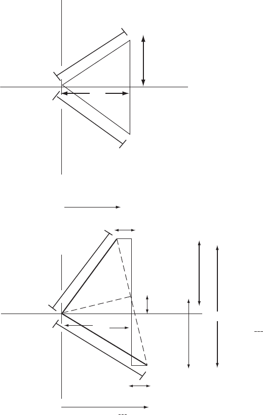
Fig. 10.7 Effects of anomalous scattering on F values.
The top diagram shows the structure factor vectors for F(hkl)andF(−h, −k, −l)inthe
absence of anomalous scattering and the lower diagram shows the effect of anom-
alous scattering on such a diagram. When there is anomalous scattering, A(hkl) =
A(−h, −k, −l). The same occurs for B values, as shown (see Chapter 5 and especially
Figure 5.1 for the fundamentals of such diagrams).
will. As a result it is possible to measure the diffraction pattern of a
macromolecular crystal with X-radiation of wavelengths near and also
far from the absorption edges of any anomalous scatterers present. Two
data sets can be measured, one near and one far from any absorption
edge of atoms in the crystal. The integrity of the crystal during so much
radiation exposure is maintained by flash freezing. For example, the
162 Anomalous scattering and absolute configuration
90º
-F
H⬘
F
P
a
P
F
H⬙
F
PH(+)
F
PH(–)
PH(–)
PH(+)
P
P
83º
0º
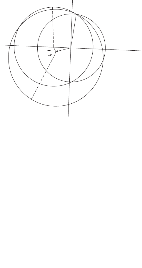
Fig. 10.8 Isomorphous replacement plus anomalous scattering (noncentrosymmetric).
The effect of anomalous scattering by an atom M, introduced to replace another atom,
may be used to resolve the ambiguity in phase-angle determination by the isomorphous
replacement method. The effect of anomalous scattering (Appendix 11) is to introduce
a phase shift, which means in effect, to change the atomic scattering factor of atom M
from a “real” quantity, f ,toa“complex”one,(f + f
)+if
. Suppose A and B refer to
that part of the structure that does not scatter anomalously, and A
and B
to the total
structure without any f
component; then A
= G( f + f
)+A and B
= H( f + f
)+B,
where G and H refer to geometric components for the anomalous scatterer, M. A
and
B
are components of the structure when anomalous scattering is present, and A
M
and
B
M
are components for the anomalous scatterer M alone. Then
A
M
= G( f + f
+if
)=A
M
+ Gf
+iGf
B
M
= H( f + f
+if
)=B
M
+ Hf
+iHf
Then, for the entire structure, including anomalous-scattering effects, we have
A
= A+ Gf + Gf
+iGf
= A
+iGf
B
= B + Hf + Hf
+iHf
= B
+iHf
As shown in Appendix 11, we have for the entire structure with anomalous scattering (by
separating and squaring the “real” and “imaginary” components)
F(hkl)
=
(A
− Hf
)
2
+(B
+ Gf
)
2
F(
¯
h
¯
k
¯
l)
=
(A
+ Hf
)
2
+(B
− Gf
)
2
(see Figure 10.4). Therefore, |F (hkl)| and |F (
¯
h
¯
k
¯
l)| are different; the intensity of each
reflection is measured to see which is the greater, as shown in Appendix 11.
A diagram to illustrate the determination of a phase angle for a macromolecule (that is,
·
P
) by the combination of isomorphous replacement and anomalous scattering is shown.
This diagram is constructed in a similar way to Figure 9.9. Circles of radii |F
PH(+)
| and
|F
PH(−)
| (for reflections of the heavy-atom derivative with indices h, k, l and −h, −k,
−l, respectively) are drawn with centers at −(F
H
+ F
H
)and−(F
H
− F
H
), respectively.
There are now three circles, radii |F
P
|, |F
PH(+)
|,and|F
PH(−)
|, and these intersect at a
phase angle of ·
P
(83
◦
). This is probably the phase angle of this reflection, h, k, l,forthe
native protein.
Sine-Patterson map 163
method of anomalous scattering may be combined with isomorphous
replacement in protein structure determination. Three data sets are
needed for this. One involves the protein crystal, and one a heavy-atom
derivative of this protein. A third data set is measured with X rays of a
wavelength that will cause maximal anomalous scattering by the heavy
atom. The heavy-atom position is located from the first two data sets,
and phase information is aided by the nonequivalent Friedel pairs of
Bragg reflections; these remove ambiguities in phase determination (see
Figures 10.7 and 10.8). This makes it possible to obtain approximate
phases from just one heavy-atom derivative.
The multiwavelength anomalous dispersion (MAD) method, sug-
gested by Wayne Hendrickson, is now a method of choice for phase
determination (Hendrickson, 1991). Generally proteins that are used
have been biologically expressed in a medium that contains only
selenomethionine. As a result the protein contains selenomethionine in
its sequence where methionine would normally be expected. Therefore
the strong anomalous signal of selenium can be used to derive phases.
X-ray diffraction data are measured near the absorption edge (where
f
has a maximum value, 1.15 electrons), and also at one or two wave-
lengths remote from any absorption edge. Only one crystal is needed,
and the data are generally measured at a synchrotron source.
In the single-wavelength anomalous dispersion (SAD) method, dif-
fraction data for one wavelength of radiation are measured on a heavy-
atom-containing protein, not necessarily near an absorption edge. Since
when heavy atoms are soaked into a crystal they may attach to various
side chains in a disordered manner, the strategy has been to generate a
protein with a heavy atom, such as that in iodophenylalanine, chemi-
cally incorporated into one amino acid (Dauter, 2004). The value of f
for iodine is 6.91 electrons for CuKα radiation. Alternatively, chromium
Kα radiation (Î =2.2909 Å) may be used to locate sulfur, which has
an anomalous signal ( f
) of 1.14 electrons for CrKα radiation, twice
that for CuKα radiation (Yang et al., 2003). This means that naturally
occurring protein side chains such as those of methionine or cysteine are
sufficient to provide phasing with CrKα radiation. Also, the data collec-
tion can be done in a laboratory and no tuning of radiation wavelength
is needed; a simple X-ray tube can be used. The phase ambiguity that
comes from measuring just one data set can be aided by direct methods
or by density modification. Obviously crystallographers are now exper-
imenting with different wavelengths of radiation and different possible
anomalous scatterers, and the trend is to study a crystal that contains
only the molecule under study (and not variations such as heavy-atom
derivatives).
Sine-Patterson map
A modified Patterson map can be used to determine the absolute con-
figuration of a structure provided Bijvoet pairs of reflections have been

164 Anomalous scattering and absolute configuration
measured and correctly indexed. The map is calculated with a func-
tion with {|F(hkl)|
2
+ |F (
¯
h
¯
k
¯
l)|
2
} as coefficients and a cosine term; this
gives peaks corresponding to Eqn. (9.1), that is, vectors between atoms.
Another function, with {|F(hkl)|
2
−|F (
¯
h
¯
k
¯
l)|
2
} as coefficients and a sine
term, known as the sine-Patterson map,
P
s
(u,v,w)=
1
V
c
all hkl
F (hkl)
2
−
F (
¯
h
¯
k
¯
l)
2
sin 2π(hu + kv + lw)
(10.8)
will have only vectors between anomalously and nonanomalously scat-
tering atoms, and these peaks are positive if the vector is from an anom-
alously scattering atom to a normal atom, and negative if the vector
is in the other direction. This map is asymmetric. Thus the absolute
configuration of the structure may be determined from such a map
(Okaya et al., 1955).
What effect does anomalous scattering have on the calculated elec-
tron density, since a term in the scattering factor now has an “imag-
inary” component? The answer is that the calculated electron den-
sity must be real, and, to obtain this, any effect of anomalous scat-
tering (which involves a complex scattering factor) must be removed
(as described in Appendix 11) (Patterson, 1963). This, of course, also
removes any means of distinguishing one enantiomorph from the other;
such information is contained only in the anomalous-scattering data.
Summary
If an atom in the crystal appreciably absorbs the X rays used, there will
be a phase change for the X rays scattered by that atom relative to the
phase of the X rays scattered by a nonabsorbing atom at the same site.
This phase change on absorption leads to anomalous scattering and,
for a noncentrosymmetric structure, results in differences in values of
|F (hkl)|
2
and |F(
¯
h
¯
k
¯
l)|
2
that are not found in the absence of anomalous
scattering. If the structure contains only one enantiomorph of a mole-
cule, its absolute configuration may be determined by a comparison of
the signs of the observed and calculated values of (|F (hkl)|
2
−|F (
¯
h
¯
k
¯
l)|
2
).

STRUCTURE
REFINEMENT
AND STRUCTURAL
INFORMATION
Part
III
This page intentionally left blank

Refinement of the trial
structure
11
When approximate positions have been determined for most, if not
all, of the atoms, it is time to begin the refinement of the structure.
In this process the atomic parameters are varied systematically so as
to give the best possible agreement of the observed structure factor
amplitudes (the experimental data) with those calculated for the pro-
posed trial structure. Common refinement techniques involve Fourier
syntheses and processes involving least-squares or maximum likeli-
hood methods. Although they have been shown formally to be nearly
equivalent—differing chiefly in the weighting attached to the experi-
mental observations—they differ considerably in manipulative details;
we shall discuss them separately here.
Many successive refinement cycles are usually needed before a struc-
ture converges to the stage at which the shifts from cycle to cycle
in the parameters being refined are negligible with respect to their
estimated errors. When least-squares refinement is used, the equations
are, as pointed out below, nonlinear in the parameters being refined,
which means that the shifts calculated for these parameters are only
approximate, as long as the structure is significantly different from the
“correct” one. With Fourier refinement methods, the adjustments in
the parameters are at best only approximate anyway; final parameter
adjustments are now almost always made by least squares, at least for
structures not involving macromolecules.
Fourier methods
As indicated earlier (Chapters 8 and 9, especially Figure 9.8 and the
accompanying discussion), Fourier methods are commonly used to
locate a portion of the structure after some of the atoms have been
found—that is, after at least a partial trial structure has been identified.
Initially, only one or a few atoms may have been found, or maybe an
appreciable fraction of the structure is now known. Once approximate
positions for at least some of the atoms in the structure are known, the
phase angles can be calculated. Then an approximate electron-density
167