Cook R.A., Stewart B. Colour Atlas of Anatomical Pathology
Подождите немного. Документ загружается.

ALIMENTARY
SYSTEM
Fig. 4.34
Fig. 4.31
Left-sided
congenital
diaphragmatic
hernia.
M/neonate. Abdominal contents
are
present
in the
thorax. There
is
hypoplasia
of the
left
lung
and the
mediastinum
is
pushed
to
the
right. Death occurred shortly
after
delivery.
Fig. 4.32 Duodenal
atresia.
Neonatal death.
The
pylorus
has
been
opened
and
ends blindly.
The
second part
of the
duodenum
has
been opened
and
terminates blindly
at
both
ends.
Fig. 4.33 Annular
pancreas.
M/4
weeks.
The
head
of the
pancreas
has
wrapped around
the
first
and
second parts
of the
duodenum, causing pyloric obstruction.
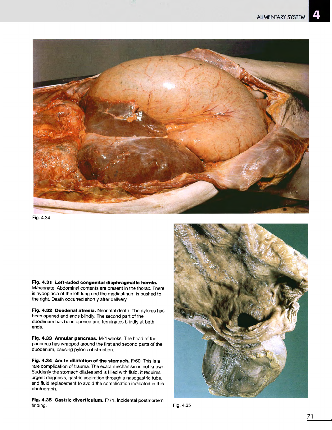
Fig. 4.34 Acute
dilatation
of the
stomach.
F/60. This
is a
rare
complication
of
trauma.
The
exact mechanism
is not
known.
Suddenly
the
stomach dilates
and is
filled with fluid.
It
requires
urgent diagnosis, gastric aspiration through
a
nasogastric tube,
and
fluid replacement
to
avoid
the
complication
indicated
in
this
photograph.
Fig. 4.35 Gastric
diverticulum.
F/71. Incidental postmortem
finding. Fig. 4.35
71
ALIMENTARY
SYSTEM
Fig.
4.37
72
Fig.
4.36
ALIMENTARY
SYSTEM
Fig.
4.38
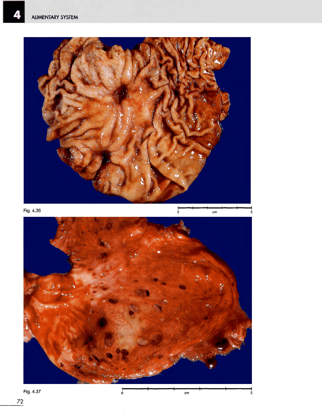
Fig. 4.36
Chronic
peptic
ulcer.
M/44.
A
partial gastrectomy
was
performed because
of
haematemesis. There
was a
bleeding
artery
in the
base
of the
ulcer.
Fig. 4.37
Acute
gastric
erosions.
M/78. These occurred just
prior
to
death.
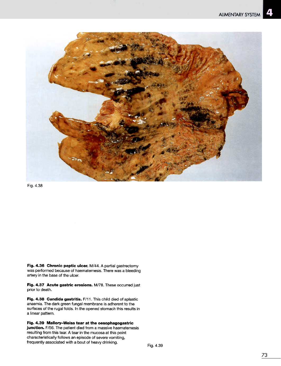
Fig. 4.38
Candida
gastritis.
F/11. This child died
of
aplastic
anaemia.
The
dark green fungal membrane
is
adherent
to the
surfaces
of the
rugal folds.
In the
opened stomach this results
in
a
linear pattern.
Fig. 4.39
Mallory-Weiss
tear
at the
oesophagogastric
junction.
F/56.
The
patient died from
a
massive haematemesis
resulting from this tear.
A
tear
in the
mucosa
at
this point
characteristically follows
an
episode
of
severe
vomiting,
frequently associated with
a
bout
of
heavy
drinking.
Fig.
4.39
73
ALIMENTARY
SYSTEM
Fig. 4.40
Fig. 4.41
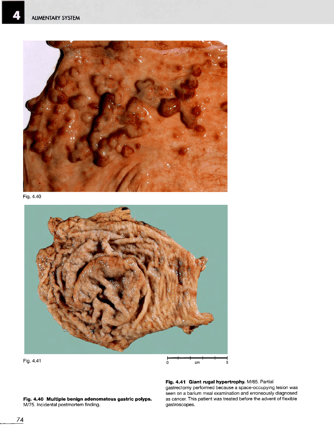
Fig. 4.40
Multiple
benign
adenomatous
gastric
polyps.
M/75. Incidental postmortem finding.
Fig. 4.41
Giant
rugal
hypertrophy.
M/65. Partial
gastrectomy performed because
a
space-occupying lesion
was
seen
on a
barium meal examination
and
erroneously diagnosed
as
cancer. This patient
was
treated before
the
advent
of
flexible
gastroscopes.
74
ALIMENTARY
SYSTEM
Fig.
4.43
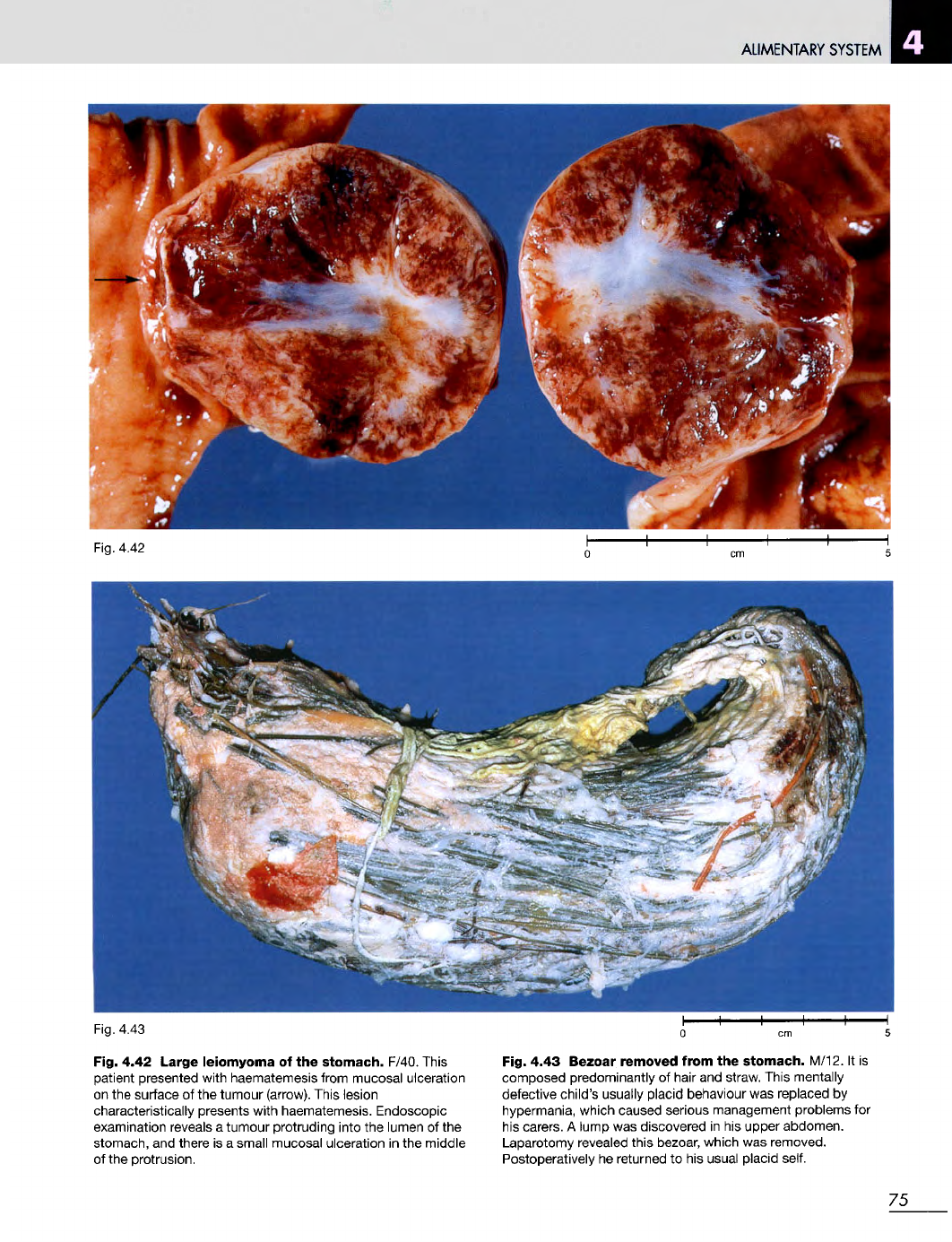
Fig. 4.42 Large
leiomyoma
of the
stomach.
F/40. This
patient presented with haematemesis from mucosal ulceration
on the
surface
of the
tumour (arrow). This lesion
characteristically presents with haematemesis. Endoscopic
examination reveals
a
tumour protruding into
the
lumen
of the
stomach,
and
there
is a
small mucosal ulceration
in the
middle
of
the
protrusion.
Fig. 4.43 Bezoar removed
from
the
stomach.
M/12.
It is
composed predominantly
of
hair
and
straw. This mentally
defective
child's usually placid behaviour
was
replaced
by
hypermania,
which caused serious management problems
for
his
carers.
A
lump
was
discovered
in his
upper abdomen.
Laparotomy revealed this bezoar, which
was
removed.
Postoperatively
he
returned
to his
usual placid self.
75
Fig.
4.42

ALIMENTARY
SYSTEM
Fig.
4.45
Fig.
4.46
76
Fig.
4.44
ALIMENTARY
SYSTEM
Fig.
4.47
Fig. 4.44 Polypoid adenocarcinoma
of the
stomach. F/86.
The
patient
was
treated
by
partial gastrectomy.
Fig. 4.45 Ulcerated adenocarcinoma
of
stomach. M/32.
The
patient
was
treated
by
partial gastrectomy.
The
probe
is in
the
pylorus, which
was
partially obstructed.
Fig. 4.46 Ulcerating adenocarcinoma
at the
oesophagogastric
junction.
M/50.
The
tumour
was
locally
resected.
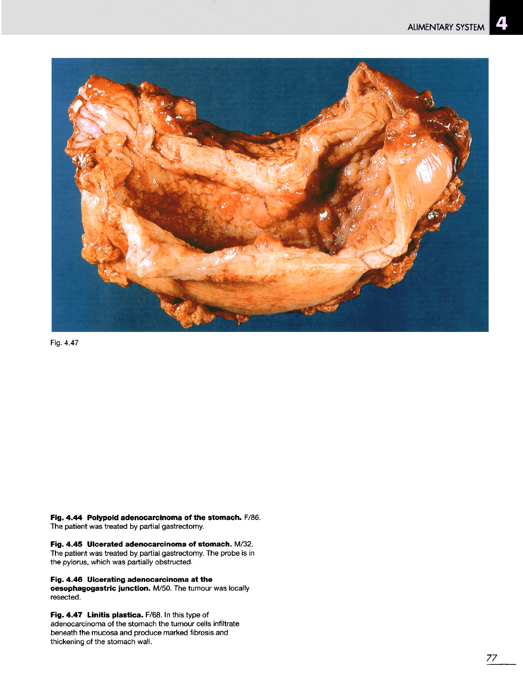
Fig. 4.47 Linitis plastica. F/68.
In
this type
of
adenocarcinoma
of the
stomach
the
tumour cells
infiltrate
beneath
the
mucosa
and
produce marked fibrosis
and
thickening
of the
stomach
wall.
77
ALIMENTARY
SYSTEM
Fig. 4.49
78
Fig. 4.48

ALIMENTARY
SYSTEM
Fig. 4.50
x16
Fig. 4.51
x16
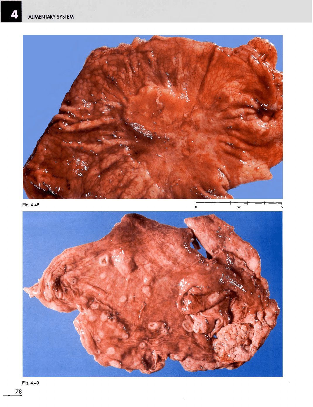
Fig. 4.48 Early
gastric
cancer
(superficial
adenocarcinoma
of the
stomach).
F/38.
The
patient
was
treated
by
partial
gastrectomy.
Note
the
firm,
plateau-like
area
on
the
stomach mucosa, with loss
of the
rugal folds.
The
abnormal area
in
such cases
may be
better seen
by
holding
the
specimen
up to the
light.
This
patient
had
rather vague,
non-specific
upper abdominal symptoms,
and
diagnosis
was
made
by
gastroscopy
and
biopsy.
Fig. 4.49
Malignant
lymphoma
of the
stomach. M/60.
Note
the
multiple
areas
of
creamy tumour
on the
stomach
mucosa.
The
mucosa
is
ulcerated
over
some
of the
deposits.
Fig. 4.50
Normal
small
intestinal
mucosa.
F/66. This tissue
was
obtained
by
means
of a
small bowel biopsy capsule
and
was
examined under
a
dissecting microscope.
The
intestinal
villi
appear
as
fingers, leaves
and
ridges. This
is a
very
much bigger
piece
of
tissue than that obtained
by
present-day fibreoptic
flexible
endoscopes.
Fig. 4.51
Flat
mucosa
(total
villus
atrophy)
in
coeliac
disease.
F/31.
The
patient
had had
lifelong
mild
diarrhoea.
The
jejunal
biopsy
was
performed because
her
child
was
diagnosed
as
having malabsorption syndrome caused
by
coeliac disease.
The
atrophic mucosal pattern
is
well seen
in
this large, capsule
biopsy. Using present-day fibreoptic flexible endoscopes,
the
flat mucosa
of
coeliac disease
can be
visualized
directly
(see
Figure
4.51
a).
Fig. 4.51
a
Coeliac
disease.
The
flat mucosal pattern
of the
duodenum seen through
a
gastroscope.
Fig. 4.51
a
79
ALIMENTARY SYSTEM
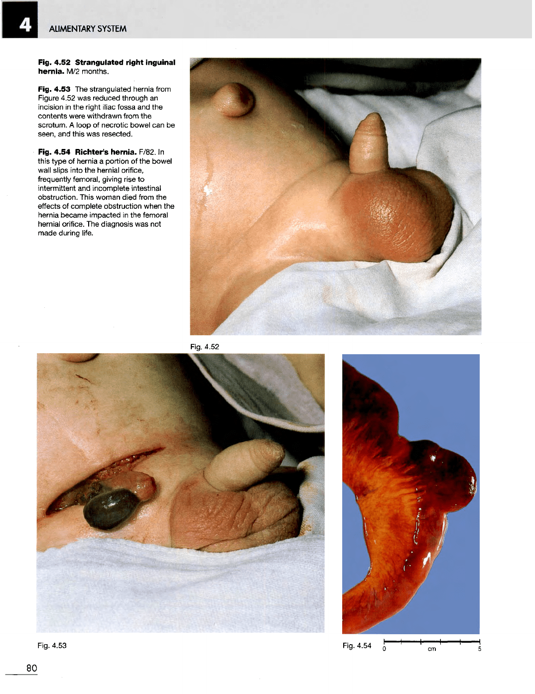
Fig. 4.52
Strangulated
right
inguinal
hernia.
M/2
months.
Fig. 4.53
The
strangulated hernia from
Figure 4.52
was
reduced through
an
incision
in the
right iliac fossa
and the
contents
were
withdrawn
from
the
scrotum.
A
loop
of
necrotic bowel
can be
seen,
and
this
was
resected.
Fig. 4.54
Richter's
hernia.
F/82.
In
this type
of
hernia
a
portion
of the
bowel
wall
slips into
the
hernial orifice,
frequently femoral, giving rise
to
intermittent
and
incomplete
intestinal
obstruction. This woman died from
the
effects
of
complete obstruction when
the
hernia became impacted
in the
femoral
hernial
orifice.
The
diagnosis
was not
made
during life.
Fig. 4.53 Fig. 4.54
80