Cook R.A., Stewart B. Colour Atlas of Anatomical Pathology
Подождите немного. Документ загружается.

NERVOUS SYSTEM
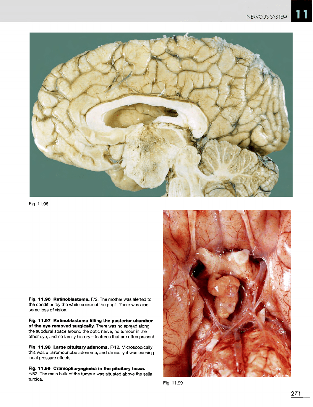
Fig. 11.98
Fig. 11.96 Retinoblastoma. F/2.
The
mother
was
alerted
to
the
condition
by the
white colour
of the
pupil.
There
was
also
some
loss
of
vision.
Fig. 11.97 Retinoblastoma
filling
the
posterior chamber
of
the eye
removed surgically.
There
was no
spread along
the
subdural space around
the
optic nerve,
no
tumour
in the
other
eye,
and no
family history
-
features that
are
often present.
Fig. 11.98 Large
pituitary
adenoma. F/12. Microscopically
this
was a
chromophobe adenoma,
and
clinically
it was
causing
local
pressure effects.
Fig. 11.99 Craniopharyngioma
in the
pituitary
fossa.
F/52.
The
main bulk
of the
tumour
was
situated
above
the
sella
turcica.
Fig. 11.99
271
NERVOUS SYSTEM
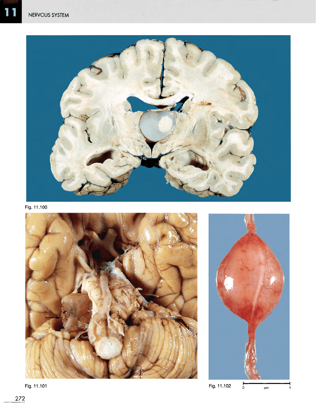
Fig.
11.100
Fig.
11.101
Fig.
11.102
272

NERVOUS SYSTEM
Fig.
11.103
Fig.
11.100
Colloid
cyst
of the
third
ventricle.
M/16.
The
patient presented with intermittent headaches, particularly
in the
head-down
position
(a
history which
is
characteristic
of
this
lesion).
Lumbar puncture
was
performed
in a
country hospital
and the
pressure shift resulted
in
herniation
of the
cerebellar
tonsils (coning)
and
death.
Fig.
11.101
Neurilemmoma
(acoustic neuroma) arising from
the
right eighth nerve. M/50.
An
incidental postmortem finding.
It
usually
presents
as
deafness. This
is a
very
large tumour. Since
the
introduction
of CT and MRI
scanning these tumours
are
identified
at a
very early stage, when they
are
small
and
easily
amenable
to
surgical removal.
Fig.
11.102
Neurilemmoma
arising
on a
peripheral
nerve.
M/20. Presented
as a
subcutaneous tumour which
was
excised.
Fig.
11.103
Huntington's
chorea.
F/54. Note
the
gross
atrophy
of
both
caudate nuclei
and
both
basal ganglia. There
is
marked compensatory hydrocephalus resulting from
the
cortical
and
basal ganglia atrophy.
273
NERVOUS SYSTEM
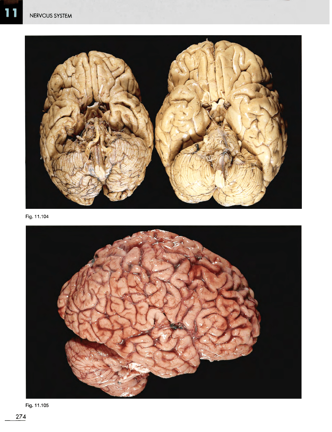
Fig. 11.104
Fig. 11.105
274

NERVOUS SYSTEM
Fig.
11.106 Fig. 11.108
Fig.
11.104
Alzheimer's
disease.
F/53.
The
atrophic brain
on the
left
is
compared with that
of a
normal 50-year-old
on the
right. Alzheimer's disease
is the
commonest form
of
presenile
dementia.
Fig.
11.105
Alzheimer's
disease.
F/57. Note
the
gross
atrophy
of the
cerebral gyri
and
widening
of the
sulci
in all
lobes,
but
particularly
in the
frontal lobe.
Fig.
11.106
Parkinson's
disease.
M/65.
The
transverse
section
of the
cerebral peduncles
in the
upper specimen shows
loss
of
pigment
in the
substantia nigra.
The
lower specimen
for
comparison shows normal substantia nigra.
Fig.
11.107
Atrophy
of the
posterior
columns
of the
spinal
cord.
Three conditions cause this appearance: tabes
dorsalis, vitamin
B
12
deficiency (subacute combined
degeneration)
and
Friedreich's ataxia.
Fig.
11.108
Central
pontine
myelinolysis.
M/61.
The
area
of
demyelination
is
shown
as a
brownish discoloration.
The
patient
was an
alcoholic. This condition
was
first described
in
alcoholics,
but it is now
known
to be
associated with
hypokalaemia, especially when this
has
been rapidly corrected.
Fig.
11.109
Multiple
sclerosis.
M/33. This diagnosis
was
made
6
years before death, when
the
patient first complained
of
ataxia.
The
ataxia
was
followed
by
increasing weakness
of the
legs
and
arms.
For a few
years before death from pneumonia
he
was
confined
to a
wheelchair. This brain slice shows
a
large grey
area
of
demyelination
in the
characteristic periventricular
distribution (A). There
is
another large area
of
demyelination
in
the
white matter
of the
temporal lobe (B). Fig. 11.109
275
Fig.
11.107
NERVOUS
SYSTEM
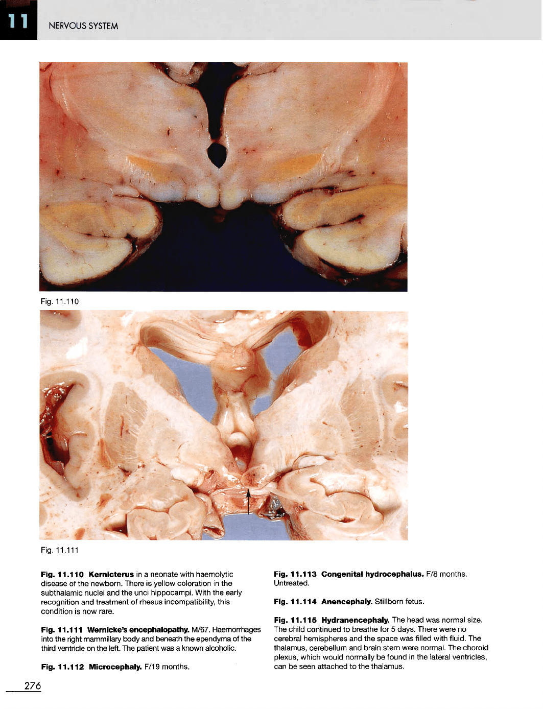
Fig.
11.111
Fig.
11.110
Kernicterus
in a
neonate with haemolytic
disease
of the
newborn. There
is
yellow coloration
in the
subthalamic nuclei
and the
unci hippocampi. With
the
early
recognition
and
treatment
of
rhesus incompatibility, this
condition
is now
rare.
Fig.
11.111
Wernicke's
encephalopathy.
M/67. Haemorrhages
into
the
right mammillary body
and
beneath
the
ependyma
of the
third ventricle
on the
left.
The
patient
was a
known alcoholic.
Fig.
11.112
Microcephaly.
F/19 months.
Fig.
11.113
Congenital
hydrocephalus.
F/8
months.
Untreated.
Fig.
11.114
Anencephaly.
Stillborn fetus.
Fig.
11.115
Hydranencephaly.
The
head
was
normal
size.
The
child continued
to
breathe
for 5
days. There were
no
cerebral hemispheres
and the
space
was
filled with fluid.
The
thalamus, cerebellum
and
brain stem were normal.
The
choroid
plexus, which would normally
be
found
in the
lateral ventricles,
can
be
seen attached
to the
thalamus.
276
Fig.
11.110
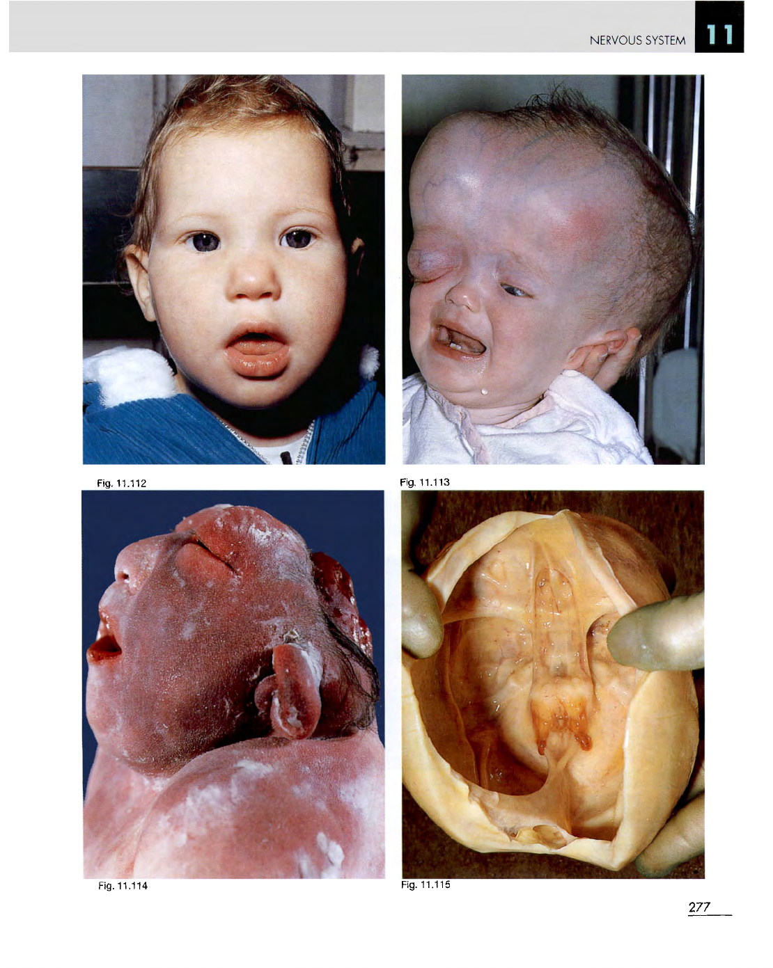
Fig.
11.114
Fig. 11.115
277
Fig. 11.112
Fig. 11.113
NERVOUS
SYSTEM
NERVOUS
SYSTEM
Fig. 11.116
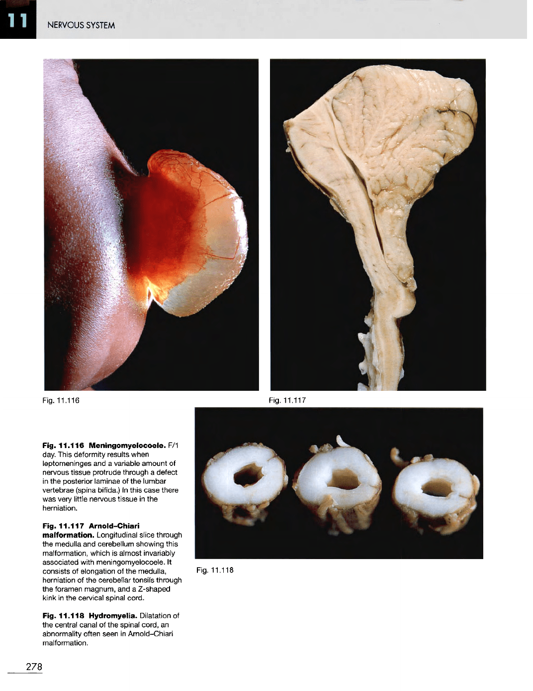
Fig.
11.116
Meningomyelocoele.
F/1
day.
This deformity results when
leptomeninges
and a
variable amount
of
nervous
tissue protrude through
a
defect
in
the
posterior laminae
of the
lumbar
vertebrae
(spina
bifida.)
In
this
case there
was
very
little nervous tissue
in the
herniation.
Fig.
11.117
Arnold-Chiari
malformation.
Longitudinal slice through
the
medulla
and
cerebellum showing this
malformation, which
is
almost invariably
associated with meningomyelocoele.
It
consists
of
elongation
of the
medulla,
herniation
of the
cerebellar tonsils through
the
foramen magnum,
and a
Z-shaped
kink
in the
cervical spinal cord.
Fig.
11.118
Hydromyelia. Dilatation
of
the
central canal
of the
spinal cord,
an
abnormality often seen
in
Arnold-Chiari
malformation.
Fig. 11.117
Fig.
11.118
278
NERVOUS
SYSTEM
Fig.
11.119
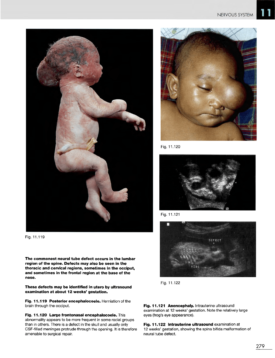
The
commonest
neural
tube
defect
occurs
in the
lumbar
region
of the
spine.
Defects
may
also
be
seen
in the
thoracic
and
cervical
regions,
sometimes
in the
occiput,
and
sometimes
in the
frontal
region
at the
base
of the
nose.
These
defects
may be
identified
in
utero
by
ultrasound
examination
at
about
12
weeks'
gestation.
Fig.
11.119
Posterior
encephalocoele.
Herniation
of the
brain
through
the
occiput.
Fig.
11.120
Large
frontonasal
encephalocoele.
This
abnormality appears
to be
more frequent
in
some racial groups
than
in
others. There
is a
defect
in the
skull
and
usually only
CSF-filled
meninges protrude through
the
opening.
It is
therefore
amenable
to
surgical repair.
Fig. 11.122
Fig.
11.121
Anencephaly.
Intrauterine ultrasound
examination
at 12
weeks' gestation. Note
the
relatively large
eyes
(frog's
eye
appearance).
Fig.
11.122
Intrauterine
ultrasound
examination
at
12
weeks' gestation, showing
the
spina bifida malformation
of
neural
tube defect.
279
Fig. 11.120
Fig. 11.121
NERVOUS SYSTEM
Fig. 11.124
280
Fig.
11.123
