Cook R.A., Stewart B. Colour Atlas of Anatomical Pathology
Подождите немного. Документ загружается.

NERVOUS
SYSTEM
Fig.
11.29
Fig.
11.30
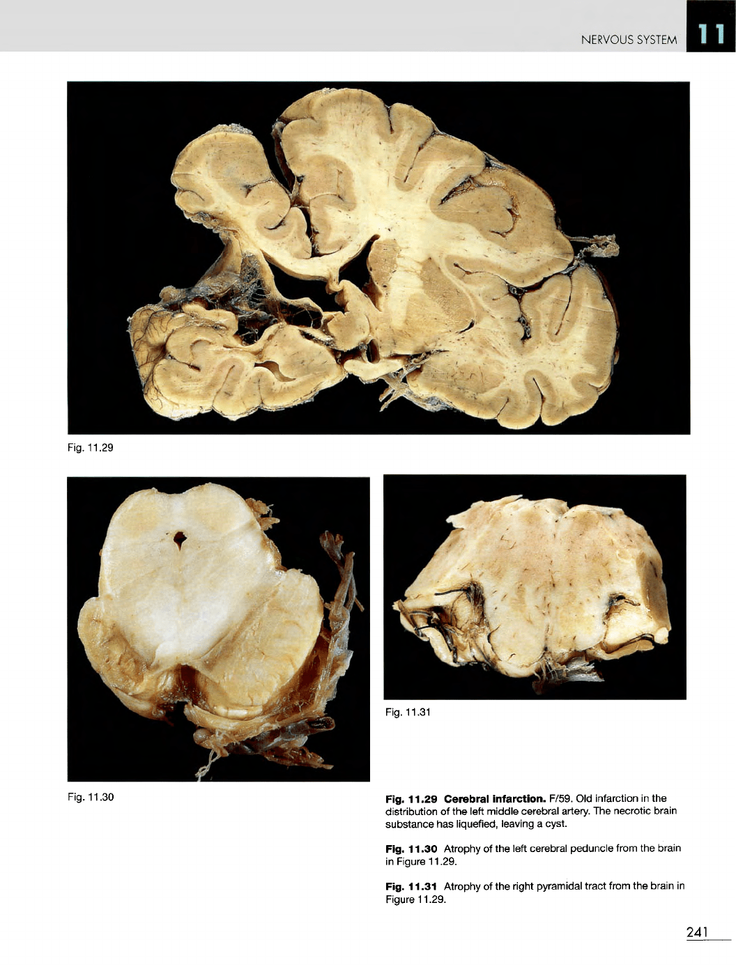
Fig. 11.29 Cerebral infarction. F/59.
Old
infarction
in the
distribution
of the
left middle cerebral artery.
The
necrotic brain
substance
has
liquefied, leaving
a
cyst.
Fig. 11.30
Atrophy
of the
left cerebral peduncle from
the
brain
in
Figure 11.29.
Fig. 11.31
Atrophy
of the
right pyramidal tract from
the
brain
in
Figure
11.29.
241
Fig.
11.31
NERVOUS
SYSTEM
Fig.
11.32
Fig.
11.33
Fig.
11.34
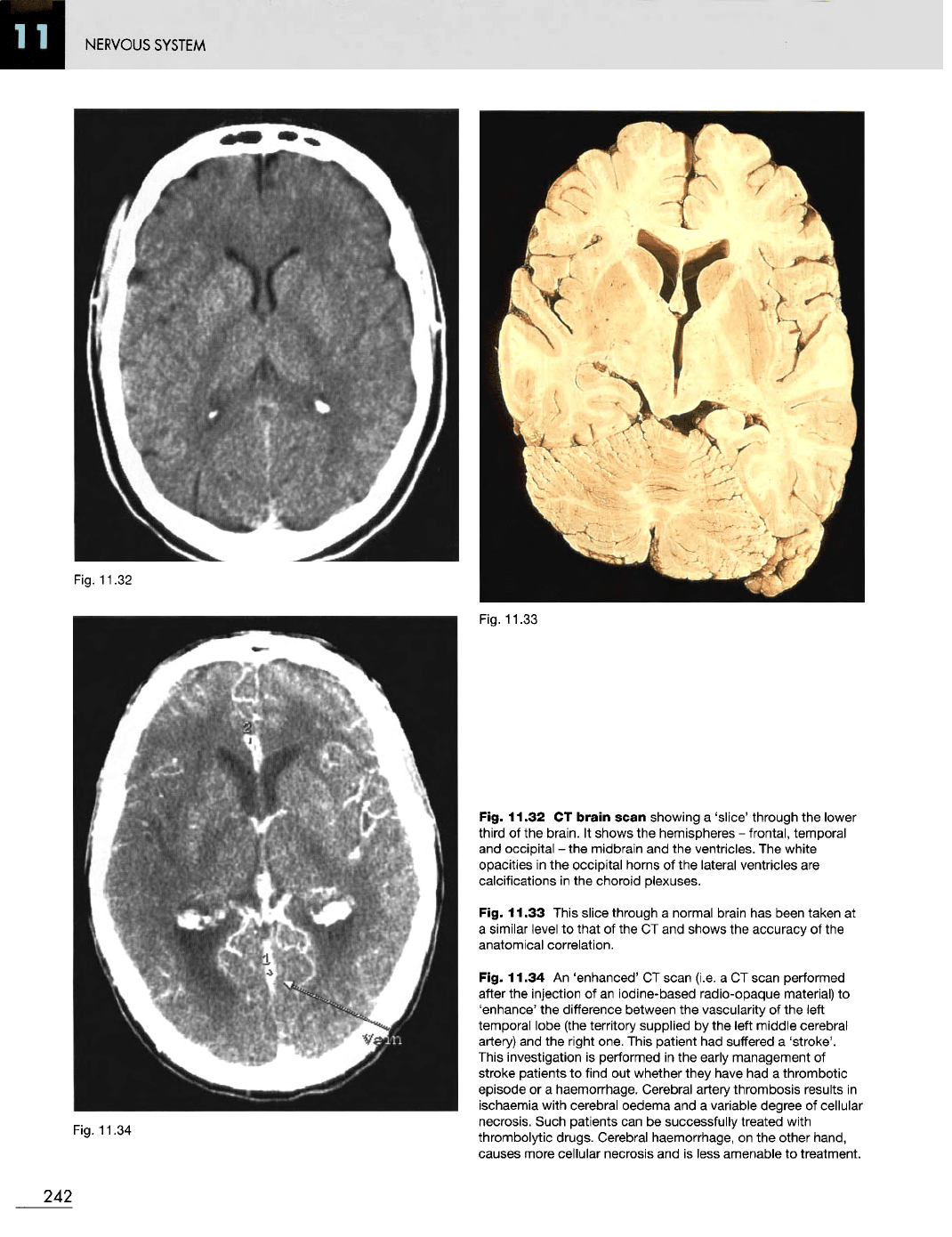
Fig. 11.32
CT
brain
scan showing
a
'slice' through
the
lower
third
of the
brain.
It
shows
the
hemispheres
-
frontal, temporal
and
occipital
- the
midbrain
and the
ventricles.
The
white
opacities
in the
occipital
horns
of the
lateral ventricles
are
calcifications
in the
choroid plexuses.
Fig. 11.33 This slice through
a
normal brain
has
been taken
at
a
similar
level
to
that
of the CT and
shows
the
accuracy
of the
anatomical correlation.
Fig. 11.34
An
'enhanced'
CT
scan (i.e.
a CT
scan performed
after
the
injection
of an
iodine-based radio-opaque material)
to
'enhance'
the
difference between
the
vascularity
of the
left
temporal lobe (the territory supplied
by the
left middle cerebral
artery)
and the
right one. This patient
had
suffered
a
'stroke'.
This
investigation
is
performed
in the
early
management
of
stroke
patients
to
find
out
whether they have
had a
thrombotic
episode
or a
haemorrhage. Cerebral artery thrombosis results
in
ischaemia with cerebral oedema
and a
variable degree
of
cellular
necrosis. Such patients
can be
successfully treated with
thrombolytic drugs. Cerebral haemorrhage,
on the
other hand,
causes more cellular necrosis
and is
less amenable
to
treatment.
242
NERVOUS
SYSTEM
Fig.
11.36
Fig.
11.35
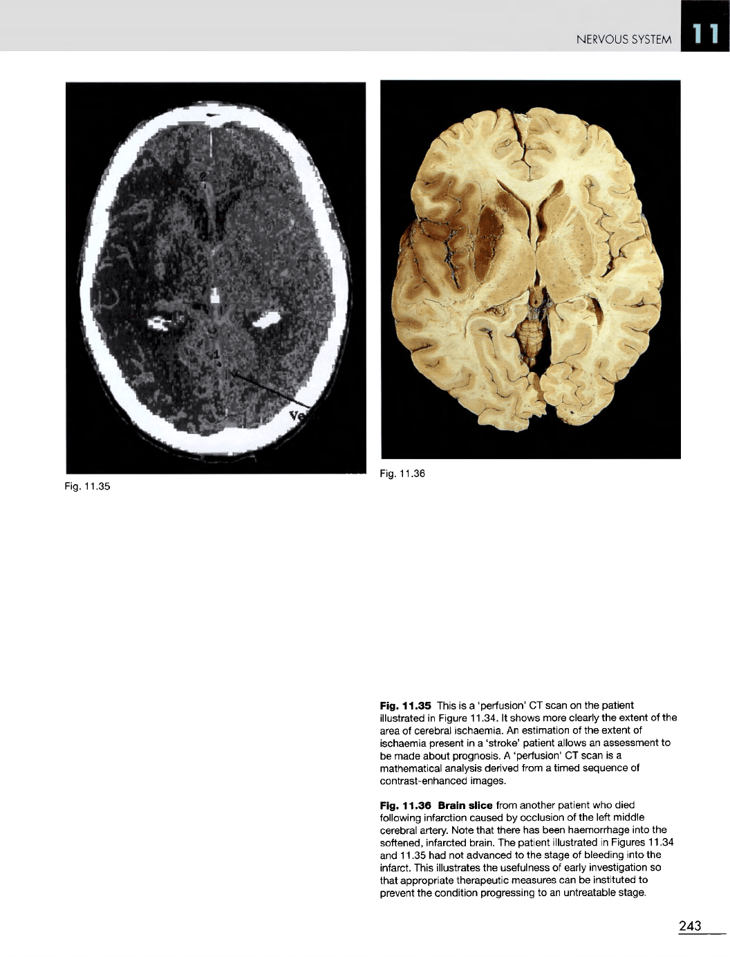
Fig. 11.35 This
is a
'perfusion'
CT
scan
on the
patient
illustrated
in
Figure 11.34.
It
shows more clearly
the
extent
of the
area
of
cerebral ischaemia.
An
estimation
of the
extent
of
ischaemia present
in a
'stroke' patient allows
an
assessment
to
be
made about prognosis.
A
'perfusion'
CT
scan
is a
mathematical analysis derived from
a
timed sequence
of
contrast-enhanced images.
Fig. 11.36
Brain
slice
from another patient
who
died
following infarction caused
by
occlusion
of the
left middle
cerebral artery. Note that there
has
been haemorrhage into
the
softened, infarcted brain.
The
patient illustrated
in
Figures 11.34
and
11.35
had not
advanced
to the
stage
of
bleeding into
the
infarct. This illustrates
the
usefulness
of
early investigation
so
that appropriate therapeutic measures
can be
instituted
to
prevent
the
condition
progressing
to an
untreatable stage.
243
NERVOUS
SYSTEM
Fig. 11.38
Fig. 11.39
244
Fig. 11.37
NERVOUS SYSTEM
Infarction
of
specific
areas
of the
brain
-
Figures
11.37
to
11.43
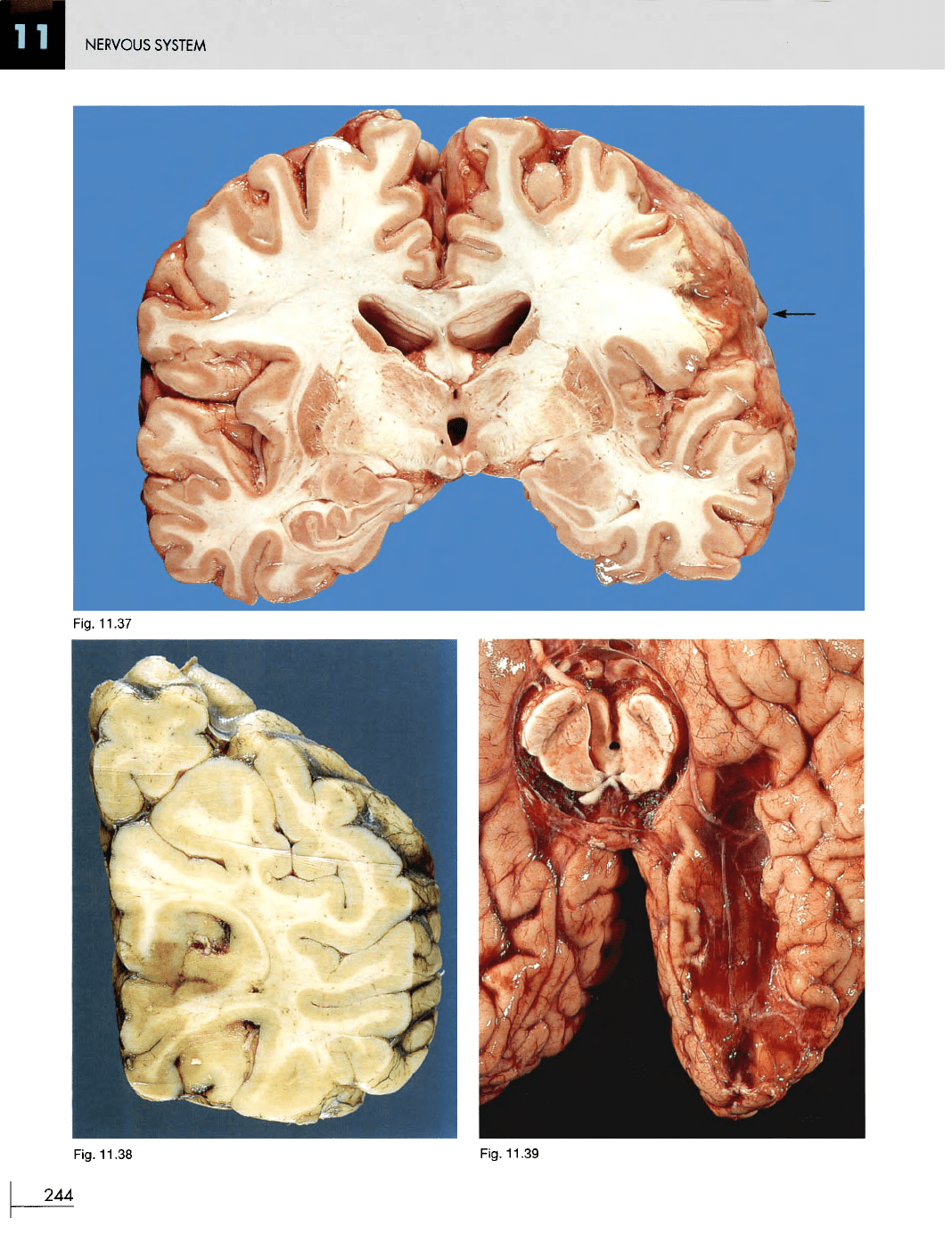
Fig. 11.37
Infarction
of the
motor
speech
area.
F/78.
The
patient
can
hear
and
understand,
but
cannot speak properly.
There
is
infarction
of a
localized area
of
cortex
and
underlying
white matter
at the
posteroinferior
end of the
left frontal lobe.
The
abnormal area shows
as a gap in the
surface grey matter,
with loss
of
some
of the
underlying white matter.
Fig. 11.38
Recent
infarction
of the
inferomedial
aspect
of
the
right
occipital
lobe
(the visual
cortex)
owing
to
occlusion
of the
right
posterior
cerebral
artery.
F/69. This
caused blurring
of her
vision.
The
infarcted area
is a
darker colour
than
the
adjacent normal cerebral cortex.
It is
oedematous,
and
softer
than
the
adjacent normal brain.
The
junction between
normal
and
infarcted brain
is
sometimes easier felt than seen.
Fig. 11.39
Cystic
area
in the
distribution
of the
left
posterior
cerebral
artery.
M/80.
The
result
of an old
infarct.
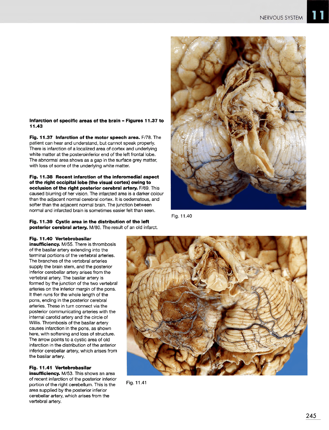
Fig. 11.40
Vertebrobasilar
insufficiency.
M/55. There
is
thrombosis
of
the
basilar artery extending into
the
terminal portions
of the
vertebral arteries.
The
branches
of the
vertebral arteries
supply
the
brain stem,
and the
posterior
inferior
cerebellar artery arises from
the
vertebral artery.
The
basilar artery
is
formed
by the
junction
of the two
vertebral
arteries
on the
inferior margin
of the
pons.
It
then runs
for the
whole length
of the
pons, ending
in the
posterior cerebral
arteries.
These
in
turn connect
via the
posterior communicating arteries with
the
internal
carotid artery
and the
circle
of
Willis.
Thrombosis
of the
basilar artery
causes infarction
in the
pons,
as
shown
here,
with softening
and
loss
of
structure.
The
arrow
points
to a
cystic
area
of old
infarction
in the
distribution
of the
anterior
inferior
cerebellar artery, which arises from
the
basilar artery.
Fig. 11.41
Vertebrobasilar
insufficiency.
M/53. This shows
an
area
of
recent infarction
of the
posterior inferior
portion
of the
right cerebellum. This
is the
area
supplied
by the
posterior inferior
cerebellar artery, which arises from
the
vertebral artery.
Fig. 11.40
Fig. 11.41
245
NERVOUS
SYSTEM
Fig.
11.42
Fig.
11.43
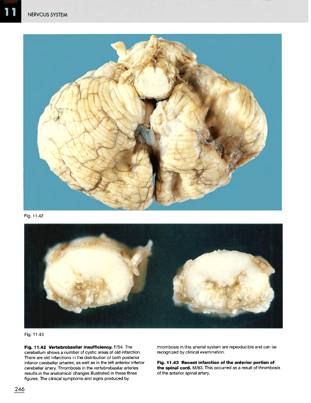
Fig. 11.42
Vertebrobasilar
insufficiency.
F/54.
The
cerebellum shows
a
number
of
cystic
areas
of old
infarction.
There
are old
infarctions
in the
distribution
of
both posterior
inferior
cerebellar arteries,
as
well
as in the
left anterior inferior
cerebellar
artery. Thrombosis
in the
vertebrobasilar arteries
results
in the
anatomical changes illustrated
in
these three
figures.
The
clinical symptoms
and
signs produced
by
thrombosis
in
this arterial system
are
reproducible
and can be
recognized
by
clinical examination.
Fig. 11.43
Recent
infarction
of the
anterior
portion
of
the
spinal
cord.
M/83. This occurred
as a
result
of
thrombosis
of
the
anterior spinal artery.
246
NERVOUS
SYSTEM
Fig. 11.44
Neonatal
intracranial
haemorrhage
resulting
from hypoxia.
M/3
weeks.
The
brain shows
an
almost complete
absence
of
development
of the
gyri, indicating marked prematurity.
Fig. 11.45 Tear
in the
tentorium
cerebelli
which resulted
from traumatic delivery
and
caused death from haemorrhage.
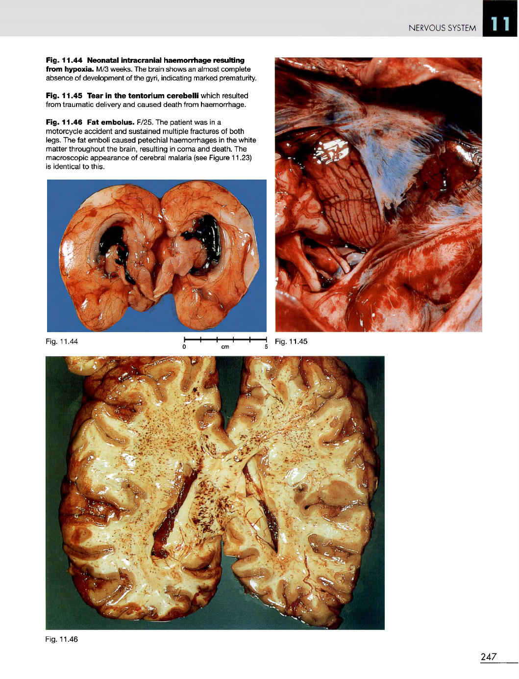
Fig. 11.46
Fat
embolus.
F/25.
The
patient
was in a
motorcycle accident
and
sustained multiple fractures
of
both
legs.
The fat
emboli caused petechial haemorrhages
in the
white
matter throughout
the
brain, resulting
in
coma
and
death.
The
macroscopic appearance
of
cerebral malaria (see Figure 11.23)
is
identical
to
this.
Fig. 11.46
247
Fig.
11.44
Fig. 11.45
NERVOUS
SYSTEM
Fig. 11.48
248
Fig. 11.47
NERVOUS
SYSTEM
Fig.
11.49
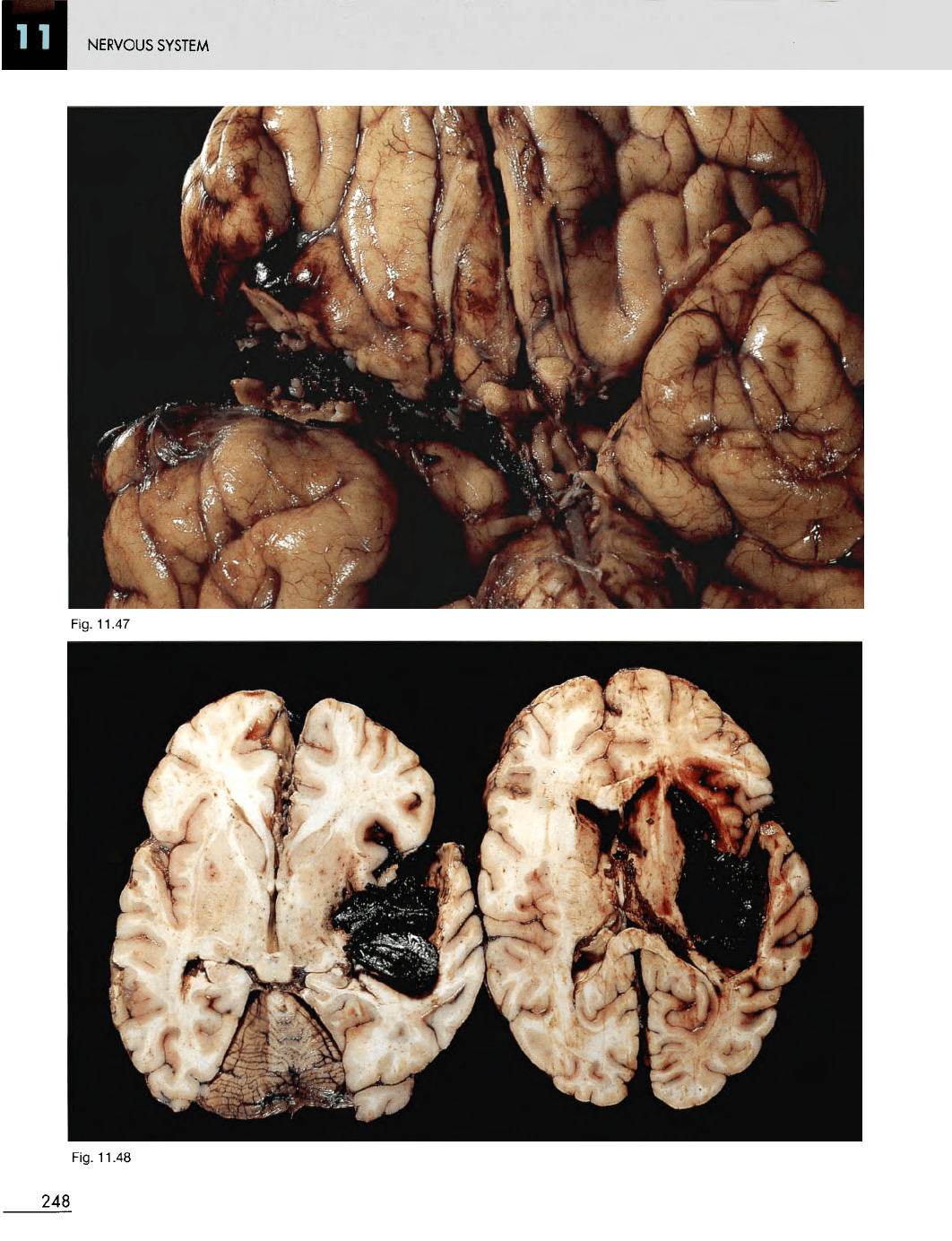
Fig. 11.47
Mycotic
embolus
occluding
the
right middle
cerebral
artery. M/13 with bacterial endocarditis.
Fig. 11.48
Cut
slices
of the
brain
in
Figure 11.47 showing
intracerebral haemorrhage resulting from rupture
of the
mycotic
aneurysm
that developed
in the
middle cerebral artery.
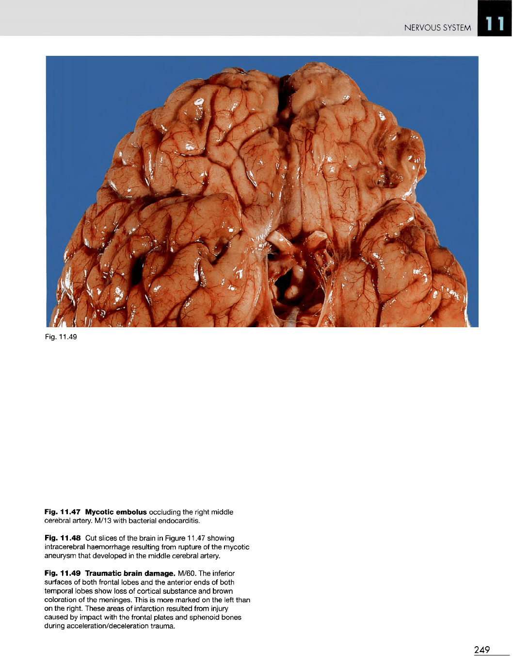
Fig. 11.49
Traumatic
brain
damage.
M/60.
The
inferior
surfaces
of
both frontal lobes
and the
anterior ends
of
both
temporal lobes show loss
of
cortical substance
and
brown
coloration
of the
meninges. This
is
more marked
on the
left
than
on the
right. These areas
of
infarction resulted from injury
caused
by
impact with
the
frontal plates
and
sphenoid bones
during acceleration/deceleration trauma.
249
NERVOUS
SYSTEM
Fig.
11.51
250
Fig.
11.50
Fig.
11.52