Cook R.A., Stewart B. Colour Atlas of Anatomical Pathology
Подождите немного. Документ загружается.

NERVOUS
SYSTEM
Fig. 11.75
Fig. 11.76 Fig. 11.77
The
following
clinical
examples
show
how
intracerebral
lesions
can be
localized
by the
clinical
effects
caused
by
pressure
on, or
stretching
of,
cranial
nerves.
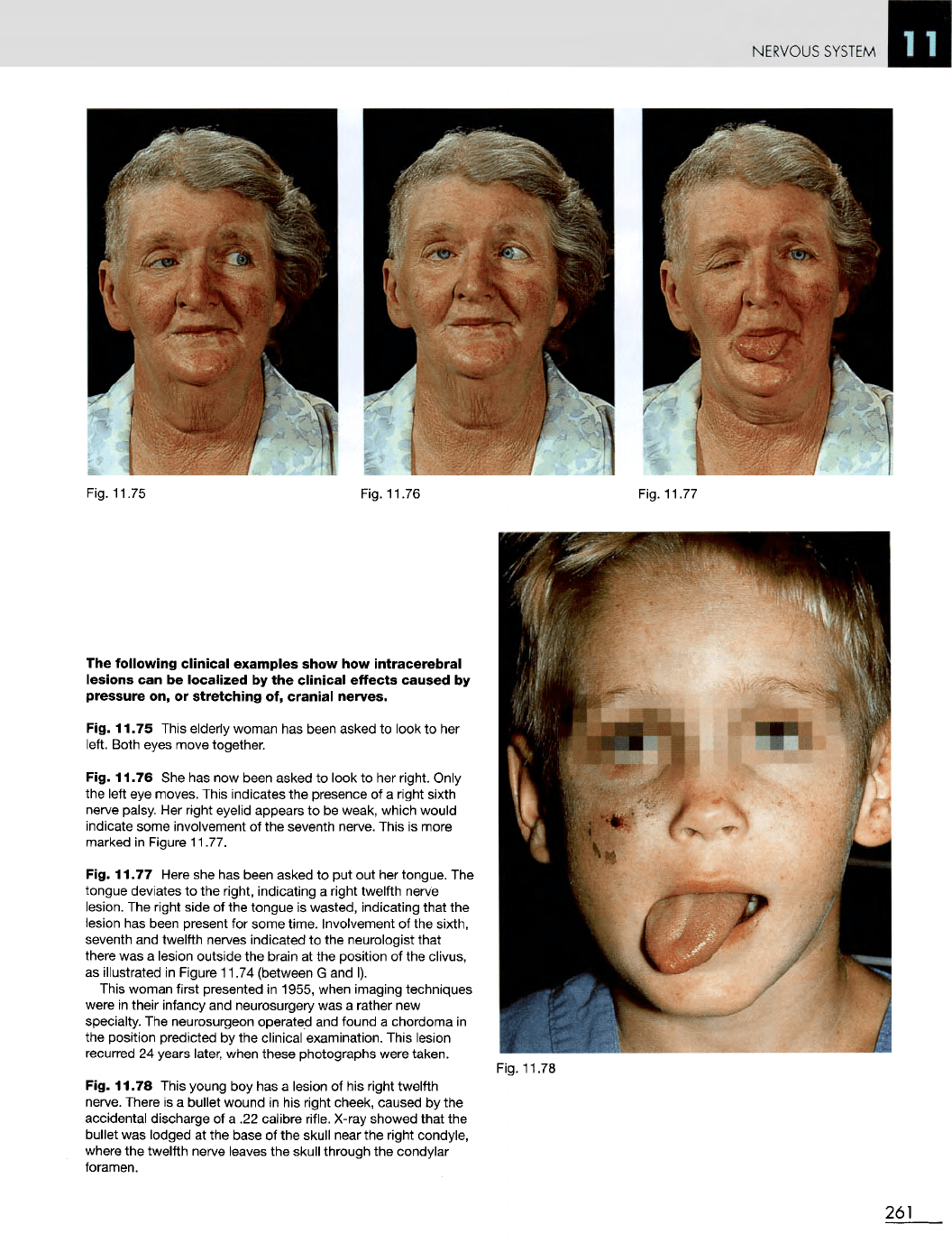
Fig. 11.75 This elderly woman
has
been asked
to
look
to her
left.
Both eyes move together.
Fig. 11.76
She has now
been asked
to
look
to her
right. Only
the
left
eye
moves. This indicates
the
presence
of a
right sixth
nerve
palsy.
Her
right eyelid appears
to be
weak, which would
indicate some involvement
of the
seventh nerve. This
is
more
marked
in
Figure 11.77.
Fig. 11.77 Here
she has
been asked
to put out her
tongue.
The
tongue deviates
to the
right, indicating
a
right twelfth nerve
lesion.
The
right side
of the
tongue
is
wasted, indicating
that
the
lesion
has
been present
for
some time. Involvement
of the
sixth,
seventh
and
twelfth nerves indicated
to the
neurologist
that
there
was a
lesion outside
the
brain
at the
position
of the
clivus,
as
illustrated
in
Figure 11.74 (between
G and I).
This
woman first presented
in
1955, when imaging techniques
were
in
their infancy
and
neurosurgery
was a
rather
new
specialty.
The
neurosurgeon operated
and
found
a
chordoma
in
the
position predicted
by the
clinical examination. This lesion
recurred
24
years later, when these photographs were taken.
Fig. 11.78 This young
boy has a
lesion
of his
right twelfth
nerve.
There
is a
bullet wound
in his
right cheek, caused
by the
accidental discharge
of a .22
calibre
rifle.
X-ray
showed that
the
bullet
was
lodged
at the
base
of the
skull near
the
right condyle,
where
the
twelfth nerve leaves
the
skull through
the
condylar
foramen.
Fig. 11.78
261
NERVOUS
SYSTEM
Fig. 11.79
Fig. 11.80
Fig. 11.79
Large
arteriovenous
malformation.
M/23.
It
involves
the
brain substance
in the
right temporal lobe
and
extends into
the
subarachnoid space.
The
bleeding caused
subarachnoid
and
intracerebral haemorrhage
and
death.
Fig. 11.80 Large
arteriovenous
malformation
in the
spinal
cord. M/31. Another cause
of
subarachnoid
haemorrhage.
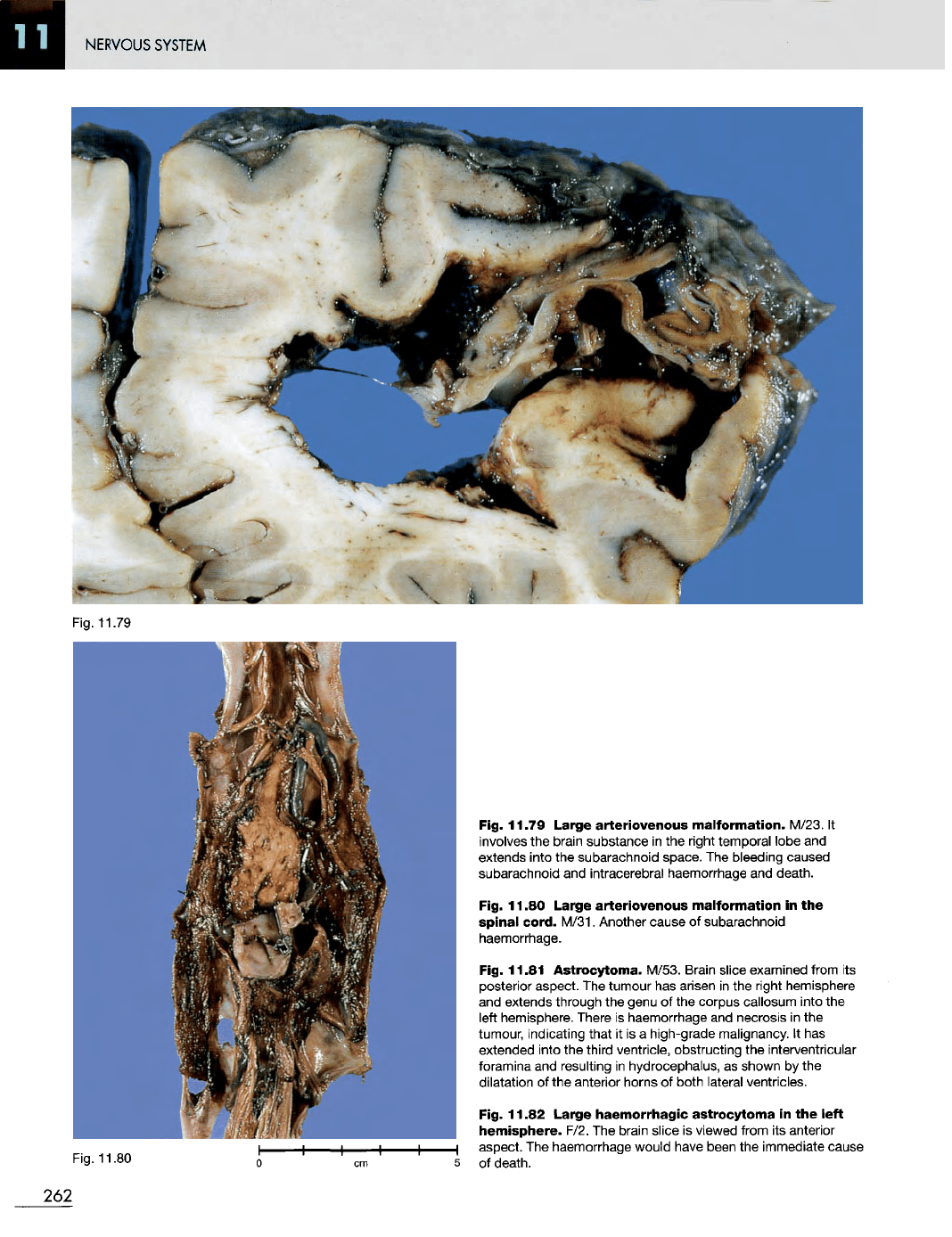
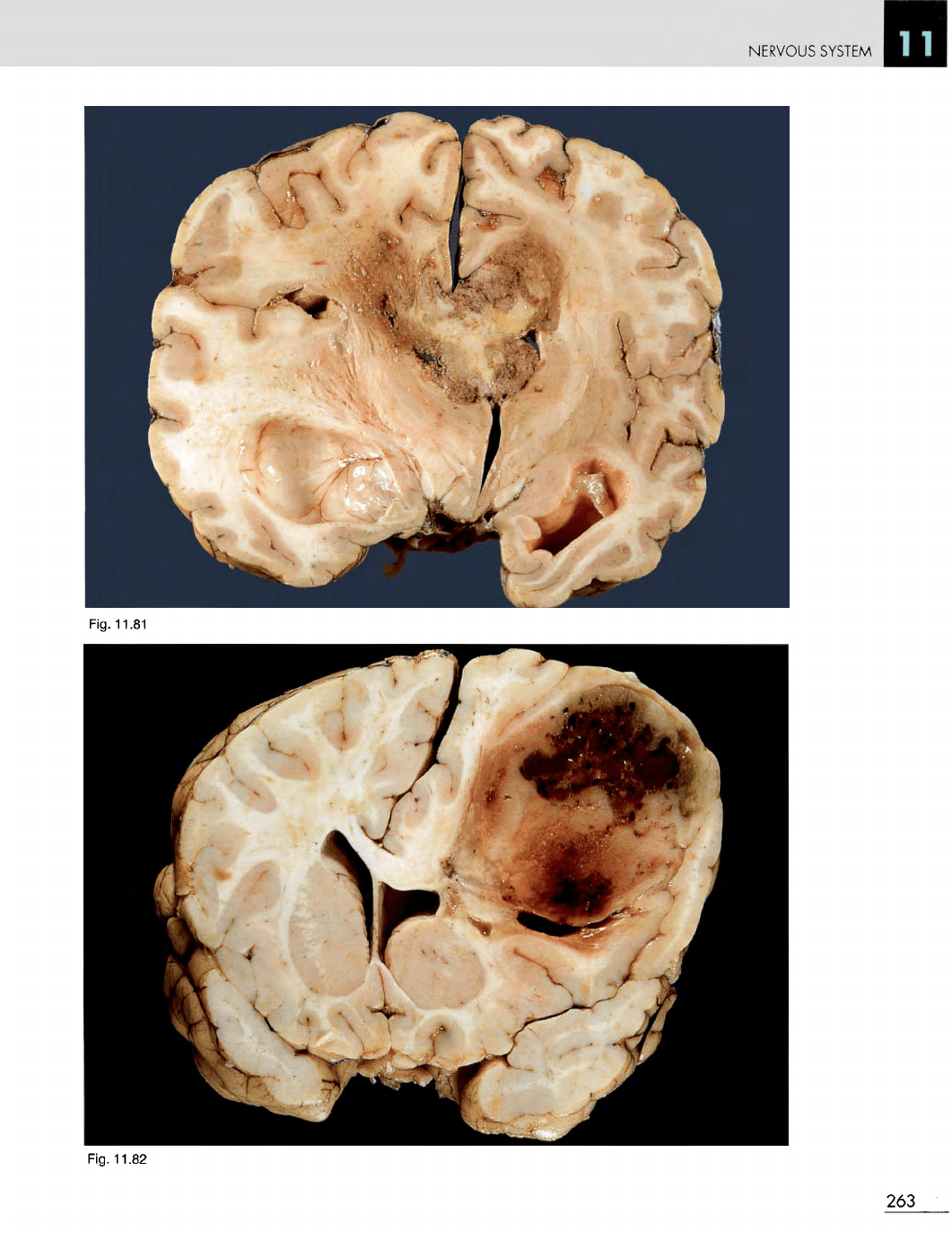
Fig. 11.81 Astrocytoma. M/53. Brain slice examined from
its
posterior aspect.
The
tumour
has
arisen
in the
right hemisphere
and
extends through
the
genu
of the
corpus callosum into
the
left
hemisphere. There
is
haemorrhage
and
necrosis
in the
tumour, indicating that
it is a
high-grade malignancy.
It has
extended into
the
third ventricle, obstructing
the
interventricular
foramina
and
resulting
in
hydrocephalus,
as
shown
by the
dilatation
of the
anterior horns
of
both lateral ventricles.
Fig. 11.82
Large
haemorrhagic
astrocytoma
in the
left
hemisphere.
F/2.
The
brain slice
is
viewed from
its
anterior
I
aspect.
The
haemorrhage would have been
the
immediate cause
5
of
death.
262
NERVOUS
SYSTEM
Fig.
11.82
263
Fig.
11.81
NERVOUS
SYSTEM
Fig.
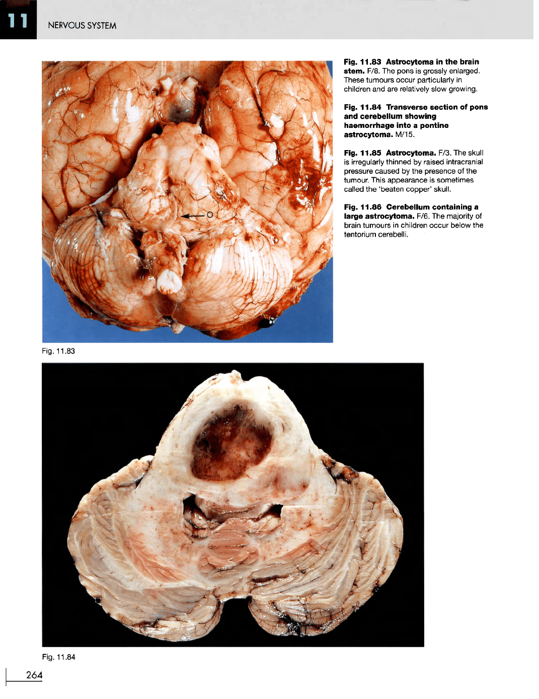
11.83
Astrocytoma
in the
brain
stem.
F/8.
The
pons
is
grossly enlarged.
These
tumours occur particularly
in
children
and are
relatively slow growing.
Fig.
11.84
Transverse section
of
pons
and
cerebellum
showing
haemorrhage
into
a
pontine
astrocytoma. M/15.
Fig.
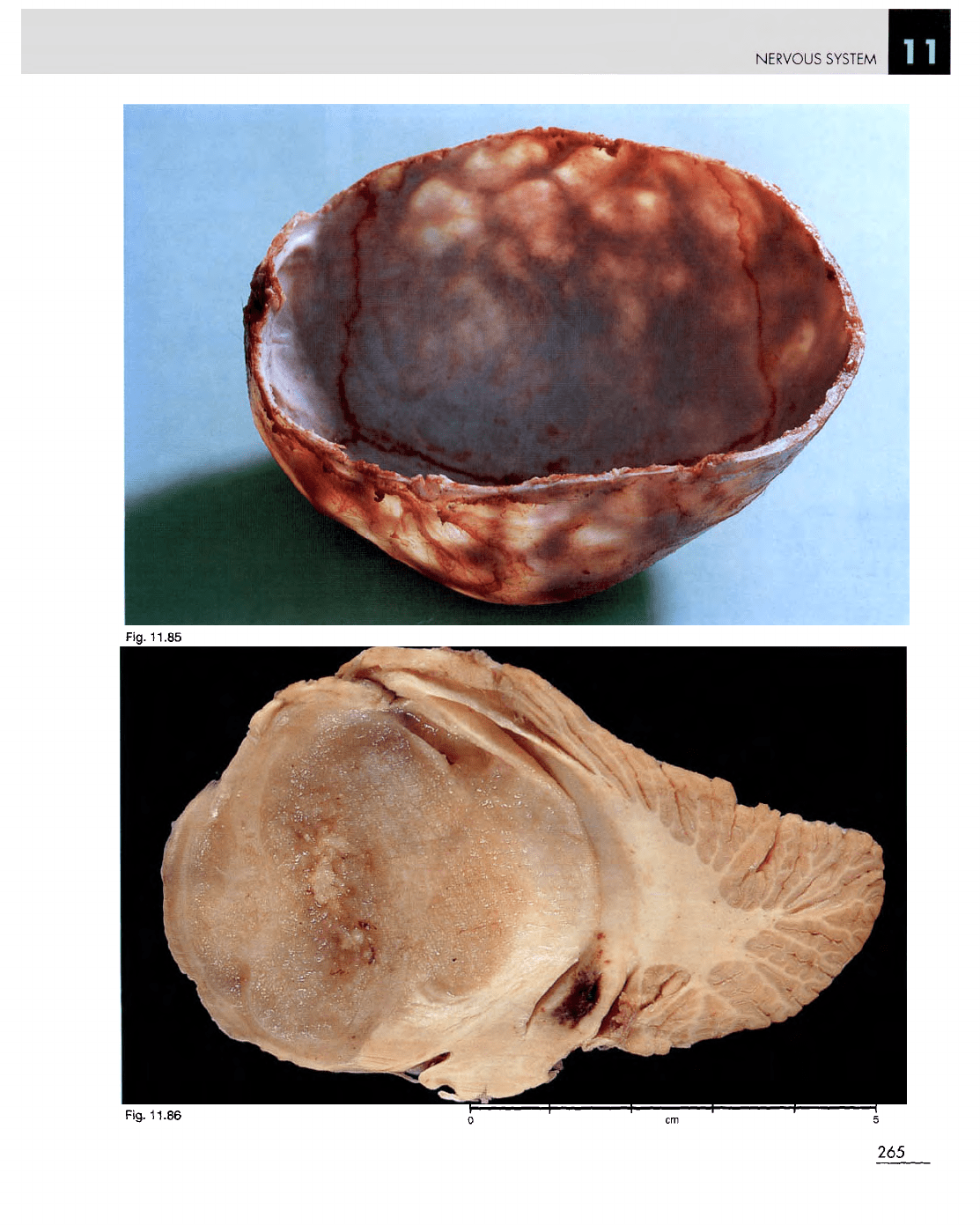
11.85 Astrocytoma. F/3.
The
skull
is
irregularly thinned
by
raised intracranial
pressure
caused
by the
presence
of the
tumour. This appearance
is
sometimes
called
the
'beaten copper' skull.
Fig.
11.86
Cerebellum
containing
a
large
astrocytoma.
F/6.
The
majority
of
i
brain tumours
in
children occur below
the
j
tentorium
cerebelli.
Fig.
11.83
Fig.
11.84
264
NERVOUS
SYSTEM
Fig.
11.86
265
Fig.
11.85
NERVOUS
SYSTEM
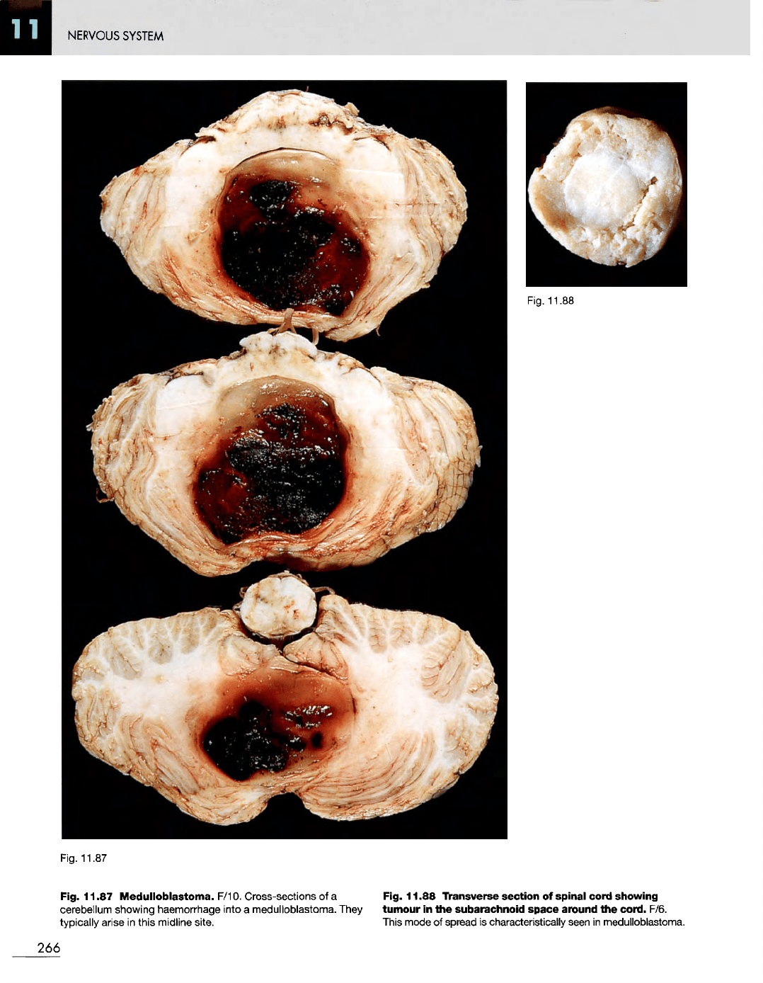
Fig. 11.87
Fig. 11.87
Medulloblastoma.
F/10. Cross-sections
of a
Fig. 11.88 Transverse section
of
spinal
cord showing
cerebellum showing haemorrhage into
a
medulloblastoma. They
tumour
in the
subarachnoid
space around
the
cord. F/6.
typically
arise
in
this midline site. This mode
of
spread
is
characteristically
seen
in
medulloblastoma.
266
Fig. 11.88
NERVOUS
SYSTEM
Fig.
11.89
Fig. 11.90
Fig. 11.89 Ependymoma
in the
right
lateral
ventricle.
F/7.
The
tumour
is
well
circumscribed
and
apparently
confined
to the
ventricle.
It has
compressed
the CSF
pathways, causing
dilatation
of the
third ventricle
and the
left lateral ventricle.
The
macroscopic appearance
of
this
tumour suggests
an
ependymoma,
but
microscopic confirmation would
be
required.
Fig. 11.90
Secondary
tumour
in the
brain.
M/49.
Secondary tumour deposits
are
usually multiple
and
well
circumscribed. Pigmented secondaries indicate secondary
melanoma
or
secondaries from
a
teratoma
of the
testis
in a
male,
and in a
female secondary melanoma
or
secondary
choriocarcinoma. There
is
marked oedema
in the
right
hemisphere, indicating
the
presence
of
raised intracranial
pressure.
267
NERVOUS
SYSTEM
Fig.
11.91
Fig.
11.92
268
NERVOUS
SYSTEM
Fig.
11.93
Fig.
11.94
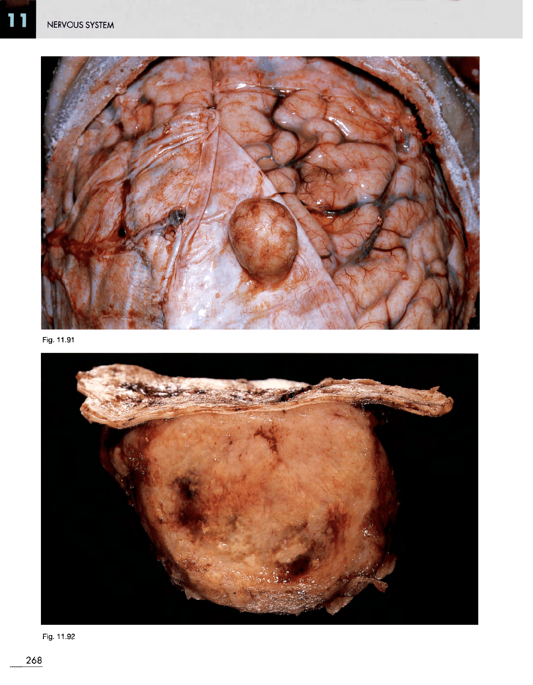
Fig. 11.91
Meningioma.
F/46. Small, asymptomatic tumour
attached
to the
dura.
Fig. 11.92 Transverse
section
of a
meningioma removed
surgically
together with
the
attached dura. M/78.
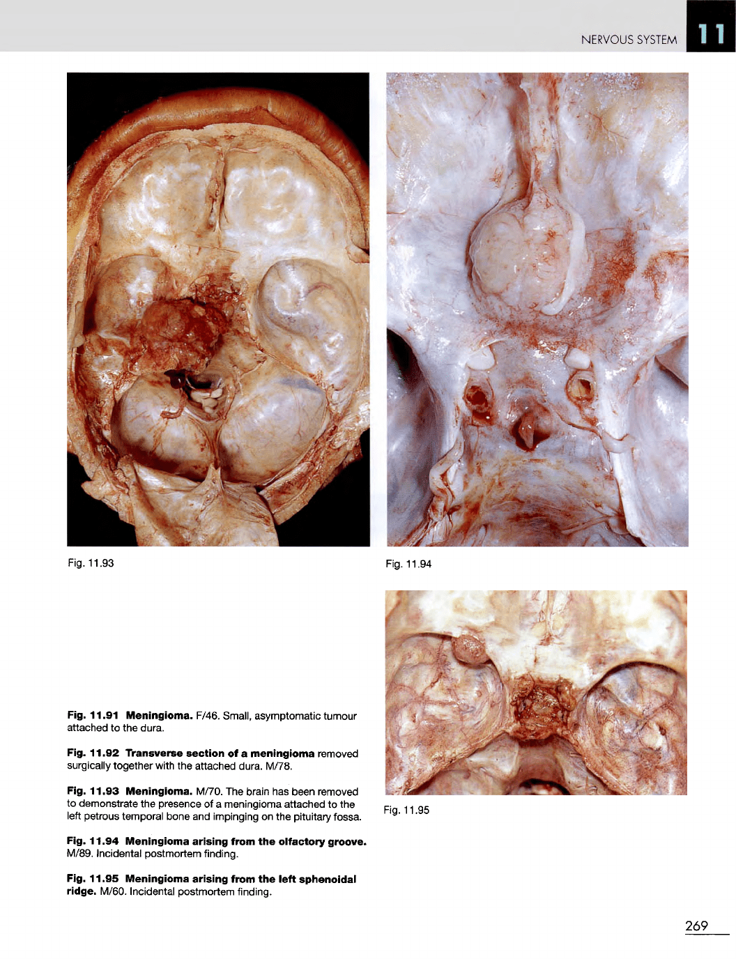
Fig. 11.93
Meningioma.
M/70.
The
brain
has
been removed
to
demonstrate
the
presence
of a
meningioma attached
to the
left
petrous temporal bone
and
impinging
on the
pituitary fossa.
Fig. 11.94
Meningioma
arising
from
the
olfactory groove.
M/89.
Incidental postmortem finding.
Fig. 11.95
Meningioma
arising
from
the
left
sphenoidal
ridge.
M/60. Incidental postmortem finding.
Fig.
11.95
269
NERVOUS
SYSTEM
Fig.
11.96
Fig.
11.97
270