Cook R.A., Stewart B. Colour Atlas of Anatomical Pathology
Подождите немного. Документ загружается.

RESPIRATORY
SYSTEM
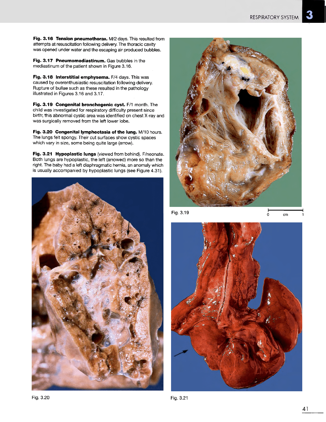
Fig. 3.16 Tension
pneumothorax.
M/2
days. This resulted from
attempts
at
resuscitation following delivery.
The
thoracic cavity
was
opened under water
and the
escaping
air
produced bubbles.
Fig. 3.17
Pneumomediastinum.
Gas
bubbles
in the
mediastinum
of the
patient shown
in
Figure 3.16.
Fig. 3.18
Interstitial
emphysema.
F/4
days. This
was
caused
by
overenthusiastic resuscitation following delivery.
Rupture
of
bullae such
as
these resulted
in the
pathology
illustrated
in
Figures 3.16
and
3.17.
Fig. 3.19
Congenital
bronchogenic
cyst.
F/1
month.
The
child
was
investigated
for
respiratory difficulty present since
birth;
this
abnormal
cystic area
was
identified
on
chest
X-ray
and
was
surgically removed from
the
left lower lobe.
Fig. 3.20
Congenital
lymphectasia
of the
lung.
M/10 hours.
The
lungs felt spongy. Their
cut
surfaces show cystic spaces
which vary
in
size, some being quite large (arrow).
Fig. 3.21
Hypoplastic
lungs
(viewed from behind). F/neonate.
Both lungs
are
hypoplastic,
the
left (arrowed) more
so
than
the
right.
The
baby
had a
left
diaphragmatic hernia,
an
anomaly which
is
usually accompanied
by
hypoplastic lungs (see Figure 4.31).
Fig. 3.20
Fig. 3.21
41
Fig. 3.19
RESPIRATORY
SYSTEM
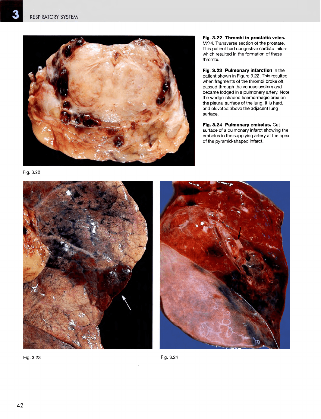
Fig. 3.22
Thrombi
in
prostatic
veins.
M/74. Transverse section
of the
prostate.
This
patient
had
congestive cardiac failure
which resulted
in the
formation
of
these
thrombi.
Fig. 3.23
Pulmonary
infarction
in the
patient shown
in
Figure 3.22. This resulted
when
fragments
of the
thrombi broke off,
passed through
the
venous system
and
became lodged
in a
pulmonary artery. Note
the
wedge-shaped haemorrhagic area
on
the
pleural surface
of the
lung.
It is
hard,
and
elevated above
the
adjacent lung
surface.
Fig. 3.24
Pulmonary
embolus.
Cut
surface
of a
pulmonary infarct showing
the
embolus
in the
supplying artery
at the
apex
of
the
pyramid-shaped infarct.
Fig. 3.22
Fig.
3.23
Fig. 3.24
42
RESPIRATORY
SYSTEM
Fig.
3.26
Fig.
3.25
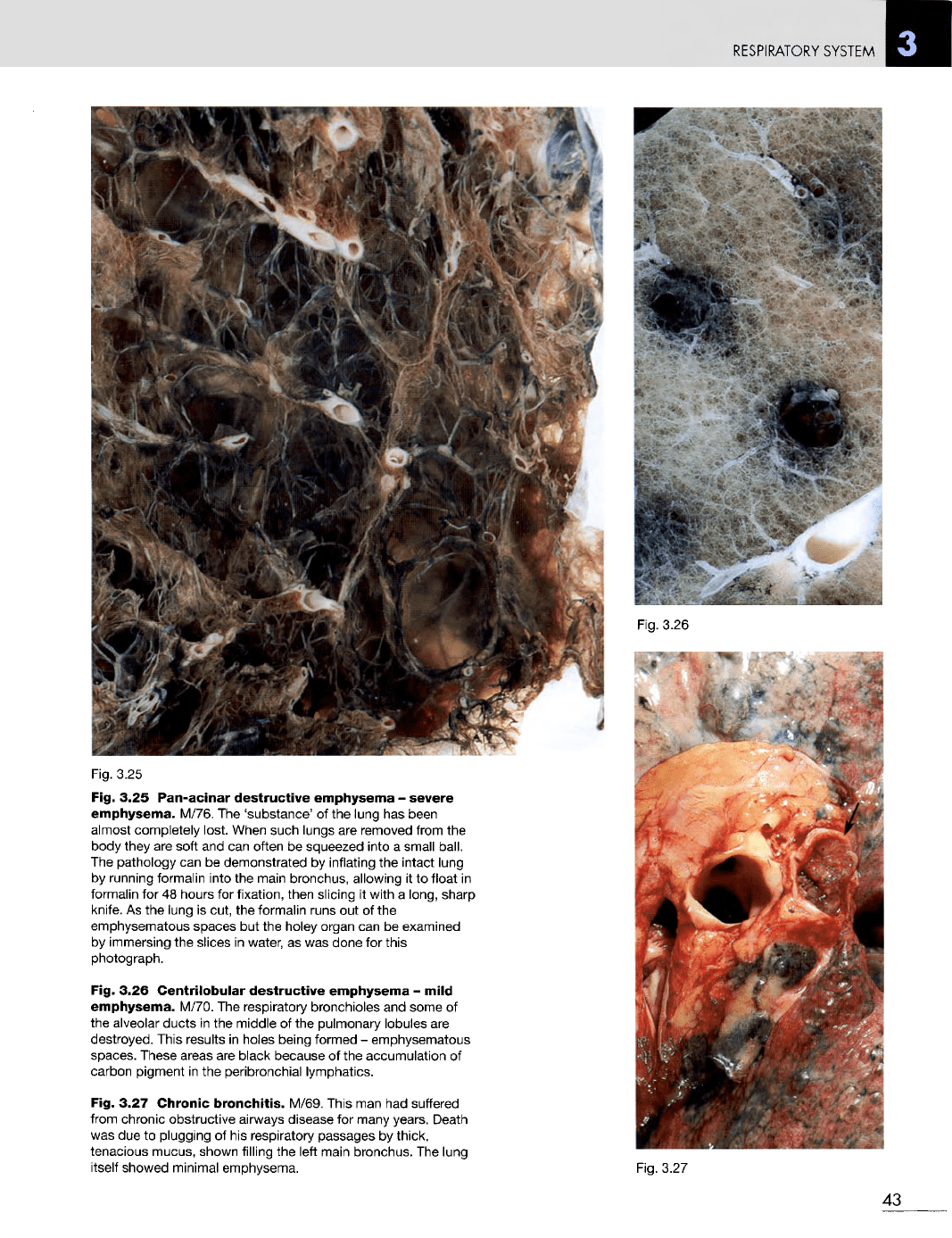
Fig. 3.25
Pan-acinar
destructive
emphysema
-
severe
emphysema.
M/76.
The
'substance'
of the
lung
has
been
almost completely lost. When such lungs
are
removed from
the
body they
are
soft
and can
often
be
squeezed into
a
small ball.
The
pathology
can be
demonstrated
by
inflating
the
intact lung
by
running formalin into
the
main bronchus, allowing
it to
float
in
formalin
for 48
hours
for
fixation, then slicing
it
with
a
long, sharp
knife.
As the
lung
is
cut,
the
formalin runs
out of the
emphysematous spaces
but the
holey organ
can be
examined
by
immersing
the
slices
in
water,
as was
done
for
this
photograph.
Fig. 3.26
Centrilobular
destructive
emphysema
-
mild
emphysema.
M/70.
The
respiratory bronchioles
and
some
of
the
alveolar ducts
in the
middle
of the
pulmonary lobules
are
destroyed.
This results
in
holes being formed
-
emphysematous
spaces. These areas
are
black because
of the
accumulation
of
carbon
pigment
in the
peribronchial lymphatics.
Fig. 3.27
Chronic
bronchitis.
M/69. This
man had
suffered
from
chronic obstructive airways disease
for
many years. Death
was
due to
plugging
of his
respiratory passages
by
thick,
tenacious mucus, shown filling
the
left
main bronchus.
The
lung
itself
showed minimal emphysema.
Fig.
3.27
43
RESPIRATORY
SYSTEM
Fig.
3.28
Fig.
3.29
0 cm
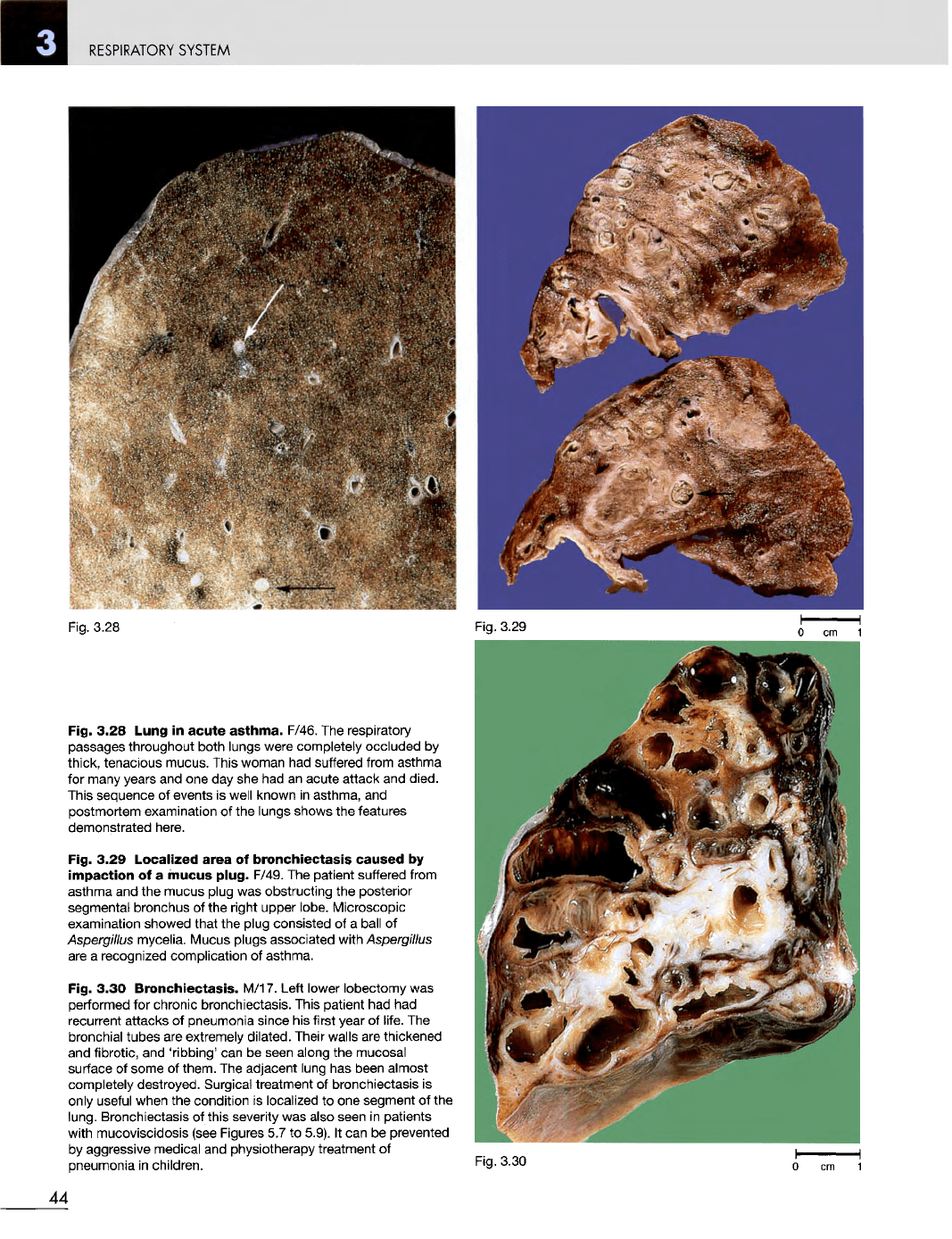
Fig. 3.28 Lung
in
acute
asthma.
F/46.
The
respiratory
passages throughout both lungs were completely occluded
by
thick, tenacious mucus. This woman
had
suffered from asthma
for
many years
and one day she had an
acute attack
and
died.
This
sequence
of
events
is
well known
in
asthma,
and
postmortem examination
of the
lungs shows
the
features
demonstrated here.
Fig. 3.29
Localized
area
of
bronchiectasis
caused
by
impaction
of a
mucus
plug.
F/49.
The
patient suffered from
asthma
and the
mucus plug
was
obstructing
the
posterior
segmental bronchus
of the
right upper lobe. Microscopic
examination showed that
the
plug consisted
of a
ball
of
Aspergillus
mycelia. Mucus plugs associated with
Aspergillus
are
a
recognized complication
of
asthma.
Fig. 3.30
Bronchiectasis.
M/17. Left lower lobectomy
was
performed
for
chronic bronchiectasis. This patient
had had
recurrent
attacks
of
pneumonia since
his
first year
of
life.
The
bronchial tubes
are
extremely
dilated.
Their walls
are
thickened
and
fibrotic,
and
'ribbing'
can be
seen along
the
mucosal
surface
of
some
of
them.
The
adjacent lung
has
been almost
completely destroyed. Surgical treatment
of
bronchiectasis
is
only
useful when
the
condition
is
localized
to one
segment
of the
lung. Bronchiectasis
of
this severity
was
also seen
in
patients
with
mucoviscidosis (see Figures
5.7 to
5.9).
It can be
prevented
by
aggressive medical
and
physiotherapy treatment
of
pneumonia
in
children.
Fig.
3.30
44
RESPIRATORY
SYSTEM
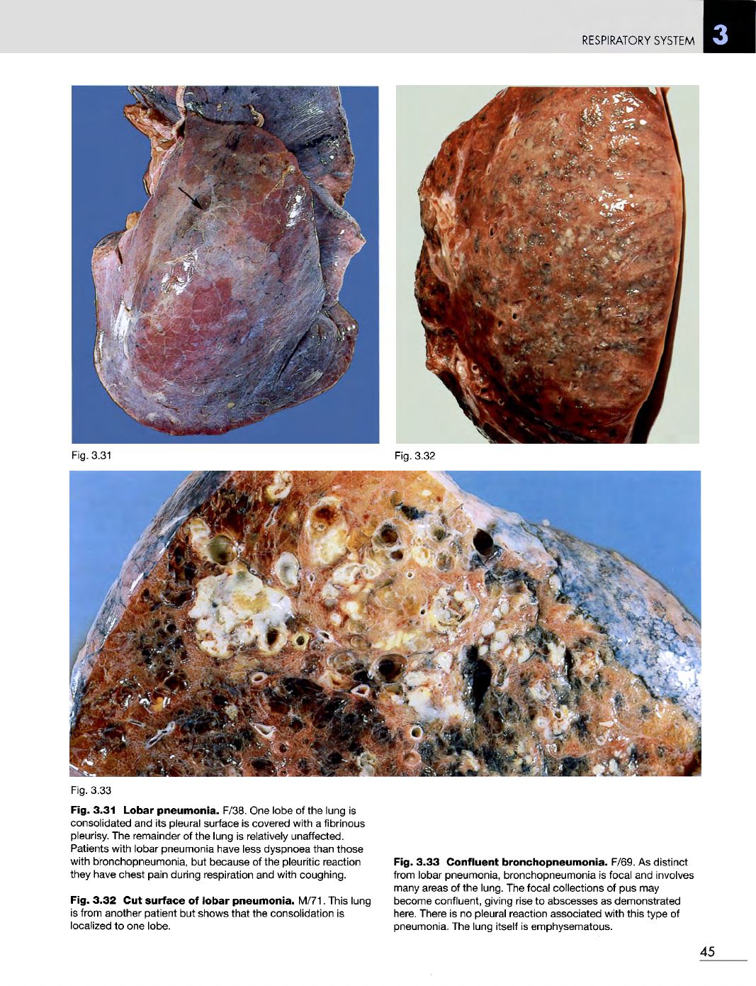
Fig. 3.31
Fig.
3.32
Fig. 3.33
Fig. 3.31 Lobar
pneumonia.
F/38.
One
lobe
of the
lung
is
consolidated
and its
pleural surface
is
covered with
a
fibrinous
pleurisy.
The
remainder
of the
lung
is
relatively unaffected.
Patients with lobar pneumonia have less dyspnoea than those
with bronchopneumonia,
but
because
of the
pleuritic reaction
they
have chest pain during respiration
and
with coughing.
Fig. 3.32
Cut
surface
of
lobar
pneumonia.
M/71. This lung
is
from another patient
but
shows that
the
consolidation
is
localized
to one
lobe.
Fig. 3.33
Confluent
bronchopneumonia.
F/69.
As
distinct
from lobar pneumonia, bronchopneumonia
is
focal
and
involves
many
areas
of the
lung.
The
focal collections
of pus may
become confluent, giving rise
to
abscesses
as
demonstrated
here.
There
is no
pleural reaction associated with this type
of
pneumonia.
The
lung itself
is
emphysematous.
45
RESPIRATORY
SYSTEM
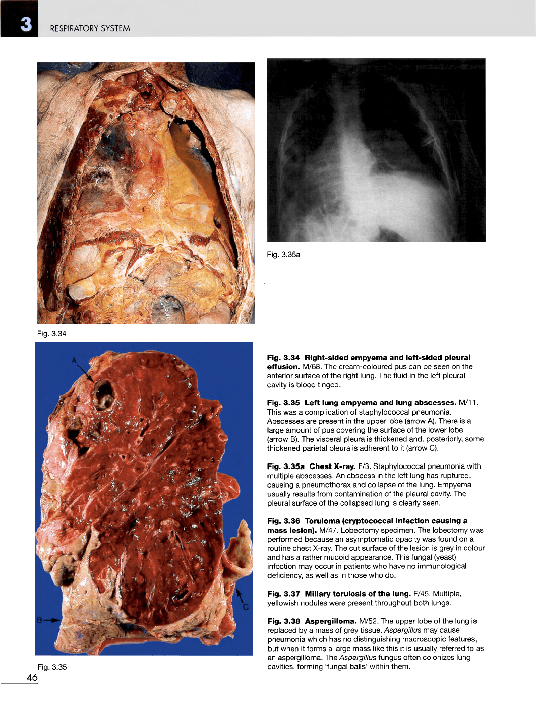
Fig. 3.34
Fig. 3.35
Fig. 3.34
Right-sided
empyema
and
left-sided
pleural
effusion.
M/68.
The
cream-coloured
pus can be
seen
on the
anterior surface
of the
right lung.
The
fluid
in the
left pleural
cavity
is
blood
tinged.
Fig. 3.35
Left
lung
empyema
and
lung
abscesses.
M/11.
This
was a
complication
of
staphylococcal pneumonia.
Abscesses
are
present
in the
upper lobe (arrow
A).
There
is a
large amount
of pus
covering
the
surface
of the
lower lobe
(arrow
B). The
visceral pleura
is
thickened and, posteriorly, some
thickened parietal pleura
is
adherent
to it
(arrow
C).
Fig.
3.35a
Chest
X-ray.
F/3. Staphylococcal pneumonia with
multiple abscesses.
An
abscess
in the
left
lung
has
ruptured,
causing
a
pneumothorax
and
collapse
of the
lung. Empyema
usually
results from contamination
of the
pleural cavity.
The
pleural
surface
of the
collapsed lung
is
clearly seen.
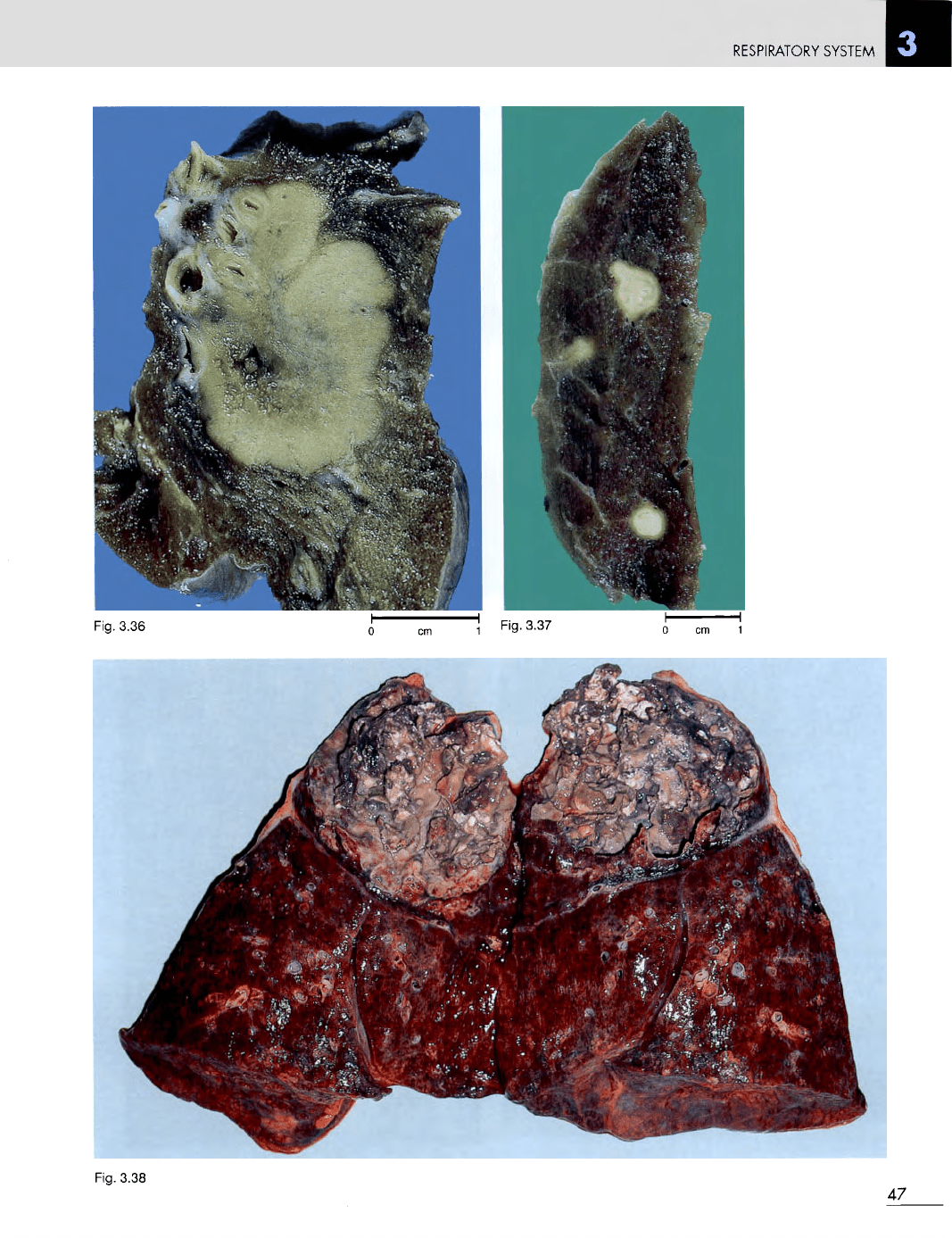
Fig. 3.36 Toruloma
(cryptococcal
infection
causing
a
mass
lesion).
M/47. Lobectomy specimen.
The
lobectomy
was
performed because
an
asymptomatic opacity
was
found
on a
routine chest
X-ray.
The cut
surface
of the
lesion
is
grey
in
colour
and has a
rather mucoid appearance. This fungal (yeast)
infection
may
occur
in
patients
who
have
no
immunological
deficiency,
as
well
as in
those
who do.
Fig. 3.37
Miliary
torulosis
of the
lung.
F/45.
Multiple,
yellowish nodules were present throughout both lungs.
Fig. 3.38
Aspergilloma.
M/52.
The
upper lobe
of the
lung
is
replaced
by a
mass
of
grey tissue.
Aspergillus
may
cause
pneumonia which
has no
distinguishing macroscopic features,
but
when
it
forms
a
large mass like this
it is
usually referred
to as
an
aspergilloma.
The
Aspergillus
fungus often colonizes lung
cavities,
forming 'fungal balls' within them.
46
Fig. 3.35a
RESPIRATORY
SYSTEM
Fig. 3.38
47
Fig. 3.37
Fig. 3.36
RESPIRATORY
SYSTEM
Fig. 3.40
Fig. 3.41
48
Fig. 3.39
RESPIRATORY
SYSTEM
Fig.
3.43
Fig.
3.42
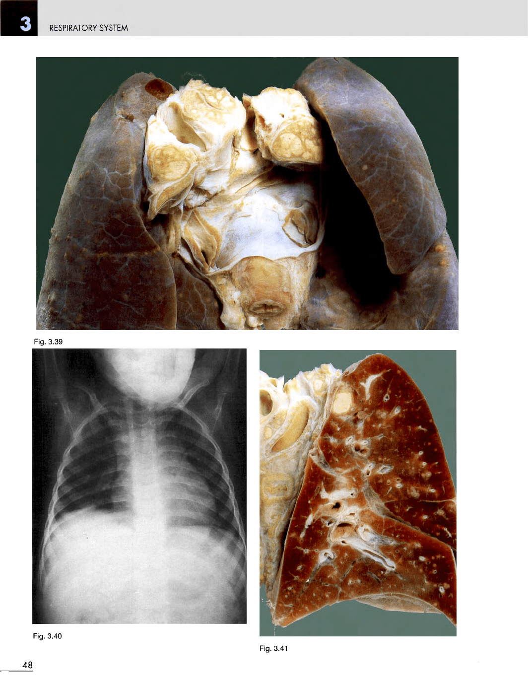
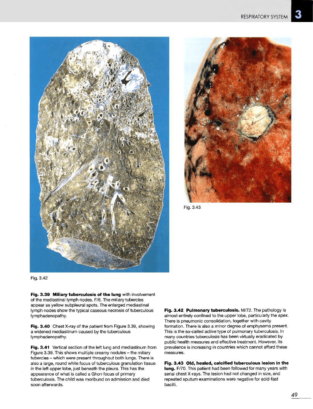
Fig. 3.39
Miliary
tuberculosis
of the
lung
with involvement
of
the
mediastinal lymph nodes. F/6.
The
miliary tubercles
appear
as
yellow subpleural
spots.
The
enlarged mediastinal
lymph nodes show
the
typical caseous necrosis
of
tuberculous
lymphadenopathy.
Fig. 3.40 Chest
X-ray
of the
patient from Figure 3.39, showing
a
widened mediastinum caused
by the
tuberculous
lymphadenopathy.
Fig. 3.41 Vertical section
of the
left lung
and
mediastinum from
Figure
3.39. This shows multiple creamy nodules
- the
miliary
tubercles
-
which were present throughout both lungs. There
is
also
a
large, round white focus
of
tuberculous granulation tissue
in
the
left upper lobe, just beneath
the
pleura. This
has the
appearance
of
what
is
called
a
Ghon focus
of
primary
tuberculosis.
The
child
was
moribund
on
admission
and
died
soon afterwards.
Fig. 3.42
Pulmonary
tuberculosis.
M/72.
The
pathology
is
almost
entirely confined
to the
upper lobe, particularly
the
apex.
There
is
pneumonic consolidation, together with cavity
formation. There
is
also
a
minor degree
of
emphysema present.
This
is the
so-called active type
of
pulmonary tuberculosis.
In
many
countries tuberculosis
has
been virtually eradicated
by
public health measures
and
effective treatment. However,
its
prevalence
is
increasing
in
countries which cannot afford these
measures.
Fig. 3.43 Old,
healed,
calcified
tuberculous
lesion
in the
lung.
F/70. This patient
had
been followed
for
many years with
serial
chest X-rays.
The
lesion
had not
changed
in
size,
and
repeated sputum examinations were negative
for
acid-fast
bacilli.
49
RESPIRATORY
SYSTEM
Fig.
3.44
Fig.
3.45
Fig.
3.46
Fig.
3.47
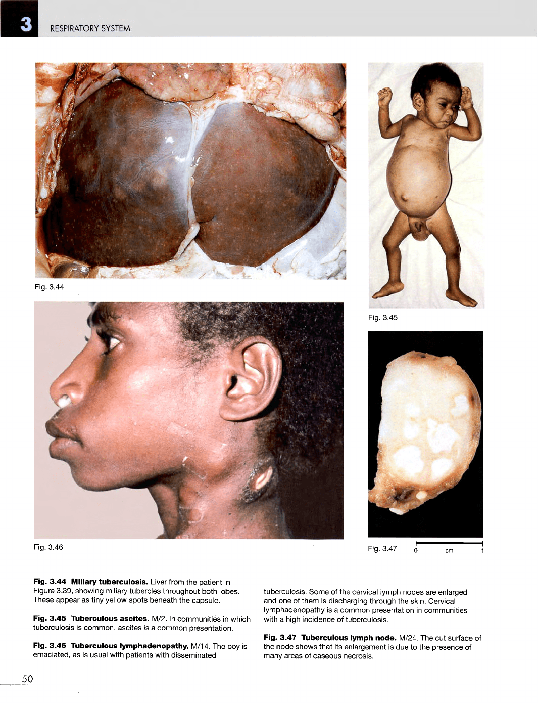
Fig. 3.44
Miliary
tuberculosis.
Liver from
the
patient
in
Figure
3.39, showing
miliary
tubercles throughout both lobes.
These
appear
as
tiny
yellow
spots beneath
the
capsule.
Fig. 3.45
Tuberculous
ascites.
M/2.
In
communities
in
which
tuberculosis
is
common, ascites
is a
common presentation.
Fig. 3.46
Tuberculous
lymphadenopathy.
M/14.
The boy is
emaciated,
as is
usual with patients with disseminated
tuberculosis. Some
of the
cervical lymph nodes
are
enlarged
and one of
them
is
discharging through
the
skin. Cervical
lymphadenopathy
is a
common presentation
in
communities
with
a
high incidence
of
tuberculosis.
Fig. 3.47
Tuberculous
lymph
node.
M/24.
The cut
surface
of
the
node shows that
its
enlargement
is due to the
presence
of
many
areas
of
caseous necrosis.
50
