Cook R.A., Stewart B. Colour Atlas of Anatomical Pathology
Подождите немного. Документ загружается.


CARDIOVASCULAR
SYSTEM
Fig.
1.50
Fig. 1.46
Organizing
fibrinous
pericarditis.
M/56.
The
parietal layer
of the
pericardium
has
been separated from
the
visceral layer with some difficulty.
The
surfaces
of
both
layers
are
covered
by
shaggy, organizing fibrinous pericarditis which made
the two
layers
adherent
to one
another.
Fig. 1.47
Pericarditis
resulting from deposits
of
secondary
cancer
in the
pericardium
and
myocardium. M/60. Lung cancer
is
the one
most likely
to
invade
the
pericardium (see Figure. 3.61).
Fig. 1.48
Air
embolus.
M/39.
The
patient died suddenly when
a
large amount
of air was
accidentally introduced during
complicated intravenous therapy.
The
presence
of the air
embolus
was
demonstrated
by
filling
the
pericardium with water
and
making
an
incision
through
the
water into
the
ventricular
cavity.
Bubbles
of air
then escaped.
Fig. 1.49
Acute
rheumatic
vegetations
on the
aortic
valve
cusps.
F/11.
The
patient died from acute rheumatic
carditis.
Fig. 1.50
A
Lambl's excrescence
on the
aortic
valve.
M/74. These
are
aggregations
of
fibrin covered
by
endothelium.
They
must
be
distinguished from true vegetations.
21
Fig. 1.49
CARDIOVASCULAR
SYSTEM
Fig.
1.51
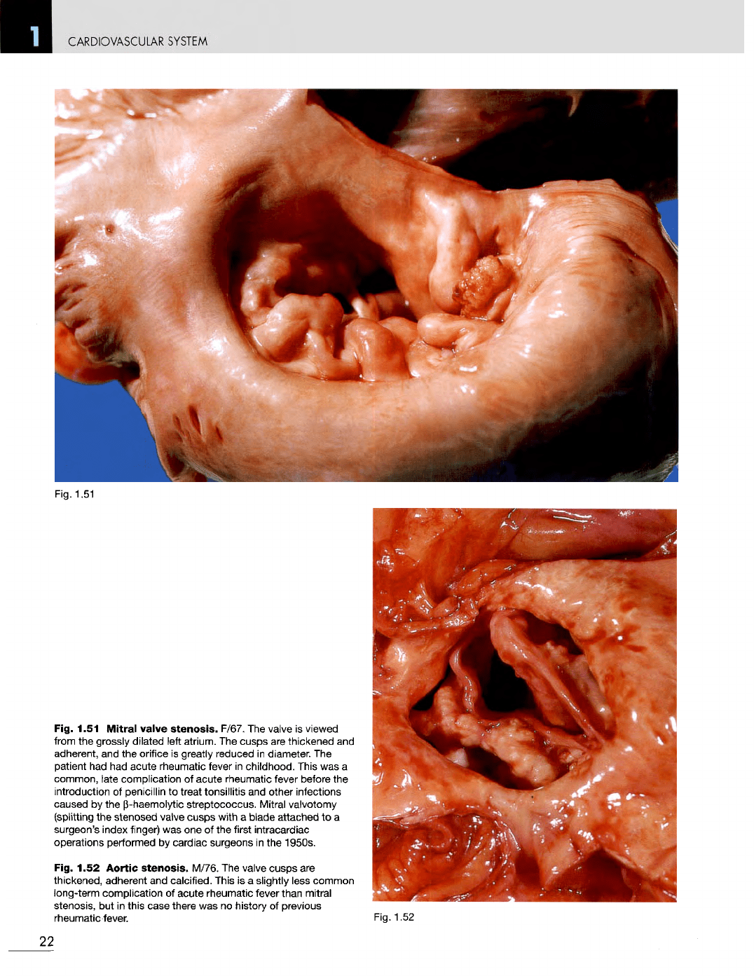
Fig. 1.51
Mitral
valve
stenosis.
F/67.
The
valve
is
viewed
from
the
grossly
dilated
left atrium.
The
cusps
are
thickened
and
adherent,
and the
orifice
is
greatly reduced
in
diameter.
The
patient
had had
acute rheumatic fever
in
childhood. This
was a
common, late complication
of
acute rheumatic
fever
before
the
introduction
of
penicillin
to
treat tonsillitis
and
other infections
caused
by the
b-haemolytic streptococcus. Mitral valvotomy
(splitting
the
stenosed
valve
cusps
with
a
blade
attached
to a
surgeon's index finger)
was one of the
first intracardiac
operations performed
by
cardiac surgeons
in the
1950s.
Fig. 1.52 Aortic
stenosis.
M/76.
The
valve
cusps
are
thickened, adherent
and
calcified. This
is a
slightly less common
long-term complication
of
acute rheumatic
fever
than mitral
stenosis,
but in
this case there
was no
history
of
previous
rheumatic
fever.
Fig.
1.52
22

Fig. 1.53
CARDIOVASCULAR
SYSTEM
Fig. 1.53
Ball
thrombus
in the
left
atrium.
M/66. This
is a
complication
of
mitral stenosis
and
auricular fibrillation,
and a
source
of
peripheral emboli;
however,
in
this case there appears
to be
very
little
abnormality
of the
mitral valve.
Fig. 1.54 Thrombus
in the
left
auricular
appendage.
F/85.
Thrombus
at
this site
is a
complication
of
auricular fibrillation,
and may be
a
source
of
peripheral emboli.
Fig. 1.55 Vegetations
on the
mitral
valve
in
subacute
bacterial
endocarditis.
M/41. These
are
also
a
source
of
peripheral emboli.
Fig. 1.54
Fig. 1.55
23
CARDIOVASCULAR
SYSTEM
Fig.
1.58
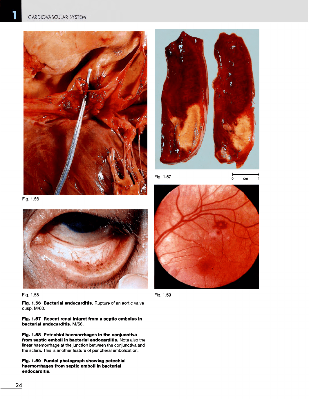
Fig. 1.56 Bacterial endocarditis.
Rupture
of an
aortic
valve
cusp.
M/60.
Fig. 1.57 Recent renal infarct from
a
septic embolus
in
bacterial endocarditis. M/56.
Fig. 1.58 Petechial haemorrhages
in the
conjunctiva
from
septic emboli
in
bacterial endocarditis. Note
also
the
linear
haemorrhage
at the
junction
between
the
conjunctiva
and
the
sclera.
This
is
another
feature
of
peripheral embolization.
Fig. 1.59 Fundal photograph showing
petechial
haemorrhages from septic emboli
in
bacterial
endocarditis.
Fig.
1.59
24
Fig.
1.56
Fig.
1.57
CARDIOVASCULAR
SYSTEM
Fig.
1.60
Fig.
1.61
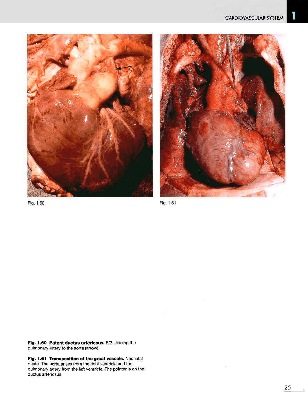
Fig. 1.60
Patent
ductus
arteriosus.
F/3. Joining
the
pulmonary artery
to the
aorta (arrow).
Fig. 1.61 Transposition
of the
great
vessels. Neonatal
death.
The
aorta arises from
the
right ventricle
and the
pulmonary artery from
the
left
ventricle.
The
pointer
is on the
ductus arteriosus.
25
CARDIOVASCULAR
SYSTEM
Fig.
1.62
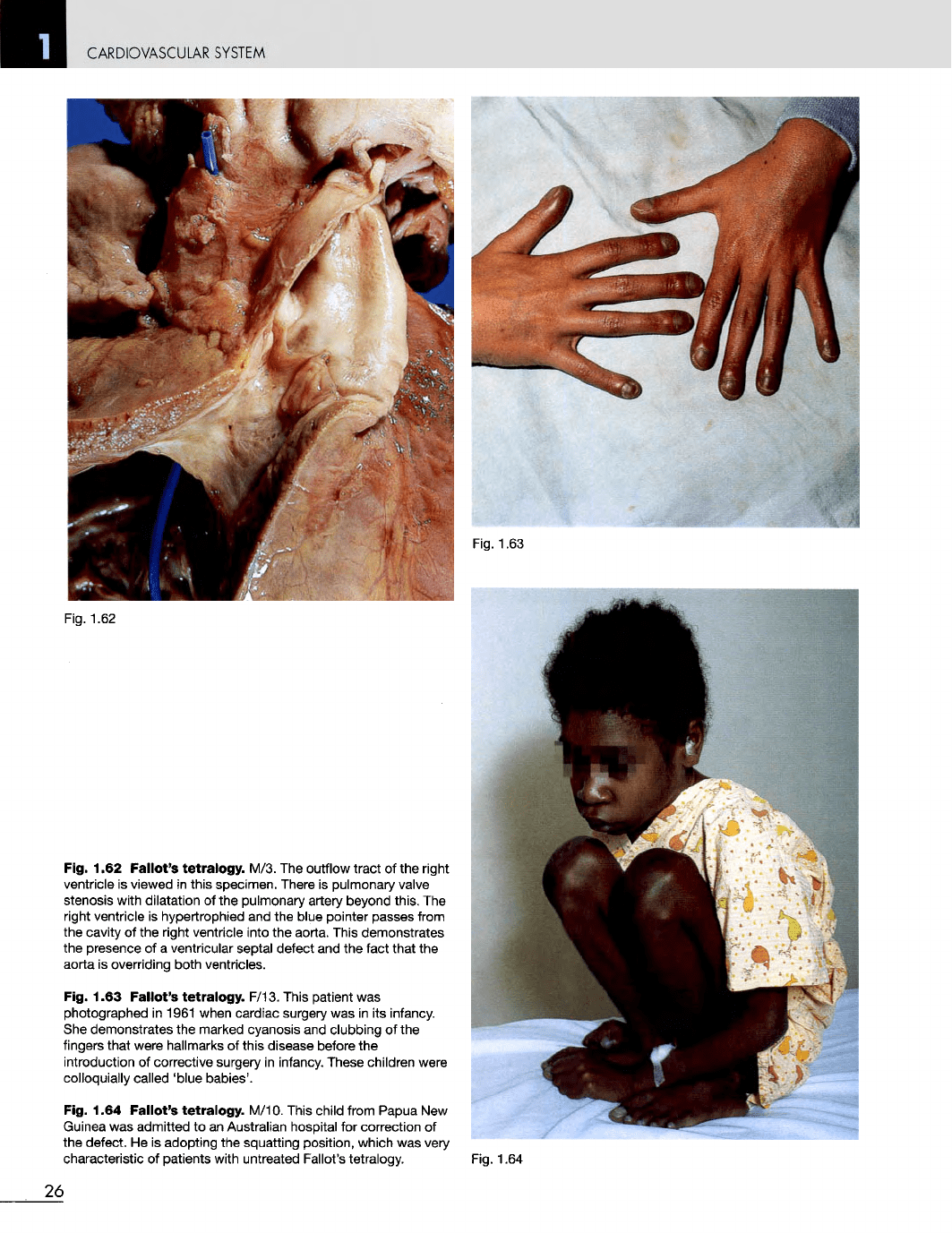
Fig. 1.62
Fallot's
tetralogy.
M/3.
The
outflow tract
of the
right
ventricle
is
viewed
in
this specimen. There
is
pulmonary
valve
stenosis with dilatation
of the
pulmonary artery beyond this.
The
right ventricle
is
hypertrophied
and the
blue pointer passes from
the
cavity
of the
right ventricle into
the
aorta. This demonstrates
the
presence
of a
ventricular septal defect
and the
fact that
the
aorta
is
overriding both ventricles.
Fig. 1.63
Pallet's
tetralogy.
F/13. This patient
was
photographed
in
1961 when cardiac surgery
was in its
infancy.
She
demonstrates
the
marked cyanosis
and
clubbing
of the
fingers
that were hallmarks
of
this disease before
the
introduction
of
corrective surgery
in
infancy. These children were
colloquially
called 'blue babies'.
Fig. 1.64
Fallot's
tetralogy.
M/10. This child from Papua
New
Guinea
was
admitted
to an
Australian hospital
for
correction
of
the
defect.
He is
adopting
the
squatting position, which
was
very
characteristic
of
patients with untreated Fallot's tetralogy. Fig. 1.64
26
CARDIOVASCULAR SYSTEM
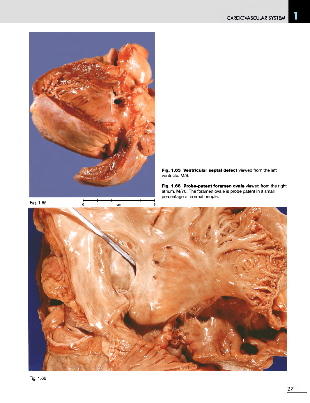
Fig. 1.65 Ventricular septal defect
viewed from
the
left
ventricle.
M/9.
Fig. 1.66 Probe-patent foramen
ovale viewed from
the
right
atrium.
M/70.
The
foramen ovale
is
probe patent
in a
small
percentage
of
normal people.
Fig.
1.66
27
CARDIOVASCULAR
SYSTEM
Fig.
1.67
Fig.
1.68
Fig.
1.69
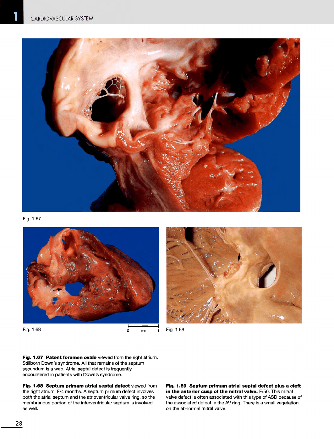
Fig. 1.67
Patent
foramen ovale viewed from
the
right atrium.
Stillborn Down's syndrome.
All
that remains
of the
septum
secundum
is a
web. Atrial septal defect
is
frequently
encountered
in
patients with Down's syndrome.
Fig. 1.68
Septum
primum
atrial
septal
defect
viewed from
the
right atrium.
F/4
months.
A
septum primum defect involves
both
the
atrial septum
and the
atrioventricular
valve
ring,
so the
membranous portion
of the
interventricular septum
is
involved
as
well.
Fig. 1.69
Septum
primum
atrial
septal
defect
plus
a
cleft
in
the
anterior
cusp
of the
mitral
valve. F/50. This mitral
valve
defect
is
often associated with this type
of ASD
because
of
the
associated defect
in the AV
ring. There
is a
small vegetation
on
the
abnormal mitral valve.
28
CARDIOVASCULAR
SYSTEM
Fig.
1.70
Fig.
1.71
Fig.
1.72
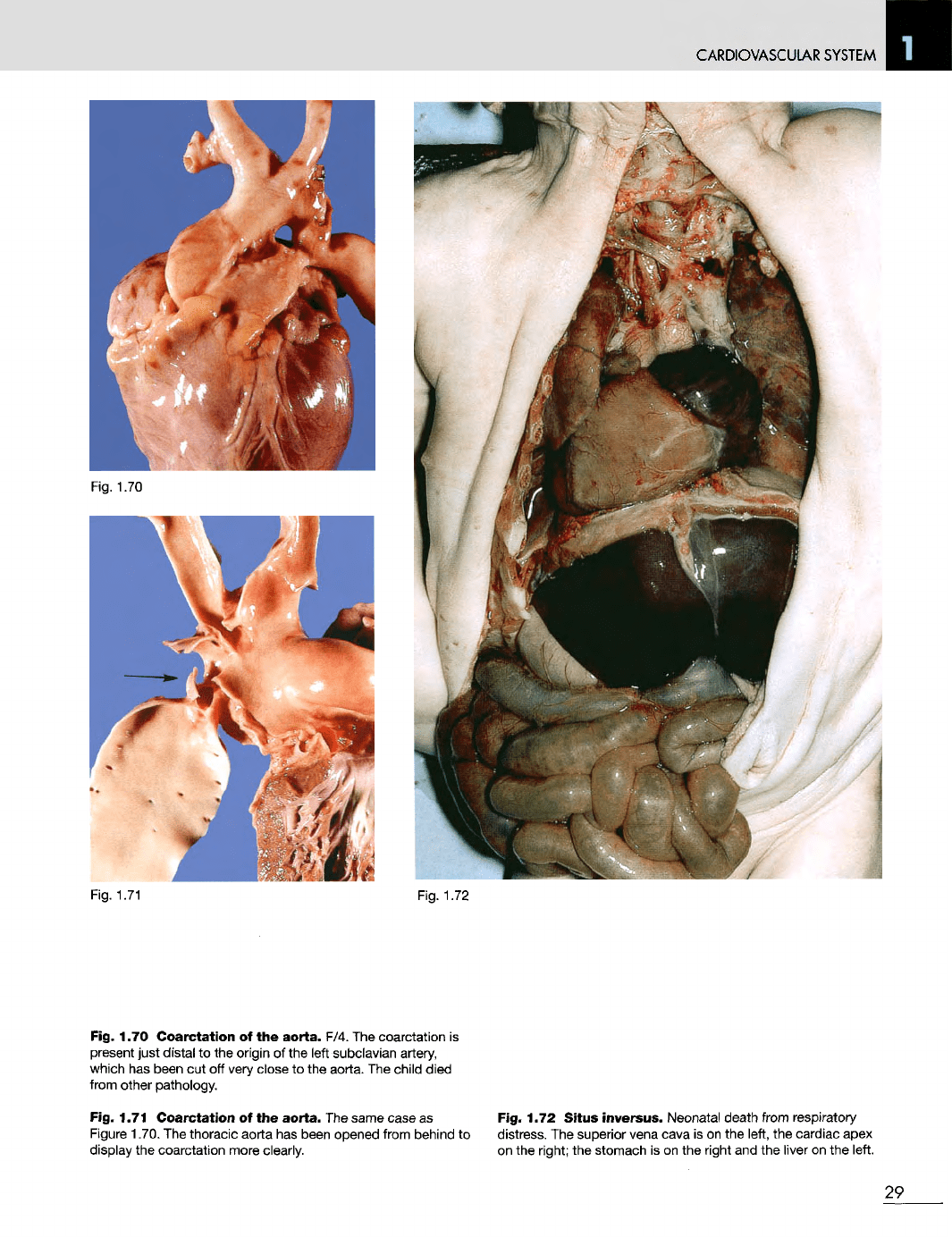
Fig. 1.70
Coarctation
of the
aorta.
F/4.
The
coarctation
is
present just
distal
to the
origin
of the
left subclavian artery,
which
has
been
cut off
very
close
to the
aorta.
The
child died
from
other pathology.
Fig. 1.71
Coarctation
of the
aorta.
The
same case
as
Figure
1.70.
The
thoracic aorta
has
been opened from behind
to
display
the
coarctation more clearly.
Fig. 1.72
Situs
inversus.
Neonatal
death
from respiratory
distress.
The
superior vena cava
is on the
left,
the
cardiac apex
on the
right;
the
stomach
is on the
right
and the
liver
on the
left.
29
CARDIOVASCULAR SYSTEM
Fig.
1.73
Fig.
1.74
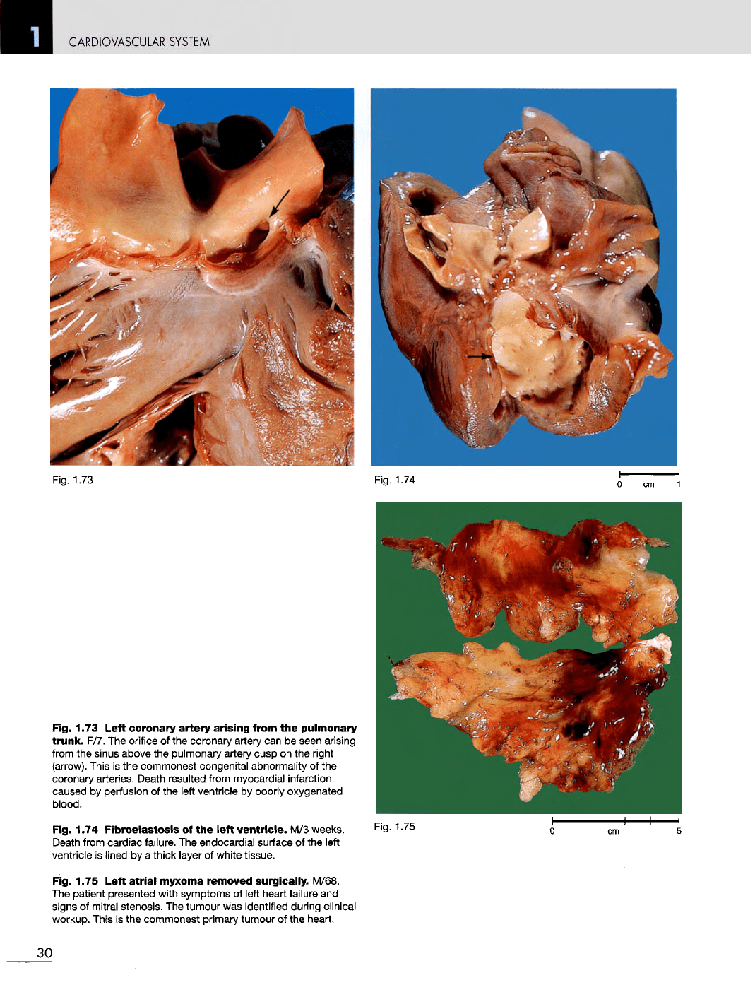
Fig. 1.73 Left coronary artery
arising
from
the
pulmonary
trunk.
F/7.
The
orifice
of the
coronary artery
can be
seen arising
from
the
sinus above
the
pulmonary artery cusp
on the
right
(arrow).
This
is the
commonest congenital abnormality
of the
coronary
arteries. Death resulted from myocardial infarction
caused
by
perfusion
of the
left ventricle
by
poorly oxygenated
blood.
Fig. 1.74
Fibroelastosis
of the
left
ventricle.
M/3
weeks.
Death
from cardiac failure.
The
endocardial surface
of the
left
ventricle
is
lined
by a
thick layer
of
white tissue.
Fig. 1.75
Left
atrial
myxoma removed
surgically.
M/68.
The
patient presented with symptoms
of
left heart failure
and
signs
of
mitral stenosis.
The
tumour
was
identified during clinical
workup. This
is the
commonest
primary
tumour
of the
heart.
Fig. 1.75
30
