Журнал - Проблемы криобиологии 2010 №2
Подождите немного. Документ загружается.


165
problems
of cryobiology
Vol. 20, 2010, №2
проблемы
криобиологии
Т. 20, 2010, №2
носными сосудами, главным образом, синусоид-
ного типа (рис. 3, а). Это свидетельствовало о нор-
мальном кровенаполнении органа. В расположен-
ных между синусами пульпарных тяжах выявлены
очаги плазмоцитогенеза. Лимфоидная ткань (белая
пульпа селезенки) располагалась в адвентиции ее
артерий в виде шаровидных скоплений или удли-
ненных лимфатических влагалищ (лимфатические
фолликулы). В них проходили центральные артерии,
которые располагались эксцентрично. От лим-
фатических фолликулов отходили гемокапилляры
по направлению к краевым синусам красной пуль-
пы. В лимфатических фолликулах можно различить
три нечетко разграниченные зоны: периартериаль-
ную (центр размножения), мантийный слой и крае-
вую (маргинальную).
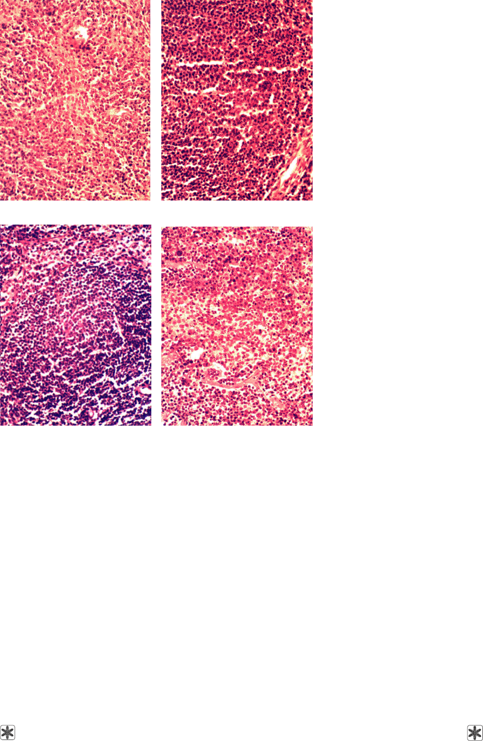
Через 7 суток в селезенке мышей группы 2,
зараженных вирусом гриппа, при гистологическом
исследовании выявлены характерные для острых
инфекций изменения: полнокровие, экссудация и
инфильтрация лейкоцитами пульпы селезенки, про-
лиферация В-лимфобластов в центрах размноже-
ния фолликулов (рис. 3, б); скопления макрофагов
с фагоцитированными лимфоцитами или их фраг-
ментами в виде хромофильных телец; дегенера-
тивные и некротические изменения со стороны тка-
невых элементов пульпы и фолликулов.
В селезенке мышей группы 3 на 7-е сутки белая
пульпа преобладала над красной, что свидетельст-
вует о раздражении лимфоидной ткани антигенами.
В многочисленных лимфатических фолликулах
различались: периартериальные зоны (центры раз-
множения), занимающие небольшие участки фол-
ликула около артериолы; мантийный слой со слоис-
тым расположением малых Т- и В-лимфоцитов,
которые образуют “корону”, расслоенную циркуляр-
но направленными толстыми ретикулярными
волокнами; краевая зона, представляющая собой
переходную область между белой и красной пуль-
пой (рис. 3, в). Центры размножения фолликулов
состояли из ретикулярных клеток и пролифери-
рующих В-лимфобластов. Здесь же обнаружены
небольшие скопления макрофагов. Красная пульпа,
занимающая относительно небольшую площадь,
содержала большое количество гемокапилляров.
При исследовании селезенки мышей группы 3
на 14-е сутки установлена нормализация ее строе-
ния. Трабекулы селезенки, отходящие от соедини-
тельнотканной капсулы внутрь и в глубоких ее
частях анастомозирующие между собой, предс-
тавлены преимущественно эластическими волок-
нами, так как капсула и трабекулы составляют
опорно-сократительный аппарат селезенки. Крас-
ная пульпа преобладала над белой. Основу пульпы
составляла ретикулярная ткань, образующая ее
строму. В адвентиции артерий, пронизывающих
which had small centers consisting of predominantly
young cells of lymphopoietic series with basophilic cy-
toplasm, were found in lymph node cortex. That is why
these centers looked darker. Solitary cells dividing
mitotically were revealed in them. A “corona” from
small lymphocytes, which is typical for the stage of
relative quiescence, was visible (Fig. 2d). No erythro-
cytes were noted in blood vessels and capillaries in-
side lymph node medullar tissue. Reticuloendothelial
Kupffer's cells forming sinus walls were plainly distin-
guished.
Thus, protective activity of the preparation “Cryo-
cell-Haemocord” manifests itself as intensification of
regenerative processes in lymph nodes of mice infected
with the influenza virus and as normalization of the
structure of these organs as early as by the 14
th
day.
Spleen. Histological analysis of spleens of the mice
from group 1, which had been introduced with the prepa-
ration “Cryocell-Haemocord” 6 months earlier, showed
their normal structure. Spleen trabecules anastomos-
ing among themselves in deep parts of spleen stretched
from the connective tissue capsule. The ratio between
red and white pulps was shifted towards red one con-
sisting of reticular tissue with blood cells and numer-
ous blood vessels, mainly of sinusoid type, in it (Fig. 3a).
That attested to a normal blood filling of the organ.
Loci of plasmocytogenesis were revealed in pulpal
strands located between sinuses. Lymphoid tissue
(spleen white pulp) was situated in adventitia of its ar-
teries in the form of sphere-like clusters or in elon-
gated lymph sheaths (lymph follicles). Central arteries
located eccentrically stretched in them. Hemocapilla-
ries stretched from lymph follicles towards red pulp
marginal sinuses. Three unclearly discriminated zones
could be distinguished: periarterial one (proliferation cen-
ter), mantle layer and marginal zone.
On the 7
th
day histological analysis revealed typical
for acute infections changes in spleens of the mice
infected with the influenza virus (group 2): plethora,
exudation and infiltration of spleen pulp with leukocytes,
proliferation of B-lymphoblasts in follicle proliferation
centers (Fig. 3b); clusters of macrophages with phago-
cytized lymphocytes or their fragments as chromophilic
corpuscles; degenerative and necrotic changes in pulp
tissue elements and follicles.
On the 7
th
day in spleens of the mice from group 3
white pulp dominated over red one, that attests to irri-
tation of lymphoid tissue by antigens. In numerous lymph
follicles one can distinguish periarterial zones (prolif-
eration centers) holding little areas of the follicle around
the arteriole; a mantle layer with lamellar disposition
of small T- and B-lymphocytes, which formed a “co-
rona” stratified with circularly directed thick reticular
fibers; a marginal zone constituting transitional area
between red and white pulps (Fig. 3c). Follicle proli-

166
problems
of cryobiology
Vol. 20, 2010, №2
проблемы
криобиологии
Т. 20, 2010, №2
chi. Alveolar bronchioles were acinus areas lined with
cubical epithelium alternating with thin-walled alveolar
protrusions. No muscles were noted in alveole walls.
Most of lung sections were occupied by incisions of
alveolar ducts and unequally expanded terminal
alveolae. Alveolar macrophages were found on the in-
ner surface of alveolae and in their cavities.
On the 7
th
day in lung parenchyma of the mice from
group 2 spongioid structure only remained in small ar-
селезенку, определялась лимфоидная ткань в виде
округлых или овальных скоплений – лимфатических
фолликулов (рис. 3, г).
Легкие. Гистологическое исследование парен-
химы легких мышей группы 1 показало их нормаль-
ное строение. Ткань легких на препаратах имела
ажурный вид вследствие того, что основную массу
ее составляли разрезы тонкостенных концевых аль-
Рис. 3. Селезенка мышей: а – через 6 месяцев после введения препарата
"Криоцелл-гемокорд"; б – зараженных вирусом гриппа (на 7-е сутки); в –
зараженных вирусом гриппа через 6 месяцев после введения препарата
(на 7-е сутки); г – зараженных вирусом гриппа через 6 месяцев после
введения препарата (на 14-е сутки). Окраска гематоксилином и эозином.
×400.
Fig. 3. Mice spleens: a – 6 months after the administration of the preparation
"Cryocell-Haemocord"; b – infected with the influenza virus (the 7
th
day); c –
infected with the influenza virus 6 months after the administration of the prepa-
ration (the 7
th
day); d – infected with the influenza virus 6 months after the
administration of the preparation (the 14
th
day). Hematoxylin and eosin stain-
ing. ×400.
а a
б b
в cг d
feration centers consisted of reticular
cells and proliferating B-lympho-
blasts. Small clusters of macropha-
ges were also revealed here. Red
pulp holding a relatively little area
comprised a lot of hemocapillaries.
On the 14
th
day in the mice from
group 3 spleen structure was estab-
lished to become normal. Spleen
trabecules stretching from the con-
nective-tissue capsule deep into the
organ and anastomosing among
themselves in deep its parts were
predominantly elastic fibers, since the
capsule and trabecules form sup-
port-contractile apparatus of spleen.
Red pulp dominated over white one.
The pulp basis was reticular tissue
forming its stroma. Lymphoid tissue
as round or oval clusters, lymph fol-
licles, was noted in adventitia arter-
ies running through spleen (Fig. 3d).
Lungs. Histological analysis of
lung parenchyma of the mice from
group 1 showed its normal structure.
In the sections lung tissue was open-
work because of its bulk consisted
of thin-walled terminal alveole inci-
sions. Small bronchi were lined with
cubical epithelium; smooth muscle
annular layer was revealed behind
bronchial tube tunic. Individual small
gland packages occurred in small
bronchus submucosal layer. As the
caliber of bronchi lessened, glands
vanished. Small bronchi were accom-
panied by bronchial arteries, incisions
of which were always visible nearby.
Lung veins containing a lot of smooth
muscles in their walls resembled ar-
teries by their structure, but were lo-
cated independently on bronchi
(Fig. 4a). Respiratory lung parts
(acini) began with alveolar bronchio-
les originated from the tiniest bron-

167
problems
of cryobiology
Vol. 20, 2010, №2
проблемы
криобиологии
Т. 20, 2010, №2
веол. Малые бронхи выстланы
кубическим эпителием, за соб-
ственной их оболочкой обнаружен
кольцевой слой гладких мышц. В
подслизистом слое малых брон-
хов встречались отдельные не-
большие пакеты желез. С умень-
шением калибра бронхов железы
исчезали. Малые бронхи сопро-
вождались бронхиальными арте-
риями, разрезы которых постоян-
но встречались возле них. Легоч-
ные вены, содержащие в своих
стенках большое количество
гладких мышц, сходны по строе-
нию с артериями, но располага-
лись независимо от бронхов
(рис. 4, а). Респираторные отделы
легких (ацинусы) начинались
альвеолярными бронхиолами, в
которые переходили самые мел-
кие бронхи. Альвеолярные брон-
хиолы представляли собой участ-
ки ацинуса, выстланные кубичес-
ким эпителием, чередующиеся с
альвеолярными выпячиваниями,
имеющими очень тонкую стенку.
В стенках альвеол мышц уже не
было. Большая часть срезов лег-
ких была занята разрезами аль-
веолярных ходов и концевых аль-
веол, растянутых в разной степе-
ни. На внутренней поверхности
альвеол и в их полости встреча-
лись альвеолярные макрофаги.
В паренхиме легких у мышей
группы 2 через 7 суток только на
небольших участках по краям
легких сохранено губчатое строе-
ние, однако альвеолы местами
растянуты, а местами – имели
разрывы стенок. Непрерывный
эндотелий альвеолярных капил-
ляров только в некоторых местах
сохранен. В большинстве случаев
он слущивался, был плохо выра-
жен, в результате чего наблюда-
eas along the lung edges, though alveolae were ex-
panded in some places and ruptured in others. Endothe-
lium in alveolar capillaries scarcely remained continu-
ous. In most cases it was desquamated, indistinct, as a
consequence of which multiple hemorrhages were
observed, and alveolae were filled with blood. In the
central parts of lungs alveolae were destructed and
“hepatized” (hemorrhagic pneumonia). In intrapulmo-
nic bronchi of various size and bronchiolae epithelium
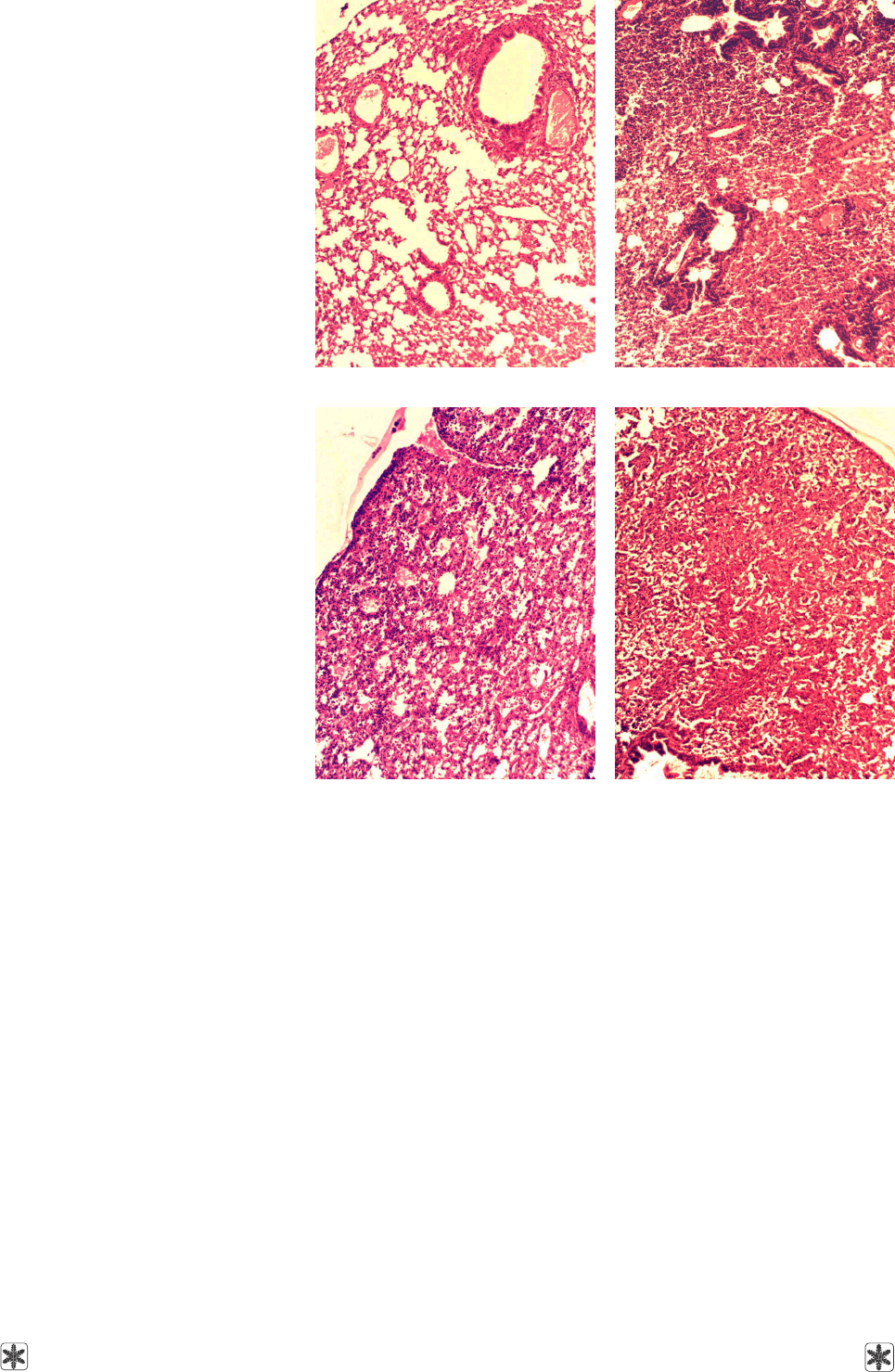
Рис. 4. Паренхима легких мышей: а – через 6 месяцев после введения
препарата "Криоцелл-гемокорд"; б – зараженных вирусом гриппа (на 7-е
сутки); в – зараженных вирусом гриппа через 6 месяцев после введения
препарата (на 7-е сутки); г – зараженных вирусом гриппа через 6 месяцев
после введения препарата (на 14-е сутки). Окраска гематоксилином и
эозином. ×200.
Fig. 4. Mice lung parenchyma: a – 6 months after the administration of the
preparation "Cryocell-Haemocord"; b – infected with the influenza virus (the
7
th
day); c – infected with the influenza virus 6 months after the administra-
tion of the preparation (the 7
th
day); d – infected with the influenza virus 6
months after the administration of the preparation (the 14
th
day). Hematoxylin
and eosin staining. ×200.
а aб b
в cг d
лись множественные кровоизлияния и заполнение
альвеол кровью. Определялись деструкция аль-
веол в центральных отделах легких и их “опечене-
ние” (геморрагическая пневмония). Во внутриле-
гочных бронхах разного калибра и бронхиолах
обнаружены десквамация эпителия, истончение
стенки бронхиальных артерий и вен, стазы крове-
носных сосудов. В паренхиме легких имела место
моноцито-лимфоцитарная инфильтрация – масса

168
problems
of cryobiology
Vol. 20, 2010, №2
проблемы
криобиологии
Т. 20, 2010, №2
was desquamated; bronchial artery and vein walls were
thinned; blood vessel stasis was noted. Monocyte-
lymphocyte infiltration, a mass of nuclear leukocytes,
among which segmented-nuclear cells were seen, oc-
curred in lung parenchyma (Fig. 4b).
On the 7
th
day the structure of lung parenchyma of
the mice from group 3 was more or less spongioid.
Alveolar saccules were narrowed in some places and
expanded in others. Alveolar capillary endothelium was
intact. Herewith “hepatized” areas of parenchyma,
where monocyte-lymphocyte infiltration was observed,
were noted. Dilated blood vessels with thinned walls
were filled with erythrocytes. Alveolar macrophages
were often seen on the inner surface of alveolae. Ter-
minal bronchiole epithelium was desquamated in some
places, nevertheless in most cases it remained its in-
tegrity (Fig. 4c).
Histological analysis of lung parenchyma of the mice
from group 3 showed that 14 days after infection its
structure was spongioid. Herewith alveolae were ex-
panded in some places and compressed in others; rup-
tures in their walls were noted somewhere. Alveolar
capillary endothelium was predominantly intact. Small
clusters of erythrocytes were observed in alveolar
ducts. Terminal bronchiole epithelium remained intact
in most cases. Monocyte-lymphocyte infiltration in lung
parenchyma was moderate. Blood vessel walls were
a little thinned; and stases were noted in some of them
(Fig. 4d).
Conclusions
Histological analysis of the immunocompetent or-
gans of the experimental animals showed that the pre-
liminary (6 months prior to the infection) administra-
tion of the preparation “Cryocell-Haemocord” com-
pletely protected them against dystrophic and necrotic
injuries caused by the influenza virus infection. It was
established that on the 14
th
day after the infection in
the animals, which had been introduced with the prepa-
ration “Cryocell-Haemocord” 6 months earlier, all the
tissue structures forming the basis of the immune pro-
tection organs became normal.
The preliminary administration of the preparation
“Cryocell-Haemocord” also prevented pathological
changes in lungs caused by the influenza virus and lead-
ing to hemorrhagic pneumonia. 14 days after the infec-
tion with the influenza virus of the animals, which had
been preliminary introduced with the preparation
“Cryocell-Haemocord”, normalization of lung paren-
chyma, preservation of alveolar capillary endothelium
and terminal bronchiole epithelium were registered in
most cases. It is possible that such protective activity
of the preparation is attributed to its influence on the
animals' immune system.
нуклеаров, среди которых встречались сегменто-
ядерные клетки (рис. 4, б).
При исследовании паренхимы легких мышей
группы 3 на 7-е сутки установлено более или менее
губчатое ее строение. Альвеолярные мешочки
местами сужены, местами – растянуты. Эндотелий
альвеолярных капилляров сохранен. При этом
выявлены участки паренхимы с “опеченением”, где
наблюдалась моноцито-лимфоцитарная инфиль-
трация. Дилатированные кровеносные сосуды с
истонченной стенкой были заполнены эритроци-
тами. На внутренней поверхности альвеол часто
встречались альвеолярные макрофаги. Эпителий
терминальных бронхиол в некоторых местах был
слущен, однако в большинстве случаев сохранял
целостность (рис. 4, в).
Гистологический анализ паренхимы легких мы-
шей группы 3 показал, что через 14 суток после ин-
фицирования она имела губчатое строение. При этом
альвеолы были то растянуты, то сжаты, в некото-
рых местах наблюдались разрывы их стенок. Эн-
дотелий альвеолярных капилляров преимущест-
венно сохранен. В альвеолярных ходах наблюда-
лись небольшие скопления эритроцитов. Эпителий
терминальных бронхиол в большинстве случаев
сохранен. В паренхиме легких установлена умерен-
ная моноцито-лимфоцитарная инфильтрация. Стен-
ки кровеносных сосудов несколько истончены, а в
некоторых из них наблюдались стазы (рис. 4, г).
Выводы
Гистологический анализ иммунокомпетентных
органов экспериментальных животных показал,
что предварительное (за 6 месяцев до инфицирова-
ния) введение препарата “Криоцелл-гемокорд” пол-
ностью защищает их от дистрофических и некроти-
ческих повреждений, которые вызывает вирус грип-
па. Установлено, что на 14-е сутки после инфици-
рования у животных, которым за 6 месяцев до этого
был введен препарат “Криоцелл-гемокорд”, норма-
лизуется строение всех тканевых структур, состав-
ляющих основу органов иммунологической защиты.
Предварительное введение препарата “Крио-
целл-гемокорд” предотвращает также вызываемые
вирусом гриппа патологические изменения в лег-
ких, приводящие к геморрагической пневмонии.
Через 14 суток после инфицирования животных ви-
русом гриппа, которым предварительно был введен
препарат “Криоцелл-гемокорд”, в большинстве
случаев установлены нормализация паренхимы
легких, сохранение эндотелия альвеолярных капил-
ляров и эпителия терминальных бронхиол. Воз-
можно, такое защитное действие препарата обус-
ловлено влиянием на иммунную систему животных.

problems
of cryobiology
Vol. 20, 2010, №2
проблемы
криобиологии
Т. 20, 2010, №2
169
Литература
Абелев Г.И. Основы иммунитета // Соросовский
образовательный журн.– 1996.– №5.– С. 4–10.
Возиянова Ж.И. Инфекционные и паразитарные болезни.
В 3-х т.– Киев: Здоров'я, 2000.– Т. 1.– С. 63-96.
Желтякова И.А, Бровко Е.В. Новый подход к профилак-
тике гриппа // Медицина третього тисячоліття: Зб. тез
міжвузівської конф. молодих вчених.– Харків, 2006.–
С. 93–94.
Липина О.В., Савченко Ю.А., Мусатова И.Б. Низкотемпе-
ратурное консервирование плазмы кордовой крови //
Биоимплантация на пороге ХХI века: Сб. тезисов
симпозиума по проблеме тканевых банков с междуна-
родным участием.– Самара, 2001.– С. 22–23.
Меркулов Г.А. Курс патологогистологической техники.–
Л.: Медгиз, 1961.– 340 с.
Цуцаєва А.О., Глушко Т.О., Лобасенко Н.П. та інш. Гемо-
корд – препарат комплексної терапії // Трансплантологія.–
2003.– Т. 4, №1.– С. 46–48.
Пат. України №31847А, МПК А01N1/02. Спосіб кріокон-
сервування кровотворних клітин кордової крові /
А.О. Цуцаєва, В.І. Грищенко, О.В. Кудокоцева та інш.;
Заявлено 05.11.98; Опубл. 15.12.2000.– Бюл. № 7.
Debieve F., Beerlandt S., Hubinont C., Thomas K. Gonado-
tropins, prolactin, inhibin A, inhibin B, and activin A in human
fetal serum from midpregnancy and term pregnancy // J. Clin.
Endocrinol. Metab.– 2000.– Vol. 85, N2.– P. 270–274.
Поступила 07.07.2009
Рецензент Н.А. Волкова
1.
2.
3.
4.
5.
6.
7.
8.
References
Abelyev G.I. Principles of immunity // Soros Educational
Journal.– 1996.– N5.– P. 4–10.
Voziyanova Zh.I. Infectious and parasitical diseases.– Kiev:
Zdorovya, 2000.– Vol. 1.– P. 63–96.
Zheltyakova I.A., Brovko Ye.V. A novel approach to the influen-
za prophylaxis // Medicine of the Third Millenium: Proceedings
of the Conference of Young Scientists.– Kharkov, 2006.–
P. 93–94.
Lipina O.V., Savchenko Yu.A., Musatova I.B. Low tempera-
ture preservation of cord blood plasma // Bioimplantation on
the Threshold of the 21st century: Proceedings of the
Symposium on Tissue Banking Issues with International
Participation.– Samara, 2001.– P. 22–23.
Merkulov G.A. Course of pathologohistological techniques. -
Leningrad: Medgiz, 1961.– 340 p.
Tsutsayeva A.O., Glushko T.O., Lobasenko N.P. et al. Hemo-
cord – a preparation for complex therapy // Transplantolo-
giya.– 2003.– Vol. 4, N1.– P. 46–48.
Patent of Ukraine N31847A, IPC A01N1/02. A method for
cryopreservation of cord blood hemopoietic cells /
A.O. Tsutsayeva, V.I. Grischenko, O.V. Kudokotseva et al.–
Filed in: 11.05.98. Published in: 12.15.2000. Bul. N7.
Debieve F., Beerlandt S., Hubinont C., Thomas K. Gonado-
tropins, prolactin, inhibin A, inhibin B, and activin A in human
fetal serum from midpregnancy and term pregnancy // J. Clin.
Endocrinol. Metab.– 2000.– Vol. 85, N2.– P. 270–274.
Accepted in 07.07.2009
1.
2.
3.
4.
5.
6.
7.
8.

170
problems
of cryobiology
Vol. 20, 2010, №2
проблемы
криобиологии
Т. 20, 2010, №2
Тезисы конференции молодых ученых “Холод в биологии и медицине – 2010”
27–28 мая 2010, г. Харьков
Артуянц А.Ю. Криоконсервирование дрожжеподобных грибов Candida albicans в условиях воздействия полиено-
вого антимикотика нистатина на цитоплазматичекую мембрану.............................................................................................
Сосимчик И.А., Черкашина Д.В. Митохондриально адресованный антиоксидант SkQ
1
снижает повреждение печени
крыс при гипотермическом хранении.................................................................................................................................................
Венцковская Е.А. Влияние различных видов ритмических холодовых воздействий на цикл сон-бодрствование крыс......
Кучков В.Н. Разработка криоскопического осмометра для криобиологических исследований................................................
Аверченко Е.А., Кавок Н.С., Боровой И.А., Погребняк Н.Л. Влияние экзогенного криопротектора на функциональное
состояние митохондрий изолированных гепатоцитов и клеток костного мозга крысы при оценке флуоресцентным
методом ....................................................................................................................................................................................................
Есипова Ю.С., Компаниец А.М., Николенко А.В. Криоконсервирование эритроцитов человека в криозащитных сре-
дах, содержащих комбинации криопротекторов...............................................................................................................................
Малюкина М.Ю., Кавок Н.С., Боровой И.А. Криовлияние экзогенного криопротектора на динамику гормон-стимули-
рованных изменений трансмембранного потенциала изолированных гепатоцитов крыс при оценке флуоресцентным
методом.......................................................................................................................................................................................................
Юрчук Т.А., Божок Г.А., Коваленко И.Ф., Бондаренко Т.П. Влияние гипертонии на объемные изменения и пространст-
венное расположение липидных капель адренокортикоцитов.........................................................................................................
Розанова С.Л. Влияние замораживания-оттаивания тканей плаценты на восстановительную активность их экстрактов...
Чернобай Н.А. Зависимость проницаемости мембран клеток надпочечников для молекул ряда криопротекторов от
температуры ................................................................................................................................................................................................
Вязовская О.В., Николенко А.В. Оценка криозащитных свойств непроникающего криопротектора оксиэтилированного
метилцеллозольва при замораживании эритроцитов донорской крови человека...................................................................
Ляшенко Т.Д. Влияние криоконсервирования на способность нервных клеток новорожденных крыс пролиферировать
и дифференцироваться ......................................................................................................................................................................
Маркова К.В., Рамазанов В.В. Антигемолитический эффект хлорпромазина при модификации цитоскелет-
мембранного комплекса эритроцитов человека в условиях холодового шока .....................................................................
Мартынюк И.Н., Гавилей О.В. Влияние амидов и диолов на интенсивность перекисного окисления липидов спермы
птиц при гипотермии .........................................................................................................................................................................
Петренко Ю.А. Мезенхимальные стромальные клетки жировой ткани взрослого человека: дифференцировочные
свойства и потенциал для низкотемпературного консервирования .........................................................................................
Бондаренко О.В. Изучение клеточных механизмов холодовой адаптации млекопитающих ..............................................
Говорова Ю.С., Зинченко А.В. Дифференциальная адиабатическая сканирующая калориметрия как метод изучения
конформационной стабильности белков в криобиологии .........................................................................................................
Димитров А.Ю., Бондарович Н.А., Сафранчук О.В., Челомбитько О.В., Останков М.В., Гольцев А.Н. Влияние криокон-
сервирования на структурно-функциональные характеристики клеток фетальной печени поздних сроков гестации ..
Зайков В.С., Труфанова Н.А., Правдюк А.И., Петренко Ю.А. Витрификация мезенхимальных стромальных клеток в
составе альгинатных микросфер ......................................................................................................................................................
Дудецкая Г.В., Божок Г.А., Гурина Т.М., Бондаренко Т.П. Влияние факторов криоконсервирования на сохранность
клеток надпочечников крыс ...............................................................................................................................................................
Поверенная Ю.А., Петренко Ю.А. Методы оценки адипогенной и остеогенной дифференцировки криоконсервиро-
ванных мезенхимальных стромальных клеток..................................................................................................................................
Robilotto A.T., J.M. Baust, R.G. Van Buskirk, Gage A.A., Baust J.G. Оценка клеточной смерти и выживаемости при
криогенном воздействии на тканеинженерной модели рака простаты человека....................................................................
Baust J. M., Klossner D. P., Van Buskirk R. G. Gage, A. A., Mouraviev V., Polascik T.J., Baust J.G. Целенаправленное
изменение экспрессии интегринов увеличивает холодовую чувствительность андроген-независимого рака
простаты................................................................................................................................................................................................
Snyder K.K., Baust J.M., Van Buskirk R.G., Baust J.G. Реакция неонатальных вентрикулярных кардиомиоцитов крысы
на кратковременные температурные воздействия ...........................................................................................................................
174
175
176
177
178
179
180
181
182
183
184
185
186
187
188
189
190
191
192
193
194
195
196
197

171
problems
of cryobiology
Vol. 20, 2010, №2
проблемы
криобиологии
Т. 20, 2010, №2
Corwin W.L., Baust J.M., Van Buskirk R.G., Baust J.G. Исследование in vitro апоптоза и некроза при холодовом хране-
нии на модели клеток дыхательных путей человека......................................................................................................................
Порожан Е.А., Останков М.В., Гольцев А.Н. Влияние криоконсервированных фетальных нервных клеток на фенотипи-
ческие характеристики Т-клеток тимуса животных с экспериментальным аллергическим энцефаломиелитом.........
Тищенко Ю.О., Кирошка В.В., Бондаренко Т.П. Стадия гистогенеза овариальной ткани как фактор, определяющий
ее морфофункциональное развитие после трансплантации.............................................................................................................
Чиж Н.А., Слета И.В., Гальченко С.Е., Шило А.В., Сандомирский Б.П. Моделирование некроза миокарда у крыс.......
Носенко Л.А., Сироус М.А., Останков М.В., Рассоха И.В., Гольцев А.Н. Влияние криоконсервированных клеток фе-
тальной печени на иммуноморфологические особенности кожи при атопическом дерматите.........................................
Трифонов В.Ю., Прокопюк В.Ю., Фалько О.В. Криоконсервированный препарат сыворотки кордовой крови в профи-
лактике акушерского антифосфолипидного синдрома...................................................................................................................
Кузнецова В.Г., Жегунов Г.Ф. Влияние экстрактов из эмбрионов кур на иммунную систему и сердце крыс..................
Кравченко М.А., Сироус М.А., Осецкий А.И., Гольцев А.Н. Оценка иммуномодулирующей активности липидного
криоэкстракта плаценты .............................................................................................................................................................................
Иванов Е.Г., Гулевский А.К. Влияние низкомолекулярной фракции (до 5 кДа) кордовой крови на морфологические
изменения в хряще коленного сустава при механической травме .................................................................................................
Бондарович Н.А., Сафранчук О.В., Останков М.В., Гольцев А.Н. Влияние криоконсервированных клеток фетальной печени
на состояние иммунной системы у мышей линии С3Н до клинического проявления рака молочной железы .....................
Бызов Д.В., Сандомирский Б.П. Применение замораживания и гамма-облучения для создания сосудистых
ксеноскаффолдов .....................................................................................................................................................................................
Сидоренко О.С., Холодный В.С., Гурина Т.М., Легач Е.И., Божок Г.А. Морфологические особенности первичной
культуры клеток надпочечников, полученной из нативных и криоконсервированных фрагментов ткани .......................
Правдюк А.И. Сохранение дифференцировочного потенциала мезенхимальных стромальных клеток в составе
альгинатных микросфер после криоконсервирования .......................................................................................................................
Гулевский А.К., Горина О.Л., Моисеева Н.Н. Изучение влияния низкомолекулярной фракции кордовой крови (до
5 кДа) на фагоцитарную и метаболическую активность деконсервированных нейтрофилов ..............................................
Лебеда Е.А., Петренко Ю.А. Разработка перфузионной термостатируемой системы для культивирования стромаль-
ных клеток в составе трехмерных пористых носителей................................................................................................................
Шевченко М.В. Особенности влияния экстрактов печени и нервной ткани новорожденных крыс на культивирование
постнатальных нервных клеток ...........................................................................................................................................................
Муценко В.В., Петренко Ю.А. Окрашивание фибробластов кожи взрослого человека карбоцианиновыми красителями
DiI и DiO........................................................................................................................................................................................................
Бабинец О.М., Гурина Т.М., Кирилюк А.Л. Вопросы криоконсервирования пробиотика Saccharomyces boulardii ......
Прокопюк В.Ю., Прокопюк О.С., Фалько О.В., Чижевский В.В., Волина В.В. Экспериментальное обоснование
эффективности мультифакторных программ криоконсервирования плацентарной ткани.....................................................
Сироус М.А., Гольцев А.Н., Рассоха И.В., Гольцев К.А. Применение криоконсервированных клеток фетальной печени
для лечения аутоиммунной гемолитической анемии........................................................................................................................
Ябланович И.Г., Жегунов Г.Ф. Влияние кардиотропных препаратов на электрофизиологические параметры сердца
крыс при гипотермии........................................................................................................................................................................................
Гольцев К.А., Кожина О.Ю., Сафранчук О.В., Грищенко В.И., Криворучко И.А. Экспериментальное обоснование
применения кордовой крови для лечения послеоперационных осложнений.............................................................................
Лебединец Д.В. , Сироус М.А., Останков М.В., Рассоха И.В., Гольцев А.Н. Влияние криоконсервированных фетальных
нервных клеток на маркеры иммунного воспаления ткани головного мозга при развитии ишемического инсульта......
198
199
200
201
202
203
204
205
206
207
208
209
210
211
212
213
214
215
216
217
218
219
220

172
problems
of cryobiology
Vol. 20, 2010, №2
проблемы
криобиологии
Т. 20, 2010, №2
Artuyants A.Yu. Cryopreservation of Candida albicans Yeast-Like Fungi Under Effect of Polyene Antimycotics Nystatin
on Cytoplasm Membrane........................................................................................................................................................................
Sosimchik I.A., Cherkashina D.V. Mitochondria-Targeted Antioxidant SkQ
1
Attenuates Rat Liver Damage During Hypo-
thermic Storage................................................................................................................................................................................................
Ventskovska O.A. Influence of Different Types of Rhythmic Cold Exposures on the Sleep-Wake Cycle in Rats........................
Kuchkov V.N. Development of Cryoscopic Osmometer for Cryobiological Studies....................................................................
Averchenko E.A., Kavok N.S., Borovoy I.A., Pogrebnyak N.L. Fluorescent Method for Estimation of Exogenous Cryopro-
tectant Influence on Functional Condition of Mitochondria of Isolated Hepatocytes and Bone Marrow Cells of Rats............
Yesipova Yu.S., Kompaniets A.M., Nikolenko A.V. Human Erythrocyte Cryopreservation in Cryoprotective Media, Contai-
ning Combinations of Cryoprotectants..............................................................................................................................................
Malyukina M.Yu., Kavok N.S., Borovoy I.A. Cryoeffect of Exogenous Cryoprotectant on Dynamics of Hormone-Stimu-
lated Changes in Transmembrane Potential of Isolated Rat's Hepatocytes Estimated with Fluorescent Method..................
Yurchuk T.A., Bozhok G.A., Kovalenko I.F., BondarenkoT.P.
Hypertonia Effect on Volumetric Changes and Spatial Distri-
bution of Lipid Drops in Adrenocorticocytes...........................................................................................................................................
Rozanova S.L. Influence of Freeze-Thawing of Placenta Tissues on Reducing Activity of Their Extracts......................................
Chernobai N.A. Temperature Dependence of Permeability of Adrenal Cortex Cell Membranes to Molecules of Some Cryo-
protectants ...............................................................................................................................................................................................
Vyazovskaya O.V., Nikolenko A.V. Assessment of Cryoprotective Properties of Non-Penetrating Cryoprotectants Oxyethy-
lated Methyl Cellosolve During Freezing of Human Erythrocytes..................................................................................................
Lyashenko T.D. Cryopreservation Effect on Ability of Newborn Rat's Nerve Cells to Proliferation and Differentiation ..........
Markova K.V., Ramazanov V.V.
Anti-Hemolytic Effect of Chlorpromazine at Modification of Cytoskeleton-Membrane
Complex of Human Erythrocyte Under Cold Shock Conditions .....................................................................................................
Martynyuk I.N., Gaviley O.V.
Effect of Amides and Diols on Intensity of Lipid Peroxidation of Avian Sperm Under Hypo-
thermia ....................................................................................................................................................................................................
Petrenko Yu.A. Adipose Tissue Derived Mesenchymal Stromal Cells: Differentiation Capacities and Potential for Low
Temperature Preservation..........................................................................................................................................................................
Bondarenko O.V. Investigation of Cell Mechanisms of Mammal Cold Adaptation......................................................................
Govorova Yu.S., Zinchenko A.V. Differential Adiabatic Scanning Calorimetry as Method of Studying Conformational
Proteins Stability in Cryobiology.................................................................................................................................................................
Dimitrov A.Yu., Bondarovich N.A., Safranchuk O.V., Chelombitko O.V., Ostankov M.V., Goltsev A.N. Cryopreservation
Effect on Structural and Functional Characteristics of Fetal Liver Cells of Late Gestation Terms.......................................................
Zaikov V.S., Trufanova N.A., Pravdyuk A.I., Petrenko Yu.A.Vitrification of Mesenchymal Stromal Cells Encapsulated in
Alginate Microspheres..........................................................................................................................................................................
Dudetskaya G.V., Bozhok G.A., Gurina T.M., Bondarenko T.P. Effect of Cryopreservation Factors on Integrity of Rat's
Adrenal Cells .........................................................................................................................................................................................
Povierenna Yu.A., Petrenko Yu.A. Assessment Methods of Adipogenic and Osteogenic Differentiation of Cryopreserved
Mesenchymal Stromal Cells ......................................................................................................................................................................
Robilotto A.T., Baust J.M., Van Buskirk R.G., Gage A.A., Baust
J.G.
Characterization of Cell Death and Survival Within
Cryogenic Lesions Using a Tissue Engineered Human Prostate Cancer Model...........................................................................
Baust J.M., Klossner D.P., Van Buskirk
R.G., Gage
A.A., Mouraviev
V., Polascik T.J., Baust
J.G.
Targeted Modulation of
Integrin Expression Increases Freeze Sensitivity of aNdrogen-Insensitive Prostate Cancer ............................................................
Snyder K.K., Baust J.M., Van Buskirk R.G., Baust J.G.
Responses of Neonatal Rat Ventricular Cardiomyocytes to Brief
Thermal Exposures...................................................................................................................................................................................
Corwin W.L., Baust J.M., Van Buskirk R.G., Baust
J.G. In Vitro Assessment of Apoptosis and Necrosis Following Cold
Storage in a Human Airway Cell Model ........................................................................................................................................................
Porozhan Ye.A., Ostankov M.V., Goltsev A.N. Effect of Cryopreserved Fetal Nerve Cells on Phenotype Characteristics of
Thymus T Cells of Animals With Experimental Allergic Encephalomyelitis ......................................................................................
Abstracts of the Conference of Young Scientists “Cold in Biology and Medicine 2010”
May, 27–28th, 2010, Kharkov, Ukraine
174
175
176
177
178
179
180
181
182
183
184
185
186
187
188
189
190
191
192
193
194
195
196
197
198
199

173
problems
of cryobiology
Vol. 20, 2010, №2
проблемы
криобиологии
Т. 20, 2010, №2
Tischenko Yu.O., Kiroshka V.V., Bondarenko
T.P.
Stage of Histogenesis of Ovarian Tissue as Factor Determining its
Morphofunctional Development After Transplantation....................................................................................................................
Chizh N.A., Sleta I.V., Galchenko S.Ye., Shilo A.V., Sandomirsky B.P. Modeling of Myocardium Necrosis in Rats.......................
Nosenko L.A., Sirous M.A., Ostankov M.V., Rassokha I.V., Goltsev A.N. Effect of Cryopreserved Fetal Liver Cells on Immu-
ne Morphological Peculiarities of Skin at Atopic Dermatitis...........................................................................................................
Trifonov V.Yu., Prokopyuk V.Yu., Falko O.V. Cryopreserved Preparation of Cord Blood Serum in Prevention of Obstetric
Antiphospholipid Syndrome...................................................................................................................................................................
Kuznetsova V.G., Zhegunov E.F. Effect of Extracts From Chicken Embryos on Immune System and Heart of Rats ........................
Kravchenko M.A., Sirous M.A., Osetsky A.I., Goltsev A.N. Assessment of Immune Modulating Activity of Lipid Placen-
tal Cryoextract................................................................................................................................................................................................
Ivanov Ye.G., Gulevsky A.K. Effect of Low Molecular Fraction (Below 5 kDa) of Cord Blood on Morphological Changes in
Knee Articular Cartilage at Mechanical Trauma ..........................................................................................................................................
Bondarovich N.A., Safranchuk O.V., Ostankov M.V., Goltsev A.N. Effect of Preventively Introduced Cryopreserved Fetal
Liver Cells on Indices of Immune System in C3H Mice Prior to Clinical Manifestation of Breast Cancer...........................................
Byzov D.V., Sandomirsky B.P. Application of Freezing and Gamma-Radiation to Create Vascular Xenoscaffolds........................
Sidorenko O.S., Kholodnyy V.S., Gurina T.M., Legach Ye.I., Bozhok G.A. Morphological Peculiarities of Primary Culture of
Adrenal cells, Derived from Native and Cryopreserved Tissue Fragments...........................................................................................
Pravdyuk A.I. Preservation of Differentiation Potential of Mesenchymal Stromal Cells Encapsulated in Alginate Microbeads
After Freeze-Thawing.............................................................................................................................................................................
Gulevsky A.K., Gorina O.L., Moiseyeva N.N. Investigation of Influence of Cord Blood Low-Molecular (Below 5 kDa)
Fraction on Phagocytic and Metabolic Activities of Frozen-Thawed Neutrophils .....................................................................
Lebeda E.A., Petrenko Yu.A. Development Thermostatted Perfusion System for Stromal Cells Culture Within Three-
Dimensional Porous Scaffolds......................................................................................................................................................................
Shevchenko M.V. Peculiarities of Effect of Liver Extracts and Nerve Tissue of Newborn Rats on Culturing of Postnatal
Nerve Cells....................................................................................................................................................................................................
Mutsenko V.V., Petrenko Yu.A. Labeling of Adult Human Skin Fibroblasts With Carbocyanine Dyes DiI and DiO .......................
Babinets O.M., Gurina T.M., Kyrylyuk A.L. Tasks of Saccharomyces boulardii Probiotics Cryopreservation .........................
Prokopyuk V.Yu., Prokopyuk O.S., Falko O.V., Chizhevsky V.V., Volina V.V. Experimental Substantiation of Efficiency of
Multifactor Cryopreservation Programs for Placental Tissue ........................................................................................................
Sirous M.A., Goltsev A.N., Rassokha I.V., Goltsev K.A. Application of Cryopreserved Fetal Liver Cells to Treat Autoimmune
Haemolytic Anemia ...................................................................................................................................................................................
Yablanovich I.G., Zhegunov G.F. Effect of Cardiotropic Preparations on Electrophysiological Parameters of Rat's Heart
Under Hypothermia..............................................................................................................................................................................
Goltsev K.A., Kozhina O.Yu., Safranchuk O.V., Grischenko
V.I., Krivoruchko
I.A.
Experimental Substantiation of Cord
Blood Use to Treat Post-Operative Complications.................................................................................................................................
Lebedinets D.V., Sirous M.A., Ostankov M.V., Rassokha I.V., Goltsev A.N. Effect of Cryopreserved Fetal Nerve Cells on
Markers of Immune Inflammation of Brain Tissue Under Development of Ischemic Stroke................................................................
200
201
202
203
204
205
206
207
208
209
210
211
212
213
214
215
216
217
218
219
220

174
problems
of cryobiology
Vol. 20, 2010, №2
проблемы
криобиологии
Т. 20, 2010, №2
Криоконсервирование дрожжеподобных грибов Candida albicans
в условиях воздействия полиенового антимикотика “Нистатина”
на цитоплазматическую мембрану
А.Ю. АРТУЯНЦ
Институт проблем криобиологии и криомедицины НАН Украины, г. Харьков
Cryopreservation of Candida albicans Yeast-Like Fungi Under Effect
of Polyene Antimycotics Nystatin on Cytoplasm Membrane
A.YU. ARTUYANTS
Institute for Problems of Cryobiology & Cryomedicine of the National Academy of Sciences of Ukraine, Kharkov
In connection with a significant specific place in infec-
tion and human immune pathology there have been estab-
lished the collections of both different species and clinical
isolates of Candida fungi. Cryopreservation is the most ef-
fective method of long-term storage of these fungi. Yeast-
like fungi are suitable model to study the mechanisms of
cryodamage, cryoprotection and reparation of cryodamage
of biological objects.
Previously we have demonstrated that the integrity of
Candida fungi during cryopreservation is affected by cool-
ing regimens and composition of cryopreservation medium.
The feature of the cytoplasm membrane (CM) structure
of yeast-like fungi is significant (up to 10%) content of dif-
ferent styrols.
Due to the above mentioned the research aim of this stu-
dy was to investigate the effect of blocking the styrol com-
ponents of MCP on cryosensitivity of C. albicans yeast-
like fungi.
In the experiments there was used 48 hrs C. albicans cell
culture, grown with wort agar (8°B) at 30°C. Initial concen-
tration of cells was 2×10
7
cells/ml. The experiments on freez-
ing comprised two series. In the first one to the cells frozen
with no preliminary treatment with Nystatin was added ei-
ther Nystatin or wort broth, the samples were incubated at
30°C for 1 hr. Afterwards they were washed by means of se-
rial dilutions and plated. In the second series of experiment
the cells were incubated prior to freezing with Nystatin. Af-
ter thawing the cells were also washed by means of serial
dilutions and plated. Viability was examined by Koch's plate
method on colony formation.
It has been established that C. albicans strain was re-
sistant to Ketokonasol, Fluconasol, Clotrimasol and sensi-
tive to Amphotericinum B and Nystatin.
Minimal inhibiting concentration of Nystatin was 250 U/ml.
In the experiments there was used the concentration of Nys-
tatin of 200 U/ml. Under this concentration Nystatin did not
cause the cell death.
It has been established that reversible binding of styrols
of fungi CM with additional formation of pores both prior to
freezing and after thawing of frozen samples did not result in
additional death of the cells. This confirms insignificant
amount of relatively lethal impairments of CM, the disorder
of reparation of those may lead to additional death of cells.
В связи с большим значением грибов рода Candida
в инфекционной и иммунной патологии человека созда-
ются коллекции как различных видов данных микроорга-
низмов, так и клинических изолятов. Наиболее эффектив-
ным методом долгосрочного хранения этих грибов явля-
ется криоконсервирование. Дрожжеподобные грибы –
удобная модель для изучения механизмов криоповреж-
дения, криозащиты и репарации криоповреждения био-
логических объектов.
Ранее нами было показано, что на сохранность грибов
рода Candida в процессе криоконсервирования влияют
режимы охлаждения и состав среды консервирования.
Характерной особенностью строения цитоплазмати-
ческой мембраны (ЦПМ) дрожжеподобных грибов явля-
ется значительное (до 10%) содержание различных сти-
ролов.
Цель работы – изучение влияния блокирования сти-
роловых компонентов ЦПМ на криочувствительность
дрожжеподобных грибов C.albicans.
В эксперименте использовали двухсуточную куль-
туру клеток C. albicans, выращенную на сусло-агаре (8°Б)
при температуре 30°С. Исходная концентрация клеток
составляла 2×10
7
кл/мл. Опыты по замораживанию сос-
тояли из двух серий. В первой серии к клеткам, заморо-
женным без предварительной обработки “Нистатином”,
добавляли “Нистатин” или сусло-бульон, образцы инку-
бировали при 30°С в течение 1 ч. После этого отмывали
с помощью серийных разведений и высевали. Во второй
серии эксперимента клетки инкубировали перед замо-
раживанием с “Нистатином”. После отогрева клетки так-
же отмывали с помощью серийных разведений и высе-
вали. Жизнеспособность определяли “чашечным” мето-
дом Коха по колониеобразованию.
Установлено, что штамм C. albicans устойчив к “Ке-
токоназолу”, “Флюконазолу”, “Клотримазолу” и чувстви-
телен к “Амфотерицину В” и “Нистатину”.
Минимальная ингибирующая концентрация “Ниста-
тина” составляла 250 Ед/мл. В экспериментах использо-
вали концентрацию “Нистатина” 200 Ед/мл. В этой кон-
центрации “Нистатин” не вызывал гибели клеток.
Установлено, что обратимое связывание стиролов
ЦПМ грибов с дополнительным образованием пор как
до замораживания, так и после отогрева замороженных
образцов не приводили к дополнительной гибели клеток.
Это свидетельствует о незначительном количестве условно-
летальных повреждений ЦПМ, нарушение репарации ко-
торых может привести к дополнительной гибели клеток.
