Yellampalli S. (ed.) Carbon Nanotubes - Polymer Nanocomposites
Подождите немного. Документ загружается.


The Application of Carbon Nanotube to Bone Cement
377
0
10
20
30
40
50
60
70
0 100 200 300 400 500 600 700 800
Time (sec)
Temperature (C)
Fig. 12. The temperature profile of the curing acrylic bone cement without modification of
multiwall carbon nanotube
3. Conclusion
In this study, we have prepared a new type of bone cement reinforced with carbon
nanotube. In order to achieve better dispersion of carbon nanotube in bone cement, we first
fabricated PMMA/CNT composites and then ground them as powder form to be
introduced into bone cement. This kind of modified bone cement exhibits excellent material
properties such as tensile and compressive strength. The results show potential usage in
clinical applications.
4. Acknowledgment
The author would like to thank National Science Council, Taiwan for financial support
under grant contract NSC 97-2221-E-224-068-. My students, Chiao-li Huang and Randy Hsu,
did a lot of work in this study.
5. References
Basgorenay, B; Ulubayram, K.; Serbetci, K.; Onurhan, E. & Hasirci1, N. (2006). Preparation,
Modification, and Characterization of Acrylic Cements. J Appl Polym Sci Vol.99,
pp. 3631–3637
Cooper, C. A.; Ravich, D.; Lips, D.; Mayer J. & Wagner, H. D. (2002). Distribution and
alignment of carbon nanotubes and nano fibrils in a polymer matrix. Composites
Science and Technology. Vol.62, pp. 1105 –1112
Harper, E. J. & Bonfield, W. (2000). Tensile Characteristics of Ten Commercial Acrylic Bone
Cements. J Biomed Mater Res (Appl Biomater). Vol.53, pp. 605–616
Iijima, S. (1991). Helical microtubules of graphitic carbon. Nature Vol.354, pp. 56 – 58

Carbon Nanotubes – Polymer Nanocomposites
378
Jia, Z.; Wang, Z.; Xu, C.; Liang, J.; Wei B.; Wu D. & Zhu, S. (1999). Study on poly(methyl
methacrylate):carbon nanotube composites. Materials Science and Engineering A.
Vol.271, pp. 395–400
Jin, Z.; Pramoda, K. P.; Xu, G. & Goh, S. H. (2001). Dynamic mechanical behavior of melt-
processed muti-walled carbon nanotube/poly(methyl methacrylate) composites.
Chemical Physics Letters Vol.337, pp. 43-47
Kearns, J. C. & Shambaugh R. L. (2002). Polypropylene fibers reinforced with carbon
nanotubes. Journal of Applied Polymer Science. Vol.86, pp. 2079-2084
Kwon, S. Y.; Cho, E. H. & Kim, S. S. (2007). Preparation and Characterization of Bone
Cements Incorporated With Montmorillonite. J Biomed Mater Res Part B:Appl
Biomater Vol.83B, pp. 276–284
Park, J. B. & Lakes, R. S. (1992). Biomaterials: An introduction, 2nd Edition, Plenum
Publishing Corporation, ISBN 0306439921, New York, USA
Saha, S. & Pal, S. (1986). Mechanical characterization of commercially made carbon-fiber-
reinforced polymethylmethacrylate. Journal of Biomedical Materials Research,
Vol.20, pp. 817-826
Saha, S. & Pal, S. (1984). Mechanical properties of bone cement: A review. Journal of
Biomedical Materials Research, Vol.18, pp. 435-462
Stephan, C. ; Nguyen, T. P.; de la Chapelle, M. L. ; Lefrant, S. ; Journet, C. & Bernier, P.
(2000). Characterization of singlewalled carbon nanotubes-PMMA composites.
Synthetic Metals. Vol.108, pp. 139-149
Usui, Y.; Aoki, K.; Narita, N.; Murakami, N., Nakamura, I.; Nakamura, K.; Ishigaki, N.;
Yamazaki, H.; Horiuchi, H.; Kato, H.; Taruta, S.; Kim Y. A.; Endo, M. & Saito, N.
(2008). Carbon Nanotubes with High Bone-Tissue Compatibility and Bone-
Formation Acceleration Effects. Small. Vol.4, No.2, pp. 240 – 246
Vallo, C. I.; Abraham, G. A.; Cuadrado, T. R. & Roman, J. S. (2004). Influence of Cross-
Linked PMMA Beads on the Mechanical Behavior of Self-Curing Acrylic Cements. J
Biomed Mater Res Part B: Appl Biomater Vol.70B, pp. 407–416
19
Single-Walled Carbon Nanotubes as a
Molecular Heater for Thermoresponsive
Polymer Gel Composite
Tsuyohiko Fujigaya and Naotoshi Nakashima
Department of Applied Chemistry, Graduate School of Engineering, Kyushu University,
Japan
1. Introduction
The structural stiffness, effective optical absorption ranging from UV to IR region and efficient
photothermal conversion of the carbon nanotubes (CNTs) are attractive feature as a molecular
heater, which can be useful for therapy, actuator and so on. Dai et al. reported that the near-IR
(NIR) irradiation to a living HeLa cell after uptaking a CNTs/DNA composite caused death of
the cells (Kam, et al., 2005) due to the intense local heating triggered by the NIR irradiation.
We have described that pulsed-NIR irradiation of CNTs wrapped by an anthracene-
pendanted polymer allowed the dissociation of the polymer from the CNTs surface followed
by aggregation of the CNTs, in which the CNTs act as a “molecular heater” that triggers
removal of the polymer from the SWNT surface.(Narimatsu, et al., 2006)
On the other hand, thermoresponsive polymer gels, which show phase transitions, are of
interests in wide areas of science and technology from the aspects of both fundamental
(experimental and theory) and applications (Bohidar, et al., 2003). Poly(N-
isopropylacrylamide) (PNIPAM) (Hirotsu, et al., 1987; Schild, 1992) and its derivatives are
well-known thermoresponsive materials, which show a phase transition triggered by
external stimuli such as the solvent composition, (Katayama, et al., 1984) pH, (Tanaka, et al.,
1980) ionic strength, (Tanaka, et al., 1980) electric field (Tanaka, et al., 1982) and light.
(Ishikawa, et al., 1993; Juodkazis, et al., 2000; Mamada, et al., 1990; Nayak & Lyon, 2004;
Suzuki & Tanaka, 1990) Among the various light sources, NIR laser light is a fascinating
stimulus especially from a biomedical point of view, because biomedical tissues have a
slight absorption in the NIR region, (Weissleder, 2001) which enable remote stimulation of
the NIR absorbent in the body from the outside.
The combination of CNTs with PNIPAM would expect to trigger the phase transition of
PNIPAM with NIR irradiation through photothermal conversion of CNTs. In this article, we
propose the utilization of single-walled carbon nanotubes (SWNTs) as a photon antenna that
serves as an effective “molecular heater” around the NIR region.
2. Photoinduced phase transition of thermoresponsive gel
2.1 Preparation of the SWNTs/PNIPAM gel composite
SWNTs are hydrophobic materials with a high aspect ratio and are strongly bundled to each
other, thus the dispersion of the SWNTs in organic and inorganic solvents is quite difficult
Carbon Nanotubes – Polymer Nanocomposites
380
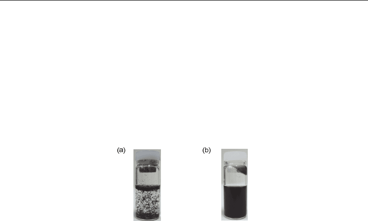
to give an aggregation in the solvent as shown in Fig. 1a.(Nakashima & Fujigaya, 2007)
Yodh et al.(Islam, et al., 2004) used a surfactant as a dispersant of the SWNTs in the
preparation of a SWNT/PNIPAM gel in an aqueous solution, and examined liquid
crystalline nature of the composite gel. However, the addition of anionic surfactants
dramatically increases the transition temperatures (T
c
) of the PNIPAM in an aqueous
system.(Kokufuta, et al., 1993) To avoid such a problem, we introduced carboxylate groups
onto the SWNTs surface by treating with strong acid to improve the dispersibility of the
SWNTs in an aqueous system. The obtained acid-treated SWNTs were dispersed in
deionized water with only mild shaking (Fig. 1b). The SWNT/PNIPAM composite gel
preparations were carried out according to the typical procedure of PNIPAM gelation
(Tanaka, 1978) in the absence of the SWNTs.
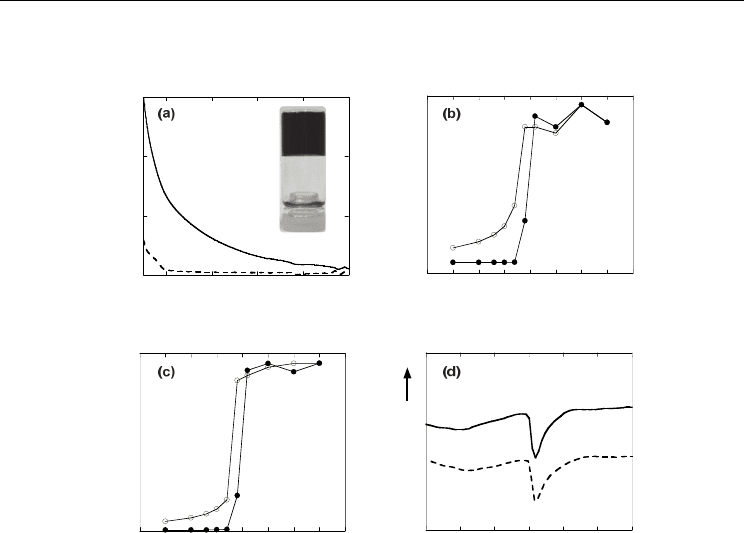
Fig. 1. SWNT dispersion solutions, (a) As-prepared SWNT dispersion in water. (b) Acid-
treated SWNT dispersion in water.
The visible-NIR spectrum of the synthesized composite gel (Fig. 2a; inset) showed absorption
over a wide range of the spectrum (solid line in Fig. 2a), while the gel without the SWNTs has
no absorption in the visible-NIR region (dotted line in Fig. 2a). The composite gel was a
transparent grey-color at temperatures below 23 °C, while upon heating in a temperature-
controlled water bath, the gel changed to opaque at around 34.6 °C, suggesting that the
transition temperature (T
c
) of the SWNT/PNIPAM composite gel was virtually identical with
that (T
c
= 34.0 ˚C) of the SWNTs/PNIPAM gel (control sample). The phase transition of the
PNIPAM gel was also investigated using UV-visible spectroscopy and differential scanning
calorimetry (DSC). By plotting the absorbance at 600 nm, the transition temperature is
determined to be ca. 33 °C (Fig. 2b). The T
c
of the SWNTs/PNIPAM gel both upon heating and
cooling were virtually identical to that of the gel without the SWNTs prepared as a control
(Fig. 2c). In DSC, the endothermic peaks were appeared at around 35 °C for both the
SWNT/PNIPAM and the PNIPAM gel (Fig. 2d), which agreed well with the results described
above. It is clear that the SWNTs in the composite gel have virtually no effect on the phase
transition temperature of the PNIPAM gel.
2.2 NIR irradiation to SWNTs/PNIPAM gel
We used the SWNTs as a “molecular heater” for the thermal phase transition of the
composite gels. NIR laser irradiation experiments for the SWNT/PNIPAM gel and the
PNIPAM gel were carried out for the gel tubes prepared and drawn from the capillary tube
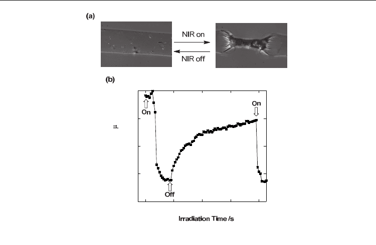
with inner diameter around 200 m. As shown in Fig. 3a, upon irradiation with the NIR
light centered at 1064 nm, the composite gel tube (initial diameter d
0
≈ 240 m) containing
the SWNTs become a smaller diameter (d ≈ 80 m) after 15 sec. As plotted in Fig. 3b that
after turning off the irradiation, the shrunken gel gradually swells and becomes around 200
Single-Walled Carbon Nanotubes
as a Molecular Heater for Thermoresponsive Polymer Gel Composite
381
m in diameter after about 67 sec. The further shrink-swell experiment upon ON/OFF
irradiations was described in following session.
0
0.5
1
1.5
400 600 800 1000 1200
Absorbance / a.u.
Wavelen
g
th / nm
15 20 25 30 35 40 45 50 55
Absorbance / a.u.
Temperature /
o
C
15 20 25 30 35 40 45 50 55
Absorbance / a.u.
Temperature /
o
C
20 25 30 35 40 45 50
Heat Flow
Temperature /
o
C
exo
Fig. 2. Characterizations of a SWNT/PNIPAM gel, (a) Visible-NIR spectra of a
SWNT/PNIPAM gel (solid line) and a PNIPAM gel (dotted line) at 20 °C. inset; photo of
SWNT/PNIPAM gel (b) Temperature dependence of the optical absorption at 600 nm of a
SWNT/PNIPAM gel was recorded for a heating process (solid circle) and a cooling process
(open circle). (c) Temperature dependence of the optical absorption at 600 nm of a PNIPAM
gel (controlled sample) upon heating process (solid circle) and a cooling process (open circle).
(d) DSC curves of the gels of SWNT/PNIPAM gel (solid line) and PNIPAM gel (dotted line).
A stronger NIR laser power (> 1.2 W) has been reported to increase the temperature through
the photothermal conversion of water.(Ishikawa, et al., 1996) However, this effect is
excluded under our experimental conditions since the 210-mW NIR irradiation to the gel
(controlled sample) prepared from the single component of PNIPAM showed no such
volume phase transition even after the NIR-laser irradiation. It is conclusive that the NIR
light irradiation to the SWNT/PNIPAM gel caused the phase transition of the PNIPAM gel
via the photothermal conversion of the SWNTs. This contrasting result between the
SWNT/PNIPAM gel and PNIPAM gel is the consequence of NIR absorption of the
SWNT/PNIPAM gel at around 1064 nm (solid line in Fig. 2a), whereas the gel without the
SWNTs has no absorption in the NIR region (dotted line in Fig. 2a). The proposed
mechanism is that the local heating of the SWNTs due to the irradiation raises the
temperature of the water around the irradiation spot in the gel over 35 °C, which induces
the phase transition of the PNIPAM gel as discussed for the visible-light induced phase
transition of the PNIPAM.(Suzuki & Tanaka, 1990) Here, the SWNTs act as a “molecular
heater” to induce the phase transition of the PNIPAM in the composite gel.
Carbon Nanotubes – Polymer Nanocomposites
382
50
100
150
200
250
0 20406080
Gel Diameter
/
m
Fig. 3. NIR-induced phase transition, (a) NIR laser-driven volume change of a
SWNT/PNIPAM gel shown by optical microscopic images. (b) Plot of the diameter of the
SWNT/PNIPAM gel plotted as a function of time.
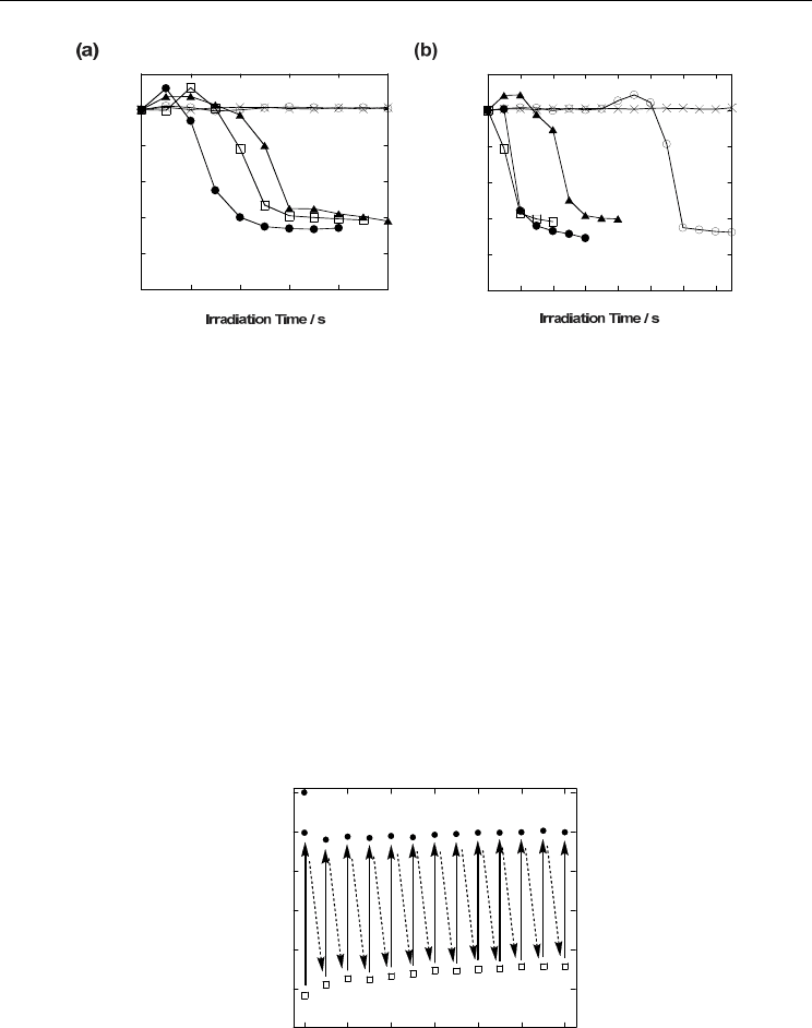
The response time of the volume change can be controlled by changing the concentration of
the SWNTs as well as the power of the NIR laser light. The d/d
0
values obtained by the 210-
mW or 390-mW irradiations of the composite gels synthesized using the different
concentrations of the acid-treated SWNTs in water (2.0, 1.0, 0.5, 0.25, and 0.13 mg/mL) are
plotted as a function of the irradiation time in Fig. 4a (210-mW irradiation) and Fig. 4b (390-
mW irradiation). In the case of the 210-mW irradiation, the NIR-driven volume change in the
gel samples synthesized at the concentrations of acid-treated aqueous SWNTs=2.0, 1.0, and 0.5
mg/mL almost finished in 4, 5, and 6 sec, respectively, whereas the gels synthesized from the
lower concentration of the SWNTs = 0.25 and 0.13 mg/mL showed no phase transition.
On the other hand, at a 390-mW irradiation, the gels synthesized at the concentration of the
SWNTs= 2.0, 1.0, 0.5, and 0.25 mg/mL shrunk within 2, 2, 5, and 12 sec, respectively, while the
gel prepared at SWNTs=0.13 mg/mL showed no change. The absence of the phase transition
observed for the gels from the SWNTs=0.25 and 0.13 mg/mL upon 210-mW irradiation and
SWNTs=0.13 mg/mL upon 390-mW irradiation suggests the importance of the initial process,
that is, the dark spot generated on the focal point enhances the absorption of the NIR light in
that region, which accelerates the heating of the gels. The rate of this initial process varies with
the concentration of the SWNTs as well as the NIR laser irradiation power. It is clear that the
higher-powered light irradiation and the higher concentrations of the SWNTs in the gels
render the composite gels a faster response. Of interest, for the gels especially with 2.0 and 1.0
mg/mL concentrations, we observed a bubble generation from the center of the irradiation
spot upon the 390-mW irradiation. This phenomenon indicates that the temperature at the
spots in the gels reached over 100 ˚C due to the photothermal conversion of the SWNTs. All
the obtained results guarantee that the phase transition of the composite gels is triggered by
photon absorption of the SWNTs in the NIR region.
Single-Walled Carbon Nanotubes
as a Molecular Heater for Thermoresponsive Polymer Gel Composite
383
0
0.2
0.4
0.6
0.8
1
1.2
0246810
d/d
0
0
0.2
0.4
0.6
0.8
1
1.2
02468101214
d/d
0
Fig. 4. Effect of SWNT concentration, The d/d
0
values of the composite gels synthesized
using the different concentrations of acid-treated aqueous SWNTs = 2.0 (•), 1.0 (□), 0.5 (▲),
0.25 (○), and 0.13 (X) mg/mL on a (a) 210-mW and (b) 390-mW NIR laser irradiation plotted
as a function of irradiation time.
2.3 Durability of SWNTs upon NIR irradiation
The SWNTs possess a robust structures arising from their rigid fused aromatic structures,
and thus the SWNT-composite gels are expected to show high durability for the repeated
ON/OFF-laser irradiation. We carried out an endurance test using the hybrid gel under
optical micrograph monitoring, in which the tests were conducted by a programmed NIR
laser operation repeated with ON irradiation for ca. 4 sec, followed by OFF irradiation for 55
sec to ensure the shrink-swell cycles (Fig. 5). Amazingly, no notable deterioration of the gel
actuation was observed even after the 1200-cycle operation. As shown in Fig. 6, the Raman
spectra of the gels before (dotted line) and after (solid line) the endurance test supports this
durability, namely, both spectra exhibit virtually identical G/D (Graphite/Defect) ratios,
which guarantee that the SWNTs remain structurally intact.
0.4
0.5
0.6
0.7
0.8
0.9
1
0 200 400 600 800 1000 1200
d/d
0
Cycle number
Fig. 5. Durability of the composite gel, The d/d
0
values of a SWNT/PNIPAM gel for ON
(open square)/OFF (solid circle) switching of the NIR laser light irradiation plotted as a
function of the cycle number. Data are collected every 100 cycles to avoid complication.
Carbon Nanotubes – Polymer Nanocomposites
384
It is readily expected that the composite gel system can tolerate additional operations of
more than 1200 cycles. We tested the gold nanorod (Au-NR)-mediated systems(Gorelikov, et
al., 2004; Sershen, et al., 2005; Sershen, et al., 2000; Sershen, et al., 2001; Shiotani, et al., 2007)
as another candidate for the NIR actuation of PNIPAM gels since Au-NR has an absorbance
in the NIR region. The extinction coefficient of the Au-NR/PNIPAM mixture at 1064 nm is
matched to that of the SWNT/PNIPAM mixture used in the endurance test for the fair
comparison. Au-NR in the Au-NR/PNIPAM gel caused an aggregation of the particles after
the 1200-cycle durability test, namely, we observed a blue shift in the peak that stemmed
from the plasmon band of Au-NR at around 800 nm. It is evident that the SWNT/NIPAM
gels have a stronger durability for the NIR irradiation.
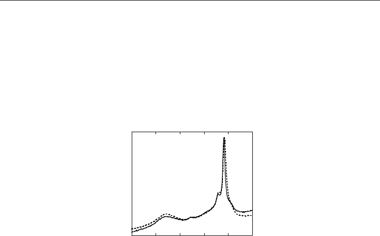
1200 1300 1400 1500 1600 1700
Intensity / a.u.
Raman Shift / cm
-1
Fig. 6. Estimation of damage for SWNTs, Raman spectra of the gel before (dotted line) and
after (solid line) the 1200-cycle irradiations.
3. Preparation and applications of SWNTs/PNIPAM gel using pristine SWNT
(Fujigaya, et al., 2011)
3.1 Preparation of the SWNTs/PNIPAM gel composite
One of the goals of the study is to design and fabricate a NIR laser-driven drug-releasing
system using isolated SWNTs as the material, in which the SWNTs having large surface
areas embedded in a polymer gel serve as an efficient drug reservoir through
physisorption of the molecules onto SWNTs. -conjugating graphitic surface induces a
molecular adsorption thorough many different interaction mode such as ,
hydrophobic and van der Waals interactions. To realize an effective adsorption of the
drug typically have a hydrophobic nature, employment of pristine SWNTs is essential
since the surface of acid-treated SWNT possess many carboxylic group and turned to
hydrophilic nature.
The preparation strategy of the composite gel are illustrated in Fig. 7. The composite gel
composed of pristine SWNTs was prepared by the gelation of NIPAM in the presence of
pristine SWNTs individually dissolved in an aqueous solution of the sodium dodecyl
benzenesulfonic acid sodium salt (SDBS). SDBS was chosen as an SWNT dispersant since it
is known to individually dissolve the SWNTs quite efficiently by encapsulating the SWNTs
into the interior of the SDBS micelle in aqueous media.(McDonald, et al., 2006) The
composite SWNT/PNIPAM gel was almost transparent similar with a PNIPAM gel without
SWNT and slightly grey-colored due to the presence of a SWNTs. The mesh size of the
SWNT/PNIPAM gel in aqueous media was determined by dynamic light scattering (DLS)
to be around 10~20 nm (data not shown) which well agreed with previous reports.(Canal &
Single-Walled Carbon Nanotubes
as a Molecular Heater for Thermoresponsive Polymer Gel Composite
385
A. Peppas, 1989; Ishidao, et al., 1997) The observation of the intense photoluminescence (PL)
signals from the composite gel guaranteed the isolation of the SWNTs even after the gelation
since the PL signals are proved to be detected only from the individually isolated SWNTs
(Fig. 8a).(O'Connell, et al., 2002)
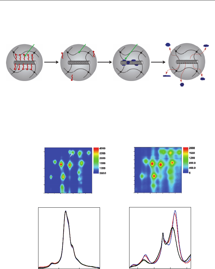
②
Surfactant
Gel
Gel network
DOX
123 4
Fig. 7. Schematic illustration showing the strategy of this study. Stage 1, Gelation carried out in
an SDBS (dark blue) solution produces an SDBS-adsorbed SWNT in the gel. Stage 2, SDBS on
the SWNT surface are removed by dipping the gel in Milli-Q water. Stage 3, The adsorption of
drug molecules (red), occurs on the “vacant” SWNT surfaces through π-π and hydrophobic
interactions. Stage 4, The drug molecules on SWNT surfaces detach from the surfaces by a pH
change or NIR laser irradiation by a photothermal conversion effect of the SWNTs.
150200250300
Intensity
Raman Shift / cm
-1
(10,5)
(10,2)
2000240028003200
Intensity
Raman Shift / cm
-1
G'
(8,3)
(7,5)
(7,6)
(a) (b)
(c) (d)
1000 13001100 1200
Emission Wavelength / nm
550
650
750
600
700
800
Excitation Wavelength / nm
1000 13001100 1200
Emission Wavelength / nm
550
650
750
600
700
800
Excitation Wavelength / nm
(7,5)
(7,6)
(8,4)
(9,4)
(10,2)
(8,6)
(8,7)
(10,5)(11,3)
(9,5)
(10,3)
Fig. 8. Isolation of SWNTs in PNIPAM gel, (a,b) 2D PL mapping of the SWNT/PNIPAM gel
before (a) and after (b) dipping in Milli-Q water for 72 h. (c,d) Raman spectra of the gel
normalized at the (10,5) peak and G’ peak for c and d, respectively; the red, blue, green and
black lines show the spectra after the immersion in water for 24, 48, and 72 h, respectively.
Carbon Nanotubes – Polymer Nanocomposites
386
In order to remove the SDBS molecules from the surfaces of the SWNTs, the composite gel
was immersed in a large amount of Milli-Q water for 72 h. The Raman spectra of the SWNTs
in the composite gel were monitored during the immersion in water by measuring the radial
breathing mode (RBM) (Fig. 8c) together with the PL signals(Jeng, et al., 2006; Moore, et al.,
2003) (Fig. 8b). We recognized that the PL peak from the (8,3) SWNT bathochromically shifts
from 2290 to 2350 cm
-1
due to the increase in the micropolarity around the SWNTs as
reported in the literature (Fig. 8d).(Jeng, et al., 2006; Moore, et al., 2003; Strano, et al., 2003)
Such a clear shift as well as the decrease in the peak intensity after the immersion in water
indicated that the SDBS molecules were removed from the SWNT surfaces and replaced by
water molecules.(Strano, et al., 2003) In order to confirm the removal of the SDBS from the
SWNT surfaces in the composite gel, we measured the X-ray photoelectron spectrum (XPS)
of the SWNT/PNIPAM gel after drying, and found that the sulfur signal almost
disappeared in the sample after the immersion in water (Fig. 9).
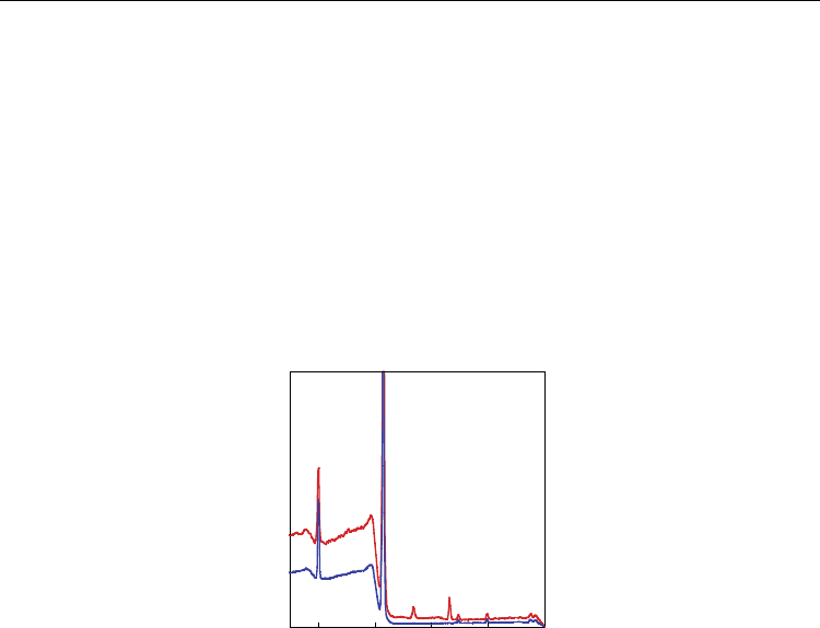
0100200300400
Binding Energy / eV
Intensity
N
1s
C
1s
S
2s
S
2p
Fig. 9. X-ray photoelectron spectra, The spectra of SDBS/SWNT/PNIPAM gel before (red
line) and after (blue line) immersion in deionized water.
Moreover, it is also important to emphasize that we observed a clear PL from the SWNTs in
the gel after the immersion, whose PL-mapping (Fig. 8b) is virtually identical with that
before the immersion (Fig. 8a). This suggests that the SWNTs remained in an isolated state
even after the removal of the SDBS molecules. The Raman data also support this, namely,
the intensities of the (10,2) RBM peak of the gel before and after immersion at around 267
cm
-1
, which is known as an indicator for the evaluation of the degree of the SWNT
aggregation,(Ericson & Pehrsson, 2005; Fujigaya, et al., 2009; Heller, et al., 2004; Kumatani &
Warburton, 2008; Luo, et al., 2006; O'Connell, et al., 2004; Strano, et al., 2003) are virtually
identical (Fig. 8c). The obtained results are in sharp contrast to those previously reported, in
which the SWNTs aggregate after removal of the dispersants.(Chen, et al., 2008; Ikeda, et al.,
2009; Ishibashi & Nakashima, 2006; Nobusawa, et al., 2008) We suggest that the SWNTs (~ 1
m) penetrated the three-dimensional gel network structure having a 10~20-nm mesh to
form the semi-interpenetrating network (semi-IPN) structure,(Gong, et al., 2003) and the
formed structure serves to prevent further assembling (aggregation) of the SWNTs even in
the absence of the dispersants. To our surprise, PL signals were also observed from the re-
swelled gel after drying in a vacuum, indicating that the isolated state of the SWNTs in the
gel is highly stable in the three-dimensional gel framework.