Yang J. (ed.) Biometrics
Подождите немного. Документ загружается.

Finger Vein Recognition
39
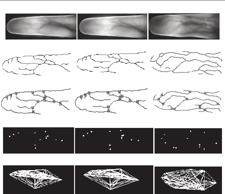
(i) (ii) (iii)
(a) The raw image of finger-vein
(b) The image after thinning
(c) The image after repairing and marking the intersecting points
(d) The image after extracting the intersecting points
(e) The fully meshed image after connecting the intersecting points
Fig. 8. The finger-vein image feature extraction process
In Fig.8, (i) and (ii) are two finger-vein images from same source, so their topology is
similar. However, (iii) is of a different source and its topology is obviously different from (i)
and (ii). Specifically, the topology expresses an integral property and peculiarity of finger-
veins, the relationship between corresponding character points is of importance.
3.1.3 Matching finger-vein images using relative distance and angles
From the thinned finger-vein image of Fig.8 (b), we can see the random finger-vein pattern
and inner structure. The inner characteristic points produced by the intersecting vein
crossings reflect the unique property of the finger-vein. However, those breakpoints may be
thought as finger-vein endpoints, which would influence recognition results. For this
reason, the more reliable intersecting points are chosen to characterize finger-veins.
Considering that different line segments are produced by intersecting points from different
finger-vein images, the two features -relative distance and angle - are combined for
matching.
Relative distance and angle are essential attributes of finger-veins, which ensure the feature
uniqueness and reflect different characteristics of finger-vein structure. Fusion of the two

Biometrics
40
features with “Logical And” make the recognition results more reliable. Thus, matching two
finger-vein images is converted into matching the similarity of topologies.
The detailed steps are as follows.
1. Calculate the relative distances and angles of finger-vein image. Suppose, there are d
points of intersection in one image, then the number of relative distance is
(1)/2dd .
The number of angles produced by the point connections is
(1)(2)/2dd d . Here a set
of finger-vein image features is defined as
(, )
mu
Rl , where l is the distance of any
two intersecting points,
is the angle produced by the point connections, m and u are
the number index respectively. Suppose,
1
(, )
mu
Rl and
2
(, )
nv
Rl are two sets of
finger-vein image features.
2. Compare m relative distances from
1
R with n relative distances from
2
R , by
calculating the number of approximately similar relative distances. If the number is
greater than the pre-defined threshold, go to next step; else, the matching is assumed to
have failed. To take care of position error of those points, we define
mn
ll e to
show the extent of similarity between any two Eigen values ( e is the allowable error
range). From experimental analysis, e =0.0005 is very appropriate.
3. Suppose there are
q
eigenvalues of approximately equivalent relative distances,
connect the
q
character points in the two sets respectively, with each other. Thus,
z angles are produced, which are denoted as
1z
and
2z
in
1
R and
2
R respectively.
On this basis, calculate the number of approximately equivalent angles. If the number
is greater than the pre-defined threshold, the matching is successful; else, the
matching is thought to have failed. Similarly,
mn
e is used to show the
relationship of two approximately equivalent. From experimental analysis,
e =0.006°
is very appropriate.
3.2 Finger vein recognition based on wavelet moment fused with PCA transform
3.2.1 Finger vein feature extraction
Different people have different finger lengths. Also, there can be variation in the image
captured for the same person due to positioning during the image capture process. Thus if
image sizes are not standardized, there is bound to be representation error which leads to a
decrease in the recognition rate. In this part, we resize the vein image into a specific image
block size to facilitate further processing. The original image is standardized to a height of
80 pixels and split along the width into 80 × 80 sub-image block size. If the image is split
evenly (given that the image width is generally about 200 pixels) there will be loss of
information that will affect recognition. Therefore, the sub-blocks are created with an
overlap of 60 pixels for every 80 × 80 image sub-block. The original image can thus be split
into 6-7sub-images, with sufficient characteristic quantities for identification.
Set a matrix
mn
A to represent the standardized images (,)fxy.
012 1
[ , , ,..., ]
mn n
AAAAA (20)
Which:
i
A is a column vector,
[0, 1]in
Here we define the sub-block of the image width
w , standardized image height h (in
experiment
w=80,h=80 ). Sub-images are extracted at interval r when (in this experiment
r =20).

Finger Vein Recognition
41
Thus can get sub-image matrix:
10 1
21
1
,...,
,...,
...
,...,
w
rwr
kkr wkr
BA A
BAA
BA A
[x] takes the maximum integer less than
x .
1
nw
k
r
(21)
Thus we get a total of
12
, ,...,
k
BB B
k sub- image, and the size of each sub-image is wh.
Then we extract features for each sub-image B
i
. The finger block, feature extraction and
recognition process shown in Fig.12.
1
v
1
B
2
B
k
B
2
v
k
v
12
; ;...;
k
Vvv v
feature vector:
1
v
1
B
2
B
k
B
2
v
k
v
12
; ;...;
k
Vvv v
feature vector:
Fig. 12. The sketch map of sub-image extraction
3.2.2 Wavelet transform and wavelet moments extraction
Wavelet moment is an invariant descriptor for image features. A wavelet moment feature is
invariant to image rotation, translation and scaling so it is successfully applied in the pattern
recognition.
For each sub-image
,
i
Bx
y
, its size is
wh. Applying two dimensional Mallat
decomposition algorithm, we can make wavelet decomposed image
,
i
Bx
y
.

Biometrics
42
Fig. 13. The sub-image and result of wavelet decomposition
Setting
22
,,()
i
fxy Bxy LR to be the analyzed sub-image vein blocks, the wavelet
decomposed layer is
123
111 1
(,)
f
x
y
ADDD
(22)
where
1
A is the scale for the low frequency component (i.e. approaching component), and
123
111
,,DDD are the scales for the horizontal, vertical and diagonal components respectively.
111
(,)
11
,,,
,(,),,
mn
A c mn mn
cmn fxy mn
(23)
11 1
(,)
11
,,,
(,) (,), (,)
kk k
mn
kk
D d mn mn
dmn fxy mn
(24)
Where 1,2,3k
1
(,)cmn is the coefficient of
1
A
1
k
d is the coefficient of the three high frequency components.
1
(,)mn is the scale function
1
(,)
k
mn is the wavelet function
Daub4 was chosen for wavelet decomposition, as it produced better identification results
from several experimental compared with other wavelets. We use the approximation
wavelet coefficients
j
c to compute the wavelet moment [4]. Set
,
pq
w expressed as (p+q)
order central moments of
(,)
f
xy . The wavelet moment approximation is:
(1)
0
,1
,
(1)
,1
,
2,
2(,)
pq j p q
pq
mn Z
pq j p q
kk
pq
mn Z
wmncmn
wmndmn
(25)
Here we access the wavelet moment
22
w .
3.2.3 PCA transformation
The advantage of using wavelet transform to reduce computation is explored here. Using
each sub-image
i
B directly without the PCA transform not only leads to poor classification
of extracted features but also huge computational cost. After the low-frequency wavelet sub-
Finger Vein Recognition
43
images compression of the original image to about one-fourth of the original size, PCA
decomposition is applied on the sub-image which greatly reduces computation.
3.2.4 The transformation matrix
Here we analyze a layer of
i
B wavelet decomposition of the low-frequency sub-image. For
PCA,
1
A is transformed to a separate / 4wh dimension of image vector
1
()Vec A .
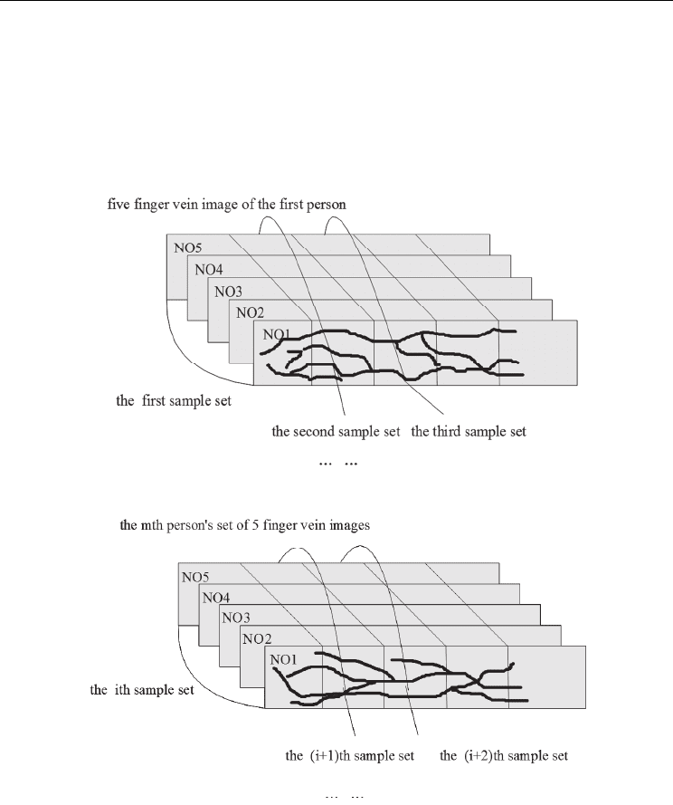
Five finger vein images per person (*same finger)
The Five finger vein images of the m
th
person
Fig. 14. The sketch map of sample classes after PCA transform
To illustrate the problem, we take finger vein samples from a total set of
c people. Each
sample of the same finger has five images as shown in Fig.14. (Note that Fig.14 is only for
illustration purpose. In practice, there is an interval of 20pixels between two adjacent sub-
blocks, and an overlap of 60pixels as earlier described).
The n-th sub-block set of the m-th person is indexed as
,mn
k ; where n = (1,2,3…., L) ;
m
L is
the total number of sub-blocks for person m.

Biometrics
44
To compare images we only use the
min
k sub-image set where
min
k
= L = min
123
(,,...)
m
LLL L
. when
min
k
= 5, then
min 1,1 1,2 1,5 ,5
min( , ,...,, ,..., )
c
kkkkk (26)
Thus, a total of
min
Cck of the available pattern classes, i.e.
12
, ,...,
C
.
Four of the corresponding samples in the i-th class (
5imk),
min
1,2,...kk is the number
of sub-image of each finger image, for simplicity, we write:
,1 ,2 ,3 ,4 ,5
,,,,
iiiii
, and all
are / 4wh dimension column vectors. The total number of training samples is 5NC.
Mean of the i-th class training sample:
5
,
1
1
5
ii
j
j
(27)
Mean of all training samples:
5
11
1
C
ij
ij
N
(28)
The scatter matrix is:
1
()( )( )
C
T
iiti
i
SP (29)
where
()
i
P
is prior probability of the i-th class of training samples. Then we can obtain the
characteristic value
12
, ,..., of
1
S (the value of these features have been lined up in
sequence by order of
12
..., ) and its corresponding eigenvector
12
, ,..., . Take d before
the largest eigenvalue corresponding to the standard eigenvectors orthogonal
transformation matrix
12
[,,...,]
d
P . For each sub-image blocks
i
B , through the wavelet
decomposition of the low-frequency sub-image
1
A ,
1
A in accordance with the preceding
method into a column vector
, to extract the features use the transformation matrix
P
obtained in the previous section, the following formula:
T
eP (30)
This
12
[,,...,]
d
eee e
is the PCA extraction of feature vectors from sub-image blocks. After
several experiments, we found that when
200d we can get a good result, and when
300d i.e a 300-dimensional compression, we get the best recognition results.
3.2.5 LDA map
In general, PCA method is the best for describing feature characteristics, but not the best for
feature classification. In order to get better classification results, we use the LDA method for
further classification of PCA features.
Each sample is transformed into a lower d -dimensional space in the post-dimensional
feature vector
12
[,,...,]
ii i
id
eee e. Using PCA projection matrix P,
1,2,...,iN is the sample
number. Our classifier design follows dimension reduction to get PCA feature vectors

Finger Vein Recognition
45
12
, ,...,
N
ee e and form the class scatter matrix
w
S and the within class scatter matrix
b
S .
Calculate the corresponding matrix
1
wb
SS of the l largest eigenvalue
eigenvectors
12
, ,...,
l
. The l largest eigenvectors corresponding to the LDA
transformation matrix
12
[,,...,]
LDA l
W . Then we use the LDA transformation matrix
LDA
W as
12
[,,...,]
ii i T
ilLDAi
zzz zWe ,
1,2,...,iN is the sample number. (31)
Thus, we can use the best classification feature z vector to replace the feature vectors e for
identification and classification.
3.2.6 Matching and recognition
Through the above wavelet decomposition and PCA transform for each sub-image
i
B , we
obtain wavelet moments
22
w and extract feature vector z of PCA and LDA.
i
B is
characterized by
22
[;]
i
vwz. Matching feature vectors of finger1
22
[;]
i
vwz and of finger 2
can be done as follows.
The first step is the length of V and
'
V
and may not be the same, that is, k and
'
k
is not
necessarily the same. Here we define:
min( , ')Kkk (32)
Taking the
K
vectors of
V
and
'V
for comparison, first analyze the corresponding sub-
image blocks
i
v and '
i
v .
22
;
i
vwz ,
22
'';'
i
vwz (33)
From several experiments, we set two threshold vectors
t
w
t
z . Euclidean distance between
i
B sub-images,
i
is defined for two feature vectors w and 'w from V and 'V . A matching
score defined for
V and 'V feature
22
w of wavelet moment of corresponding sub-image
i
B
matching score:
_
0
ti
it
t
i
w
if w
w
wmark
else
(34)
Finally, we obtain wavelet moment feature of the finger matching score:
0
__
K
i
i
wmark wmark (35)
Similarly, we create a match
V
and
'V
, of the feature vector z scores _zmark.
Finally, combining the scores:
12
___total mark s w mark s z mark (36)
12
,ss
are the share of feature matching scores, and
12
0, 0,ss
12
1ss
.
Biometrics
46
Thus, if the finger vein 1 and 2 match _total mark value score is greater than a given
threshold, the two fingers match, otherwise they do not match. A minimum distance
classifier can also be used for the recognition task.
4. Experimental results
4.1 Processing experimental results
To verify the effectiveness of the proposed method, we test the algorithm using images from
a custom finger vein image database. The database includes five images each of 300
individuals’ finger veins. Each image size is 320*240.
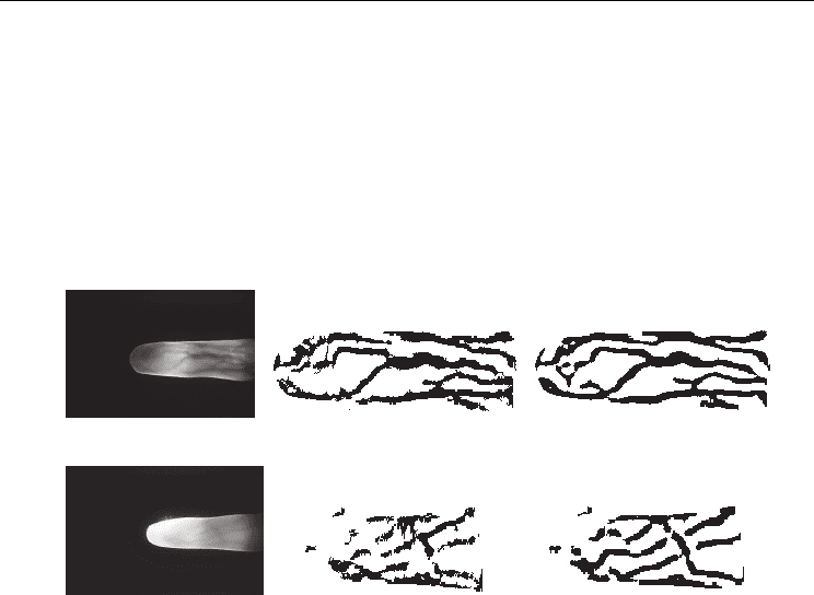
(a) Original image 1 (c) NiBlack segmentation method (d) Our method
(b) Original image 2 (e) NiBlack method (f) Our method
Fig. 15. Experimental results
We have used a variety of traditional segmentation algorithms and their improved
algorithms to segment vein image. But segmentation results of vein image by these
algorithms aren’t ideal. Because the result of NiBlack segmentation method is better than
other methods [13], we use NiBlack segmentation method as the benchmark for comparison.
Segmentation was done for all the images in our database using NiBlack segmentation
method and using our method. Experimental results show that our method has better
performance. To take full account of the original image quality factor, we select two typical
images from our database with one from high quality images and the other from poor
quality images to show the results of comparative test. Where Fig. 15(a) is the high-quality
vein image in which veins are clear and the background noise is small. Fig. 15(b) is the low-
quality vein image. The uneven illumination caused the finger vein image to be fuzzy,
which seriously affects image quality. We extract veins feature by using our method and
compare with results of the NiBlack segmentation method. Experimental results shown in
Fig. 15(c) and Fig. 15(e) are obtained from the NiBlack method applied in [9]. This algorithm
extracts smooth and continuous vein features of high-quality image. There are a few
pseudo-vein characteristics in Fig. 15(c). But in Fig. 15(e), there is much noise in the
segmentation results. Segmented image features have poor continuity and smoothness, and
there is the effect of the over-segmentation. Experimental results show that apart from
smoothness and continuity or removal of noise and pseudo-vein characteristics, the method
proposed in this paper extracts vein features effectively not only from the high-quality
Finger Vein Recognition
47
images but also from the low-quality vein images as shown in Fig.15(d) and Fig.15(f). We
show that the algorithm proposed in this paper performs better than the traditional NiBlack
method.
4.2 Relative distance and angle experimental results
Finger-vein images (size 320×240) of 300 people were selected randomly from Harbin
Engineering University finger-vein database. One forefinger vein image of each person was
acquired, so there are 300 training images.
Generally, a good recognition algorithm can be successfully trained on a small dataset to get
the required parameters and achieve good performance on a large test dataset. Therefore
four more images from the forefinger of those 300 people were acquired giving a total sum
of 1200 images to be used as verification dataset.
When matching, every sample is matched with others, so there are (300×299)/2 = 44850
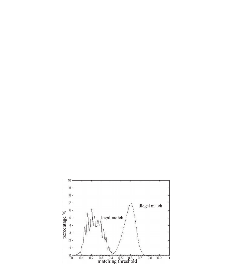
matching times; 300 of which are legal, while the others are illegal matches. Two different
verifying curves are shown in Fig.16. The horizontal axis stands for the matching threshold,
and vertical axis stands for the corresponding probability density. The solid curve is legal
matching curve, while the dashed is illegal. Both curves are similar to the Gaussian
distribution. The two curves intersect, at a threshold of 0.41. The mean legal matching
distance corresponds to the wave crest near to 0.21 on the horizontal axis, and the mean of
illegal matching distance corresponds to the wave crest near to 0.62 on the horizontal axis.
The two wave crests are far from each other with very small intersection. So this method can
recognize different finger-veins, especially when the threshold is in the range [0.09-0.38],
where the GAR is highest.
Fig. 16. Legal matching curve and illegal matching curve
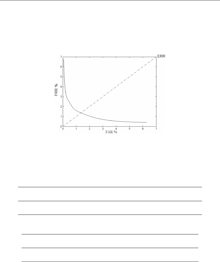
The relationship between FRR and FAR is shown in Fig.17. For this method, the closer the
ROC curve is to the horizontal axis, the higher the Genuine Acceptance Rate (GAR). Besides,
the threshold should be set suitably according to the fact, when FRR and FAR are
equivalent, the threshold is 0.47, that is to say, EER is 13.5%. In this case, GAR of the system
is 86.5%. The result indicates that this method is reasonable, giving accurate finger-vein
recognition.
The method above compares the numbers of relative distances and angles which are
approximately equivalent from two finger-vein images. In the second step, only the
Biometrics
48
intersecting points which are matched successfully on the first step are used and thus,
computation of superfluous information is avoided and only information vital to decision
making is used. Only when the two matching steps are successful is the recognition
successful. According to Theorem 3, the relative distance and angle would not change when
even after image translation and rotation. So the proposed algorithm is an effective method
for finger-vein recognition.
Fig. 17. ROC curve of the method
In 1:1 verifying mode, compare the one image out of 1200 samples in verifying set with the
image, which has the same source with the former one, in training set to verify. The
experiment result is shown in Tab.1, the times of success are 1120, and the rate of success is
93.33%. In 1:n recognition mode, compare the 1200 images with all images in training set,
360000 times in sum. The result is as Tab.2, the times of FAR is 25488, and GAR is 92.92%.
Total matching
times
Number of
successes
Number of
failures
Success rate
(%)
FRR(%)
1200 1120 80 93.33 6.67
Table 1. Test result of FRR in 1:1 mode
Total matching
times
Total false
acceptance
GAR (%) FAR(%)
360000 25488 92.92 7.08
Table 2. Test result of FAR in 1:n mode
To test the ability to overcome image translation and rotation, translate randomly in the
range
[10,10] and rotate the image randomly in the range
00
[10,10], in order to establish
the translation and rotation test sets. Then verify and recognize the two sets respectively.
The samples which have the same source are compared in a 1:1 experiment; matching each
sample from the two sets with the samples from the training set to accomplish 1:n
experiment. The result is shown in Tab.3, Tab.4, Tab.5 and Tab.6.
