Wunderlich W. (ed.). Ceramic Materials
Подождите немного. Документ загружается.

Ceramic Materials and Color in Dentistry 163
based on age, duration of exposure of the eye, fatigue or illness related to color, such as color
blindness. (Van Der Burght et al., 1990). In instrumental analysis, colorimetric instruments
objectively observe and record color. Two types of equipment have been used: colorimeters
and spectrophotometers. Colorimeters analyze the values for red, green and blue reflected
through filters that simulate sensing photoreceptor cells of the human eye. The tristimulus
X, Y and Z coordinates are automatically converted to L*, a*, b* values of the CIE L*a*b*
system. (Gliese & Petter, 2000). Spectrophotometers measure and record the amount of light
reflected or transmitted from the object through its wavelength. These devices have high
precision, sensitivity to measure absolute colors and are equipped with spectral
distributions of various illuminants. The most accurate spectrophotometers are those of
integrating spheres, called spherical optical, in which the object is exposed to light at
different angles and directions for its analysis. (Paravina, 2002).
Light source or illuminant: The illuminant may be a natural or artificial light source, which,
according to its origin, can change the perceived color of an object. For example, a white
sheet of paper may seem bluer under fluorescent light and more yellowish under a light
bulb, returning to its original color in the presence of daylight. This phenomenon is known
as metamerism. (Knispel 1991). Aiming to standardize the visual and instrumental analysis
of color, the International Commission of I'Eclairage (CIE) has classified illuminants
according to their effect on the perception of color. The CIE appointed 3 standard
illuminants: A, B and C. Later, the illuminant series D, the hypothetical illuminant E and
unofficially the illuminant series F were added (Chu et al., 2004):
Illuminant CIE definition
A Tungsten light source with an average temperature of 2,856 K, which
produces a reddish-yellow light. Generally used to simulate conditions of
incandescent light.
B Tungsten light source coupled to a liquid filter to simulate direct sunlight
with an average temperature of 4,874 K.
C Tungsten light source coupled to a liquid filter to simulate indirect
sunlight with an average temperature of 6,774 K. However, it is not a
perfect simulation of sunlight, because it contains a large amount of
ultraviolet light needed in the analysis of fluorescence.
D Series of illuminants representing different conditions of the day.
Illuminants D50 and D65 (so called due to temperatures of 5,000 K and
6,500, respectively) are generally used as pattern illuminants and match
the reflectance of blue light.
E It is a theoretical light source, in which an equal amount of energy would
be present at all wavelengths. Currently this illumination does not exist,
but this value is used as a tool for scholars of hypothetical colorimetry.
F It involves a series of fluorescent lights. Fluorescent lamps have peaks
that are evident in their spectral curves and do not fit the color
temperature, therefore, are not considered an officially illuminating
pattern. However, as the fluorescent display is widely used, the CIE
recommends these illuminations to evaluate colors observed in the
fluorescent environment.
Table 1. Series of illuminants
4.3 Colorimetric parameters
In 1936, Munsell (O'Brien et al., 1989) described the three dimensions of color to opaque
objects: hue, chroma and value. This language became known worldwide; therefore, it
became important to understand the color three-dimensional concept to perform visual and
instrumental analysis.
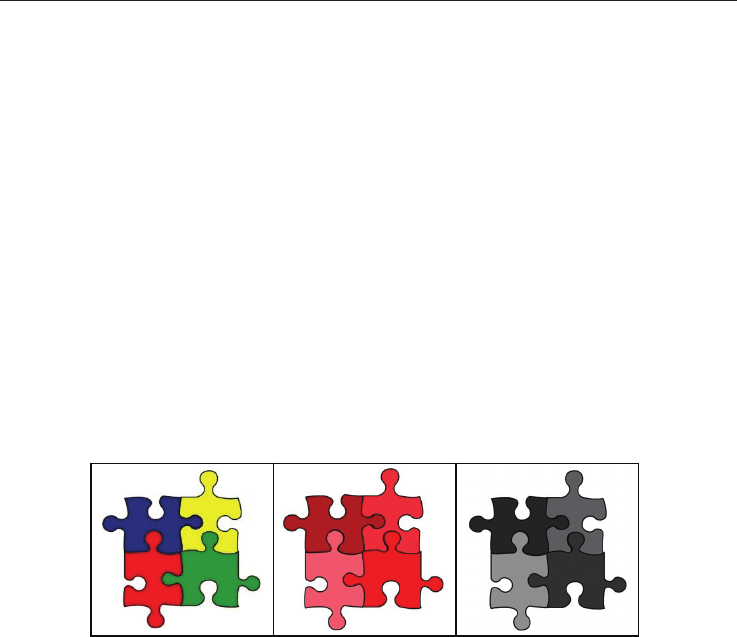
Hue or tint: the first attribute by which a color is identified and distinguished, or the
name of the color: blue, yellow, red, green, among others. It corresponds to the
wavelength of light reflected by the objects. (Fig. 3) (Sproull, 2001).
Chroma or saturation: indicates the purity of the color, quantifying its saturation. The
lighter a color is, the lower its saturation. Moreover, saturation increases as the object is
darker. For example, red is a saturated hue, while pink is the same hue, but less
saturated. (Fig. 4) (Sproull, 2001).
Value or light intensity: the property which is distinguished by the lightness or
darkness of a color. The clearer the color, the greater its value (brightness) and the
darker, the lower the value. A good example is the brightness of full white, represented
by the maximum value on the intensity scale (100), while black shows the absolute
value of 0, or the total absence of light. (Fig. 5) (Sproull, 2001).
Fig 3. Hues blue, yellow, red and green.
Fig 4. Saturation of the hue red.
Fig 5. Values of light intensities or saturations shown in Figure 3.
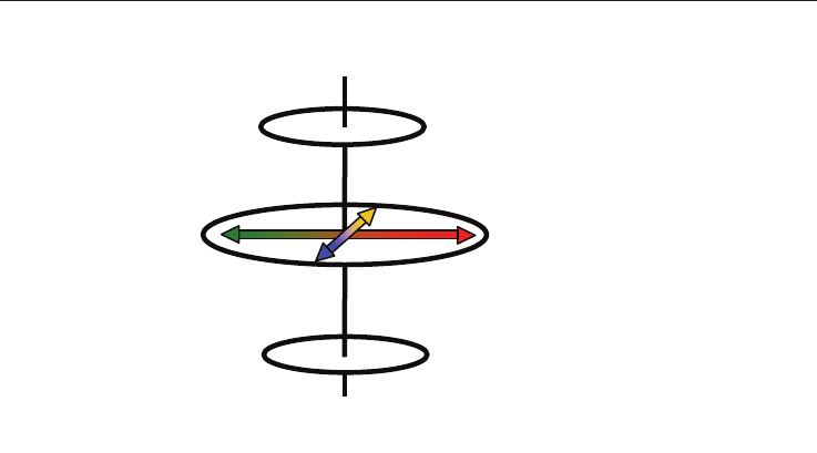
4.4 Color space CIEL*a*b*
To improve the precision in color communication of an object, the International Commission
of I'Eclairage (CIE) has developed some methods to express the spectral curves in a
numerical form. The method used in dentistry is the uniform color space, known as
CIEL*a*b*. (Fig. 6) (Rosenstiel & Johnston, 1988). Color space is a numerical area that
expresses and references the object's color. Here, L* indicates the lightness coordinate of the
object, with values from 0 (absolute black) to 100 (absolute white). The values a* and b*
indicates the chromaticity coordinates, showing the three-dimensional position of the object
in the color space and its direction. When the coordinate a* is positive (+a*), the object color
tends to red. When this coordinate is negative (-a*), the trend is green. This coordination can
range from -90 to 70 Δa*. The coordinate b* indicates the direction to yellow (+b*) or blue
(-b*), and can vary from -80 to 100 Δb*. (O'Brien et al. 1989; Barath et al., 2003).
3
4
5
Ceramic Materials 164
Fig. 6. System CIEL*a*b*
Source: Barath et al., 2003
The achromatic values of color are represented in the axis of lightness (L*), while the spatial
projection of data color is presented on the axes of chromaticity (a* and b*) allowing the
conceptualization of the components of the chromatic color changes. (Knispel, 1991). The
values of the coordinates L* a* b* are obtained from the tristimulus X, Y and Z (generated by
light reflected from the object observed) from the following equations, where the values Xn,
Yn and Zn correspond to white (Gliese & Petter, 2000):
L* = 116 (Y/Y
n
)
1/3
- 16 (1)
a* = 500[(X/X
n
)
1/3
– (Y/Y
n
)
1/3
] (2)
b* = 200[(X/X
n
)
1/3
– (Z/Z
n
)
1/3
] (3)
The space between two colorful points is calculated as a color difference (ΔE). The
magnitude of this difference can be obtained by the following equation (Gliese & Petter,
2000):
ΔE = ((ΔL *)
2
+ (Δa *)
2
+ (Δb *)
2
)
1/2
(4)
Where:
ΔL* = L*1 – L*2
(5)
Δa* = a*1 – a*2
(6)
Δb* = b*1 – b*2
(7)
Black
White
+ L
- L
+ a
- a
+ b
-b
4.5 Clinical significance
As the final judgment of colorimetric evaluation is visual, it has been necessary to establish a
relationship between visual and instrumental analysis. In the classic work of Kuehni &
Marcus (1979), color differences between samples 1ΔE were perceived and judged
acceptable by 50% of observers under ideal conditions of illumination. From this work,
Johnston & Kao (1989) assessed this relationship in dentistry. Since there is difficulty in
controlling light conditions in the oral cavity, an average difference of up to 3.7 ΔE was
considered acceptable by those authors, who suggested an extended visual rating scale
(EVRSAM) to understand the clinical significance of the numerical results of instrumental
analysis evaluation of color for dental materials.
ΔE Clinical significance.
0 Excellent esthetics with accurate color choice, not being clinically perceived,
or only with great difficulty.
2 Very slight difference in color, with very good aesthetics.
4 Obvious difference, but with an average acceptable to most patients.
6 Poor aesthetics, but within the limits of acceptability.
8 Aesthetics are very poor and unacceptable to most patients.
10 Aesthetics are totally unacceptable.
Table 2. Extended visual rating scale (EVRSAM)
Source: Johnston & Kao, 1988
5. Behavior of color on natural teeth
The color of a tooth is determined by a combination of intrinsic and extrinsic colorimetric
effects. The intrinsic properties color are associated with the reflection and absorption of
light; with the extrinsic properties related to coloring materials interacting with enamel,
such as coffee, tea, tobacco (Chu et al., 2004). When light falls on a natural tooth, four
associated phenomena can be described: the transmission of light through the tooth,
specular reflection from a tooth’s outer surface, diffuse reflection of light from the buccal
surface, absorption and scattering of light in the dental tissues. Factors such as enamel
thickness, shape, surface texture, dominant color of dentin, double layer effect and light
source may further complicate the visual perception of the various nuances of the whole
tooth. (Joiner, 2004).
6. Physical characteristics of dental ceramics
6.1 Color stability
The lower the degree of porosity evidenced by a ceramic after laboratory processing, the
higher its color stability. This means that industrially manufactured ceramic prostheses
show greater color stability when compared to hand crafted prostheses. However, variables,
such as the use of extrinsic dyes, number of firings, association with vacuum, type of
ceramic material, presence of metal framework and thickness of the ceramic materials, can
influence the color stability of ceramics (Brewer et al. 1985; Kourtis et al., 2004).

Ceramic Materials and Color in Dentistry 165
Fig. 6. System CIEL*a*b*
Source: Barath et al., 2003
The achromatic values of color are represented in the axis of lightness (L*), while the spatial
projection of data color is presented on the axes of chromaticity (a* and b*) allowing the
conceptualization of the components of the chromatic color changes. (Knispel, 1991). The
values of the coordinates L* a* b* are obtained from the tristimulus X, Y and Z (generated by
light reflected from the object observed) from the following equations, where the values Xn,
Yn and Zn correspond to white (Gliese & Petter, 2000):
L* = 116 (Y/Y
n
)
1/3
- 16 (1)
a* = 500[(X/X
n
)
1/3
– (Y/Y
n
)
1/3
] (2)
b* = 200[(X/X
n
)
1/3
– (Z/Z
n
)
1/3
] (3)
The space between two colorful points is calculated as a color difference (ΔE). The
magnitude of this difference can be obtained by the following equation (Gliese & Petter,
2000):
ΔE = ((ΔL *)
2
+ (Δa *)
2
+ (Δb *)
2
)
1/2
(4)
Where:
ΔL* = L*1 – L*2
(5)
Δa* = a*1 – a*2
(6)
Δb* = b*1 – b*2
(7)
Black
White
+ L
- L
+ a
- a
+ b
-b
4.5 Clinical significance
As the final judgment of colorimetric evaluation is visual, it has been necessary to establish a
relationship between visual and instrumental analysis. In the classic work of Kuehni &
Marcus (1979), color differences between samples 1ΔE were perceived and judged
acceptable by 50% of observers under ideal conditions of illumination. From this work,
Johnston & Kao (1989) assessed this relationship in dentistry. Since there is difficulty in
controlling light conditions in the oral cavity, an average difference of up to 3.7 ΔE was
considered acceptable by those authors, who suggested an extended visual rating scale
(EVRSAM) to understand the clinical significance of the numerical results of instrumental
analysis evaluation of color for dental materials.
ΔE Clinical significance.
0 Excellent esthetics with accurate color choice, not being clinically perceived,
or only with great difficulty.
2 Very slight difference in color, with very good aesthetics.
4 Obvious difference, but with an average acceptable to most patients.
6 Poor aesthetics, but within the limits of acceptability.
8 Aesthetics are very poor and unacceptable to most patients.
10 Aesthetics are totally unacceptable.
Table 2. Extended visual rating scale (EVRSAM)
Source: Johnston & Kao, 1988
5. Behavior of color on natural teeth
The color of a tooth is determined by a combination of intrinsic and extrinsic colorimetric
effects. The intrinsic properties color are associated with the reflection and absorption of
light; with the extrinsic properties related to coloring materials interacting with enamel,
such as coffee, tea, tobacco (Chu et al., 2004). When light falls on a natural tooth, four
associated phenomena can be described: the transmission of light through the tooth,
specular reflection from a tooth’s outer surface, diffuse reflection of light from the buccal
surface, absorption and scattering of light in the dental tissues. Factors such as enamel
thickness, shape, surface texture, dominant color of dentin, double layer effect and light
source may further complicate the visual perception of the various nuances of the whole
tooth. (Joiner, 2004).
6. Physical characteristics of dental ceramics
6.1 Color stability
The lower the degree of porosity evidenced by a ceramic after laboratory processing, the
higher its color stability. This means that industrially manufactured ceramic prostheses
show greater color stability when compared to hand crafted prostheses. However, variables,
such as the use of extrinsic dyes, number of firings, association with vacuum, type of
ceramic material, presence of metal framework and thickness of the ceramic materials, can
influence the color stability of ceramics (Brewer et al. 1985; Kourtis et al., 2004).

Ceramic Materials 166
6.2 Translucency and opacity
Paradoxically, dental ceramics should present both translucency and opacity to mimic
dental structures. The opacity is directly related to dentin, because the light passes through
enamel and reaches dentin, which, as an opaque body, reflects the light again. On the other
hand, enamel behaves like a translucent object, allowing the passage of light, which permits
visualization of the dentin while also providing the scattering of light at a wavelength of
blue through its hydroxyapatite crystals (Joiner, 2004). There is no ceramic dental product
that can simultaneously display characteristics of opacity and translucency in a single
material. Therefore, manufacturers have offered ceramics for infrastructure building,
opaque ceramic coverage for the construction of the dentin and translucent glazes to be used
in layering techniques (Heffernan et al., 2002 a, b).
6.3 Fluorescence
Some substances have capacity to absorb the energy of a non-visible light (ultraviolet,
cathode rays or X-rays) and turn it into a visible light, ie light with a greater wavelength
than the incident radiation. (Vanini, 1996). When natural teeth are exposed to ultraviolet
(UV) light, fluorescence is observed, with an emission spectrum band ranging from white to
intense blue light with a wavelength shorter than 400nm. The responsibility for this
phenomenon falls with dentin, which has a much more intense fluorescence than enamel
(three times more fluorescent), due to the presence of a greater amount of UV photosensitive
organic pigment. In order to mimic the behavior of this optical phenomenon, some dental
ceramics exhibit fluorescent characteristics similar to teeth in order to create an effect of
luminosity. Rare earth metals (europium, terbium, cerium and ytterbium) have been used as
luminophor agents in the composition of ceramic powders, because they show an intense
blue-white and yellow fluorescence. When a ceramic dental material is not fluorescent, it
tends to have an appearance of reduced vitality, presenting a grayish appearance, especially
in dark or black lights (Monsenego et al., 1993).
6.4 Opalescence and counter-opalescence
There are properties of transparent or translucent materials which gives those materials a
milky appearance, with iridescent reflections that resemble opal stone. This optical
phenomenon is attributed to the enamel, as it is a highly mineralized tissue. Enamel acts as a
filter and has the ability to selectively forward the long waves, while at the same time,
reflect the short waves. Because of this reflection, incisal enamel can be viewed as having a
bluish-white color. When long waves that were being transmitted relate to dentin and are
reflected back, they give enamel an orange glaze (an effect known counter-opalescence). In
ceramics, the effects of counter-opalescence and opalescence are obtained by using different
opalescent glazes. Some have a bluish color and others an orange color, depending on the
addition of pigments to the ceramic powder. At the time of construction of the prosthesis,
the lab tech stratifies enamel into layers, according to the optical nature of the tooth,
ensuring the combination of aesthetic effects with regards to fluorescence (Cho et al., 2009).
7. Behavior of color in dental ceramics
7.1 Optical influence of the illuminant
The light source has been identified as one of the factors that most influences the choice of
color (Dagg et al. 2004). The type of light source, its intensity and inclination are some of the
variables cited in the literature (Barna et al., 1981, Dagg et al. 2004). Due to the three-
dimensional nature of color, the behavior of light has been studied; however, its complexity
creates a variety of questions of when to use a specific light source. Metamerism is largely
responsible for distortions in the selection of color, thus adopting rules to minimize this
effect may help in the optical outcome of the ceramics used. As color selection is usually
performed under an indirect natural light source, it is prudent to involve fluorescent and
incandescent light sources for the selection of color and the evidence of ceramic prosthesis in
minimizing landmark metamerism (Volpato et al., 2009).
7.2 Optical influence of the substrate
The type and color of a substrate must be considered when selecting a restorative material,
because they influence the final shade of ceramic restorations, mainly with glassy systems.
When a substrate has a color similar to the ceramic, the recommended thickness (about 1.5
mm) can be used for vitreous systems. Ceramics, with high translucency and when using
layering techniques (such as glass-ceramics based on lithium disilicate), can be fabricated on
darkened substrates, such as posts and metal cores, since the dental preparation offers a
larger space (about 2.0 mm) (Volpato et al., 2009). However, if this space is not available,
systems with a ceramic infrastructure should be used (infiltrated, compact or machined
systems), because they are able to provide enough opacity to mask these substrates under a
reduced material thickness. (Heffernan et al. 2002b; Koutayas et al., 2003).
7.3 Optical influence of material thickness
The amount of dental reduction while performing the preparation determines the space
required for the production of ceramic prostheses. Each ceramic system should ensure that
this thickness does not compromise the emergence profile of the restorative work. However,
as mentioned above, preparations are not always made on substrates that have a similar
color as the tooth. In prostheses with favorable substrates, it is important to communicate
the color of the ceramic substrate when fabricating the prosthesis based on this background
(Dozic et al., 2003). However, if the substrate is not favorable, it is ideal to utilize more room
for the glass-ceramics or to associate infrastructures that provide a degree of opacity that
may block the arrival of light to the substrate. Achieving a deeper preparation can improve
the ability of a ceramic to hide the substrate, as increasing the thickness of a ceramic
material decreases the degree of translucency. (Vichi, Ferrari, Davisdon 2000; Carossa et al.,
2001, Nakamura et al. 2002; Dozic et al. 2003, Volpato et al., 2009). However, care must be
taken so that wear of the ceramic does not compromise the mechanical properties that are
necessary for the dental preparation.
7.4 Optical influence of material composition and manufacturing process
Based on the variety of ceramics available, it is important to understand that the
composition and manufacturing process directly influence the optical outcome of the

Ceramic Materials and Color in Dentistry 167
6.2 Translucency and opacity
Paradoxically, dental ceramics should present both translucency and opacity to mimic
dental structures. The opacity is directly related to dentin, because the light passes through
enamel and reaches dentin, which, as an opaque body, reflects the light again. On the other
hand, enamel behaves like a translucent object, allowing the passage of light, which permits
visualization of the dentin while also providing the scattering of light at a wavelength of
blue through its hydroxyapatite crystals (Joiner, 2004). There is no ceramic dental product
that can simultaneously display characteristics of opacity and translucency in a single
material. Therefore, manufacturers have offered ceramics for infrastructure building,
opaque ceramic coverage for the construction of the dentin and translucent glazes to be used
in layering techniques (Heffernan et al., 2002 a, b).
6.3 Fluorescence
Some substances have capacity to absorb the energy of a non-visible light (ultraviolet,
cathode rays or X-rays) and turn it into a visible light, ie light with a greater wavelength
than the incident radiation. (Vanini, 1996). When natural teeth are exposed to ultraviolet
(UV) light, fluorescence is observed, with an emission spectrum band ranging from white to
intense blue light with a wavelength shorter than 400nm. The responsibility for this
phenomenon falls with dentin, which has a much more intense fluorescence than enamel
(three times more fluorescent), due to the presence of a greater amount of UV photosensitive
organic pigment. In order to mimic the behavior of this optical phenomenon, some dental
ceramics exhibit fluorescent characteristics similar to teeth in order to create an effect of
luminosity. Rare earth metals (europium, terbium, cerium and ytterbium) have been used as
luminophor agents in the composition of ceramic powders, because they show an intense
blue-white and yellow fluorescence. When a ceramic dental material is not fluorescent, it
tends to have an appearance of reduced vitality, presenting a grayish appearance, especially
in dark or black lights (Monsenego et al., 1993).
6.4 Opalescence and counter-opalescence
There are properties of transparent or translucent materials which gives those materials a
milky appearance, with iridescent reflections that resemble opal stone. This optical
phenomenon is attributed to the enamel, as it is a highly mineralized tissue. Enamel acts as a
filter and has the ability to selectively forward the long waves, while at the same time,
reflect the short waves. Because of this reflection, incisal enamel can be viewed as having a
bluish-white color. When long waves that were being transmitted relate to dentin and are
reflected back, they give enamel an orange glaze (an effect known counter-opalescence). In
ceramics, the effects of counter-opalescence and opalescence are obtained by using different
opalescent glazes. Some have a bluish color and others an orange color, depending on the
addition of pigments to the ceramic powder. At the time of construction of the prosthesis,
the lab tech stratifies enamel into layers, according to the optical nature of the tooth,
ensuring the combination of aesthetic effects with regards to fluorescence (Cho et al., 2009).
7. Behavior of color in dental ceramics
7.1 Optical influence of the illuminant
The light source has been identified as one of the factors that most influences the choice of
color (Dagg et al. 2004). The type of light source, its intensity and inclination are some of the
variables cited in the literature (Barna et al., 1981, Dagg et al. 2004). Due to the three-
dimensional nature of color, the behavior of light has been studied; however, its complexity
creates a variety of questions of when to use a specific light source. Metamerism is largely
responsible for distortions in the selection of color, thus adopting rules to minimize this
effect may help in the optical outcome of the ceramics used. As color selection is usually
performed under an indirect natural light source, it is prudent to involve fluorescent and
incandescent light sources for the selection of color and the evidence of ceramic prosthesis in
minimizing landmark metamerism (Volpato et al., 2009).
7.2 Optical influence of the substrate
The type and color of a substrate must be considered when selecting a restorative material,
because they influence the final shade of ceramic restorations, mainly with glassy systems.
When a substrate has a color similar to the ceramic, the recommended thickness (about 1.5
mm) can be used for vitreous systems. Ceramics, with high translucency and when using
layering techniques (such as glass-ceramics based on lithium disilicate), can be fabricated on
darkened substrates, such as posts and metal cores, since the dental preparation offers a
larger space (about 2.0 mm) (Volpato et al., 2009). However, if this space is not available,
systems with a ceramic infrastructure should be used (infiltrated, compact or machined
systems), because they are able to provide enough opacity to mask these substrates under a
reduced material thickness. (Heffernan et al. 2002b; Koutayas et al., 2003).
7.3 Optical influence of material thickness
The amount of dental reduction while performing the preparation determines the space
required for the production of ceramic prostheses. Each ceramic system should ensure that
this thickness does not compromise the emergence profile of the restorative work. However,
as mentioned above, preparations are not always made on substrates that have a similar
color as the tooth. In prostheses with favorable substrates, it is important to communicate
the color of the ceramic substrate when fabricating the prosthesis based on this background
(Dozic et al., 2003). However, if the substrate is not favorable, it is ideal to utilize more room
for the glass-ceramics or to associate infrastructures that provide a degree of opacity that
may block the arrival of light to the substrate. Achieving a deeper preparation can improve
the ability of a ceramic to hide the substrate, as increasing the thickness of a ceramic
material decreases the degree of translucency. (Vichi, Ferrari, Davisdon 2000; Carossa et al.,
2001, Nakamura et al. 2002; Dozic et al. 2003, Volpato et al., 2009). However, care must be
taken so that wear of the ceramic does not compromise the mechanical properties that are
necessary for the dental preparation.
7.4 Optical influence of material composition and manufacturing process
Based on the variety of ceramics available, it is important to understand that the
composition and manufacturing process directly influence the optical outcome of the

Ceramic Materials 168
prosthesis produced, and are important factors for the aesthetic success of the case. For
maximum reflection and opacity, the ceramic particles should be slightly larger than the
wavelength of light and have a different refractive index of the array where it is
incorporated, as a higher refractive index of a material produces greater opacity. (Heffernan
et al., 2002a). Ceramic systems using alumina or zirconia have high refractive indices and
are therefore used as opacifying elements (Vagkopoulou et al., 2009).
8. Methods of color selecting in Dentistry
8.1 Visual analysis of color
Traditionally, the visual perception of color is the most commonly used selection method in
dentistry. This subjective method is based on standardized scales that are composed of
representative samples of the average of the colors present in the human dentition. Through
this shade guide, you can perform a visual comparison with natural teeth. (Segui et al.,
1989). Color selection using scales is extremely complex because the selection process may
be influenced by variables ranging from the interpretation of three-dimensional nature of
color, to environmental influences. Selections may be inadequate due to factors such as
fatigue, age, stress, prior exposure of eyes to light, the observer's visual individuality,
inappropriate positioning of the object and an illuminating influence from the environment,
and metamerism. Moreover, the human eye can detect small differences in color, but the
ability to communicate these differences in terms of magnitude and nature of the difference
is very limited. (Knispel, 1991). Despite being widely publicized, prefabricated scales are not
representative of the wide range of colors present in natural teeth. These colors are not
systematically distributed in the CIEL*a*b* system. Additionally: the material available for
use in the dental clinic and laboratory is not the same as the material used in the scales; the
thickness of the teeth in the scale does not simulate the clinical condition; most scales have
no metallic or ceramic infrastructure; the degree of translucency of the incisal portion; and
the characterizations and pigmentation in the cervical third of the scales all make
comparison with natural teeth very difficult. These factors may necessitate the observer to
get used to a single scale in order to avoid errors during the visual selection of color
(Sproull, 2001b).
As color scales have limitations, some factors should be observed to improve their use: the
selection of color must be performed at the beginning of the procedure, when the teeth are
not dehydrated and the professional is not tired; the color scale should be kept on the same
plane of the lower anterior teeth so that light falling on them can be reflected in the same
plane to the observer; the scale and teeth should be slightly moistened to facilitate the
reflection of light; the operator should look for a maximum of 10 to 15s at each hue and rest
by looking at a neutral gray background between observations; the patient should be
prepared with a protective clear color so that the color of their clothes does not confuse the
operator; the environment must provide the quantity and quality of indirect natural light,
usually between 11 and 14 o'clock; and finally, the optical behavior of the color chosen
should be reassessed with different artificial illuminations (eg, incandescent light and
fluorescent light), in search of a remarkable metamerism that can derail the chosen color.
(Chu et al., 2004)
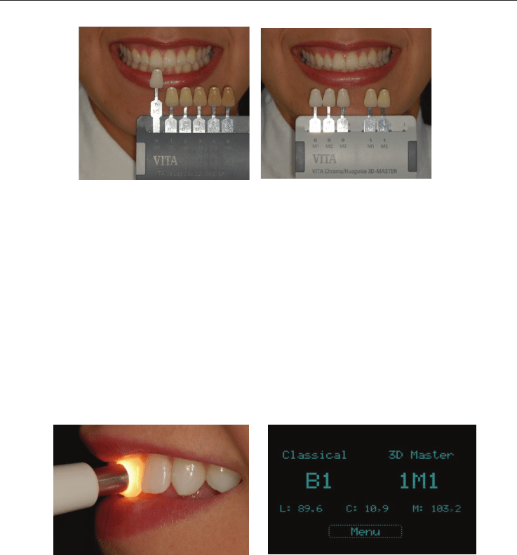
Fig. 7. Visual selection of color – value
Fig. 8. Visual selection of color - hue and chroma
8.1 Instrumental analysis of color
Colorimeters and spectrophotometers have been used in dentistry for the determination,
quantification and comparison of color in the industrial manufacture of materials.
Instruments have been designed for clinical use, in order to select the color in the mouth and
transmit the data to a laboratory, controlling the result for direct and indirect restorations,
tooth whitening, and to minimize subjective visual perception. (Sproull, 2001b). Just as in
visual analysis, care must be taken during the selection of an instrumental color: the teeth
should be clean and dry for capturing the color, as the presence of plaque and saliva may
affect the actual color registered by the equipment; the probe should perpendicularly touch
the selected area; and a minimum of three readings should be performed at the cervical,
middle and incisal thirds of dental tissues. (Chu et al., 2004)
Fig. 9. Instrumental selection of color
Fig. 10. Results obtained by oral spectrophotometer
9. Color Communication
The precise communication of color is one of the most important requirements for achieving
excellent aesthetic work. This can be achieved by joining two ways: chromatic maps and
digital photos.
9.1 Chromatic maps
Since teeth are composed of different shades of color and optical effects, it is necessary to
identify: the predominant color (middle third), areas that differ from the predominant color
9 10
7
8
Ceramic Materials and Color in Dentistry 169
prosthesis produced, and are important factors for the aesthetic success of the case. For
maximum reflection and opacity, the ceramic particles should be slightly larger than the
wavelength of light and have a different refractive index of the array where it is
incorporated, as a higher refractive index of a material produces greater opacity. (Heffernan
et al., 2002a). Ceramic systems using alumina or zirconia have high refractive indices and
are therefore used as opacifying elements (Vagkopoulou et al., 2009).
8. Methods of color selecting in Dentistry
8.1 Visual analysis of color
Traditionally, the visual perception of color is the most commonly used selection method in
dentistry. This subjective method is based on standardized scales that are composed of
representative samples of the average of the colors present in the human dentition. Through
this shade guide, you can perform a visual comparison with natural teeth. (Segui et al.,
1989). Color selection using scales is extremely complex because the selection process may
be influenced by variables ranging from the interpretation of three-dimensional nature of
color, to environmental influences. Selections may be inadequate due to factors such as
fatigue, age, stress, prior exposure of eyes to light, the observer's visual individuality,
inappropriate positioning of the object and an illuminating influence from the environment,
and metamerism. Moreover, the human eye can detect small differences in color, but the
ability to communicate these differences in terms of magnitude and nature of the difference
is very limited. (Knispel, 1991). Despite being widely publicized, prefabricated scales are not
representative of the wide range of colors present in natural teeth. These colors are not
systematically distributed in the CIEL*a*b* system. Additionally: the material available for
use in the dental clinic and laboratory is not the same as the material used in the scales; the
thickness of the teeth in the scale does not simulate the clinical condition; most scales have
no metallic or ceramic infrastructure; the degree of translucency of the incisal portion; and
the characterizations and pigmentation in the cervical third of the scales all make
comparison with natural teeth very difficult. These factors may necessitate the observer to
get used to a single scale in order to avoid errors during the visual selection of color
(Sproull, 2001b).
As color scales have limitations, some factors should be observed to improve their use: the
selection of color must be performed at the beginning of the procedure, when the teeth are
not dehydrated and the professional is not tired; the color scale should be kept on the same
plane of the lower anterior teeth so that light falling on them can be reflected in the same
plane to the observer; the scale and teeth should be slightly moistened to facilitate the
reflection of light; the operator should look for a maximum of 10 to 15s at each hue and rest
by looking at a neutral gray background between observations; the patient should be
prepared with a protective clear color so that the color of their clothes does not confuse the
operator; the environment must provide the quantity and quality of indirect natural light,
usually between 11 and 14 o'clock; and finally, the optical behavior of the color chosen
should be reassessed with different artificial illuminations (eg, incandescent light and
fluorescent light), in search of a remarkable metamerism that can derail the chosen color.
(Chu et al., 2004)
Fig. 7. Visual selection of color – value
Fig. 8. Visual selection of color - hue and chroma
8.1 Instrumental analysis of color
Colorimeters and spectrophotometers have been used in dentistry for the determination,
quantification and comparison of color in the industrial manufacture of materials.
Instruments have been designed for clinical use, in order to select the color in the mouth and
transmit the data to a laboratory, controlling the result for direct and indirect restorations,
tooth whitening, and to minimize subjective visual perception. (Sproull, 2001b). Just as in
visual analysis, care must be taken during the selection of an instrumental color: the teeth
should be clean and dry for capturing the color, as the presence of plaque and saliva may
affect the actual color registered by the equipment; the probe should perpendicularly touch
the selected area; and a minimum of three readings should be performed at the cervical,
middle and incisal thirds of dental tissues. (Chu et al., 2004)
Fig. 9. Instrumental selection of color
Fig. 10. Results obtained by oral spectrophotometer
9. Color Communication
The precise communication of color is one of the most important requirements for achieving
excellent aesthetic work. This can be achieved by joining two ways: chromatic maps and
digital photos.
9.1 Chromatic maps
Since teeth are composed of different shades of color and optical effects, it is necessary to
identify: the predominant color (middle third), areas that differ from the predominant color
9 10
7
8
Ceramic Materials 170
(c
e
ef
f
T
h
la
b
9.
2
Di
m
e
n
o
to
o
fo
r
co
l
as
s
fr
o
I
m
sh
a
q
u
of
co
n
co
n
wi
an
in
f
Fi
g
Fi
g
e
rvical and incis
a
f
ects, and mame
l
h
ese chromatic
m
b
orator
y
.
2
Digital Photos
g
ital photos can
e
thods of comm
u
o
t in the same to
w
o
th and the scale
r
the rehabilitati
o
l
or scale in posit
i
s
essed on photo
g
o
m the mouth to
s
m
a
g
e-editin
g
pro
g
a
pe and texture.
u
alit
y
(not quanti
t
its value. Thus,
n
firmin
g
the sel
e
n
trast (+50%) an
d
i
ll enable the dis
p
opalescent halo
f
ormation for the
g
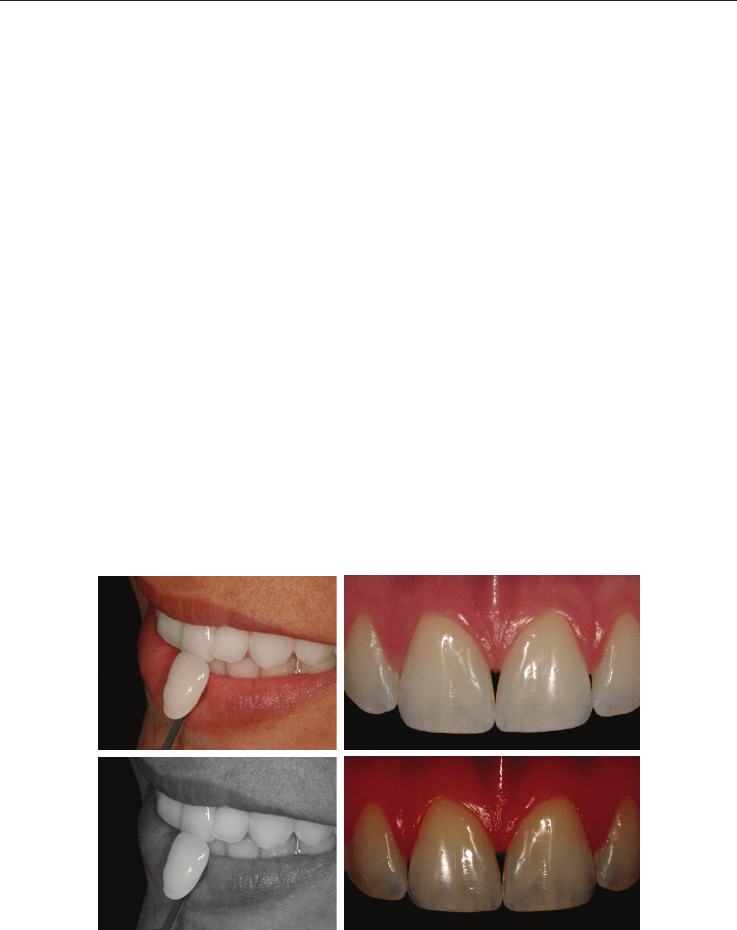
. 11a, b. Ori
g
ina
l
g
. 12a, b. Ori
g
in
a
11a
11b
a
l), and the pres
e
l
on spots. All of
m
aps are efficien
t
be used as a re
u
nication with th
e
w
n as the dentis
t
color that was c
h
o
n of a patient, it
i
on to inform the
g
raphs taken wit
h
s
trike the ename
l
g
rams can be u
s
A photo can be
ty
) of the
g
ra
y
co
it is possible to
e
ction of value.
A
d
decrease the br
p
lay of transluce
n
at the incisal ed
g
lab tech in the c
o
l
photo and
g
ra
ys
a
l photo and incr
e
e
nce of transluc
e
this informatio
n
t
documents in
ference for colo
r
e
laborator
y
, esp
e
t
and patient. Th
h
osen. If transluc
is important to
p
lab tech of the s
u
h
a black back
g
r
o
l
again.
s
ed to anal
y
ze c
di
g
itall
y
conver
t
lor, a colored ob
j
compare the va
l
A
nother process
t
i
g
htness (-50%)
o
n
t areas, the prec
i
g
e, and white s
p
o
nstruction of th
e
s
cale to view the
e
ase the contrast
a
e
nt areas, fluore
s
n
must be record
e
communicating
r
selectio
n
; there
f
e
ciall
y
when the
l
e photo
g
raph sh
c
ent ceramic is to
p
hoto
g
raph the
p
u
bstrate color. Tr
a
o
und, because it
p
olor dimensions
t
ed into
g
ra
y
sca
l
ect photo in
g
ra
y
l
ue of the scale
w
t
hat can be perfo
o
f the di
g
ital pho
i
se format of ma
m
p
ots and cracks,
b
e
prosthesis. (Mi
y
value.
a
nd brightness o
f
s
cent effects opa
l
e
d correctl
y
on
a
color variations
f
ore, they are ex
l
aborator
y
techn
i
ould be taken w
i
be used in resto
r
p
repared teeth w
a
nslucent areas a
r
p
revents reflecte
d
and characteri
s
l
e, because valu
e
y
scale will be the
w
ith the natural
rmed is to incre
a
to. The resultin
g
m
elons, the pres
e
b
ecause this is v
a
y
ashita, 2005)
f
the di
g
ital phot
o
12a
12b
l
escent
a
map.
to the
cellent
i
cia
n
is
i
th the
r
ations
ith the
r
e best
d
li
g
ht
s
tics of
e
is the
ima
g
e
tooth,
a
se the
ima
g
e
e
nce of
a
luable
o
.
10. Conclusion
Since the introduction of metal ceramic crowns, clinicians and researchers have been looking
for a restorative system that can associate beauty, strength and durability, but without the
presence of a metallic infrastructure. Indeed, dental ceramics are materials that come
aesthetically closest to natural teeth. With the improvement of ceramic systems, it has been
possible to combine the excellent aesthetic characteristics of this material with a considerable
resistance to fracture. It is imperative to understand the phenomenon of color and its
variables in the oral cavity, as well as associate different techniques of color selection and
communication, in order to get prostheses that look closer to natural dental tissues.
11. References
Barath, V.S.; Faber, F.J. ; Westland, S. & Niedermeier, W. (2003). Spectrophotometric
anallysis of all-ceramic materials and their interaction with luting agents and
different backgrounds. Adv Dent Res, Vol. 17, (Dez, 2003) pp. 55-60, ISSN 1544-0737.
Barna, G. J.; Taylor, J. W. ; King, G. E. & Pelleu, G. B. (1981). The influence of selected light
intensities on color perception within the color range of natural teeth. J Prosthet
Dent, Vol. 46, No. 4, (Oct, 1981) pp. 450-453, ISSN 0022-3913.
Brewer, J. D. ; Akers, C. K. ; Garlapo, D. A. & Sorensen, S. E. (1985). Spectrometric analysis of
the influence of metal substrates on the color of metal-ceramic restorations. J Dent
Res, Vol. 64, No. 1, (Jan, 1985) pp. 74-77, ISSN
0022-0345.
Carossa, S. ; Lombardo, S. ; Pera, P. ; Corsalini, M. ; Rastello, M.L. & Preti, G. (2001).
Influence of posts and cores on light trasmission through different all-ceramic
crowns: spectrophotometric and clinical evaluation. Int J Prosthodont, Vol. 14, No.1,
(Jan/Feb, 2001) pp. 9-14, ISSN 0893-2174.
Chevalier, J. (2006). What future for zirconia as a biomaterial? Biomaterials, Vol. 27, No. 4,
(Jan, 2006) pp. 535-43, ISSN 0142-9612.
Cho, M. S.; Yu, B. & Lee, Y. K. (2009). Opalescence of all-ceramic core and veneer materials.
Dent Mater, Vol. 25, No. 6 (Jun, 2009) pp. 695-702, ISSN 0109-564.
Chu, S. J. ; Devigus, A. & Mieleszko, A. J. (2004). Fundamentals of color: shade matching and
communication in esthetic dentistry, Quintessence, ISBN 0-86715-434-9, Chigaco.
Dagg, H. ; O’Conell, N.; Claffey, D.; Byrne, D. & Gorman, C. (2004). The influence of some
different factores on the accuracy of shade selection. J Oral Rehabil, Vol. 31, No. 9,
(Sep, 2004) pp. 900-904, ISSN 0305-182X.
Denry, I. & Kelly, J. R. (2008). State of the art of zirconia for dental applications. Dent Mater,
Vol. 24, No. 3, (Mar, 2008) pp. 299-307, ISSN 0109-564.
Dozic, A. ; Kleverlaan, C.J.; Meegdes, M.; Zel, J. & Feilzer, A.J. (2003). The influence of
porcelain layer thickness on the final shade of ceramic restorations. J Prosthet Dent,
Vol. 90, No. 6, (Dec, 2003) pp. 563-570, ISSN 0022-3913.
Heffernan, M. J.; Aquilino, S. A.; Diaz-Arnold, A. M.; Haselton, D. R.; Stanford, C. M. &
Vargas, M. A. (2002). Relative translucency of six all-ceramic systems. Part I: Core
materials. J Prosthet Dent, Vol. 88, No. 1, (Jul, 2002) pp. 4-9, ISSN 0022-3913.
Heffernan, M. J.; Aquilino, S. A.; Diaz-Arnold, A. M.; Haselton, D. R.; Stanford, C. M. &
Vargas, M. A. (2002). Relative translucency of six all-ceramic systems. Part II: Core
and venner materials. J Prosthet Dent, Vol. 88, No. 1, (Jul, 2002) pp. 10-15, ISSN
0022-3913.

Ceramic Materials and Color in Dentistry 171
(c
e
ef
f
T
h
la
b
9.
2
Di
m
e
n
o
to
o
fo
r
co
l
as
s
fr
o
I
m
sh
a
q
u
of
co
n
co
n
w
i
an
in
f
Fi
g
Fi
g
e
rvical and incis
a
f
ects, and mame
l
h
ese chromatic
m
b
orator
y
.
2
Digital Photos
g
ital photos can
e
thods of comm
u
o
t in the same to
w
o
th and the scale
r
the rehabilitati
o
l
or scale in posit
i
s
essed on photo
g
o
m the mouth to
s
m
a
g
e-editin
g
pro
g
a
pe and texture.
u
alit
y
(not quanti
t
its value. Thus,
n
firmin
g
the sel
e
n
trast (+50%) an
d
i
ll enable the dis
p
opalescent halo
f
ormation for the
g
. 11a, b. Ori
g
ina
l
g
. 12a, b. Ori
g
in
a
11a
11b
a
l), and the pres
e
l
on spots. All of
m
aps are efficien
t
be used as a re
u
nication with th
e
w
n as the dentis
t
color that was c
h
o
n of a patient, it
i
on to inform the
g
raphs taken wit
h
s
trike the ename
l
g
rams can be u
s
A photo can be
ty
) of the
g
ra
y
co
it is possible to
e
ction of value.
A
d
decrease the br
p
la
y
of transluce
n
at the incisal ed
g
lab tech in the c
o
l
photo and
g
ra
ys
a
l photo and incr
e
e
nce of transluc
e
this informatio
n
t
documents in
ference for colo
r
e
laborator
y
, esp
e
t
and patient. Th
h
osen. If translu
c
is important to
p
lab tech of the s
u
h
a black back
g
r
o
l
again.
s
ed to anal
y
ze c
di
g
itall
y
conver
t
lor, a colored ob
j
compare the va
l
A
nother process
t
i
g
htness (-50%)
o
n
t areas, the prec
i
g
e, and white s
p
o
nstruction of th
e
s
cale to view the
e
ase the contrast
a
e
nt areas, fluore
s
n
must be record
e
communicating
r
selectio
n
; there
f
e
ciall
y
when the
l
e photo
g
raph sh
c
ent ceramic is to
p
hoto
g
raph the
p
u
bstrate color. Tr
a
o
und, because it
p
olor dimensions
t
ed into
g
ra
y
sca
l
ect photo in
g
ra
y
l
ue of the scale
w
t
hat can be perfo
o
f the di
g
ital pho
i
se format of ma
m
p
ots and cracks,
b
e
prosthesis. (Mi
y
value.
a
nd brightness o
f
s
cent effects opa
l
e
d correctl
y
on
a
color variations
f
ore, they are ex
l
aborator
y
techn
i
ould be taken w
i
be used in resto
r
p
repared teeth w
a
nslucent areas a
r
p
revents reflecte
d
and characteri
s
l
e, because valu
e
y
scale will be the
w
ith the natural
rmed is to incre
a
to. The resultin
g
m
elons, the pres
e
b
ecause this is v
a
y
ashita, 2005)
f
the di
g
ital phot
o
12a
12b
l
escent
a
map.
to the
cellent
i
cia
n
is
i
th the
r
ations
ith the
r
e best
d
li
g
ht
s
tics of
e
is the
ima
g
e
tooth,
a
se the
ima
g
e
e
nce of
a
luable
o
.
10. Conclusion
Since the introduction of metal ceramic crowns, clinicians and researchers have been looking
for a restorative system that can associate beauty, strength and durability, but without the
presence of a metallic infrastructure. Indeed, dental ceramics are materials that come
aesthetically closest to natural teeth. With the improvement of ceramic systems, it has been
possible to combine the excellent aesthetic characteristics of this material with a considerable
resistance to fracture. It is imperative to understand the phenomenon of color and its
variables in the oral cavity, as well as associate different techniques of color selection and
communication, in order to get prostheses that look closer to natural dental tissues.
11. References
Barath, V.S.; Faber, F.J. ; Westland, S. & Niedermeier, W. (2003). Spectrophotometric
anallysis of all-ceramic materials and their interaction with luting agents and
different backgrounds. Adv Dent Res, Vol. 17, (Dez, 2003) pp. 55-60, ISSN 1544-0737.
Barna, G. J.; Taylor, J. W. ; King, G. E. & Pelleu, G. B. (1981). The influence of selected light
intensities on color perception within the color range of natural teeth. J Prosthet
Dent, Vol. 46, No. 4, (Oct, 1981) pp. 450-453, ISSN 0022-3913.
Brewer, J. D. ; Akers, C. K. ; Garlapo, D. A. & Sorensen, S. E. (1985). Spectrometric analysis of
the influence of metal substrates on the color of metal-ceramic restorations. J Dent
Res, Vol. 64, No. 1, (Jan, 1985) pp. 74-77, ISSN
0022-0345.
Carossa, S. ; Lombardo, S. ; Pera, P. ; Corsalini, M. ; Rastello, M.L. & Preti, G. (2001).
Influence of posts and cores on light trasmission through different all-ceramic
crowns: spectrophotometric and clinical evaluation. Int J Prosthodont, Vol. 14, No.1,
(Jan/Feb, 2001) pp. 9-14, ISSN 0893-2174.
Chevalier, J. (2006). What future for zirconia as a biomaterial? Biomaterials, Vol. 27, No. 4,
(Jan, 2006) pp. 535-43, ISSN 0142-9612.
Cho, M. S.; Yu, B. & Lee, Y. K. (2009). Opalescence of all-ceramic core and veneer materials.
Dent Mater, Vol. 25, No. 6 (Jun, 2009) pp. 695-702, ISSN 0109-564.
Chu, S. J. ; Devigus, A. & Mieleszko, A. J. (2004). Fundamentals of color: shade matching and
communication in esthetic dentistry, Quintessence, ISBN 0-86715-434-9, Chigaco.
Dagg, H. ; O’Conell, N.; Claffey, D.; Byrne, D. & Gorman, C. (2004). The influence of some
different factores on the accuracy of shade selection. J Oral Rehabil, Vol. 31, No. 9,
(Sep, 2004) pp. 900-904, ISSN 0305-182X.
Denry, I. & Kelly, J. R. (2008). State of the art of zirconia for dental applications. Dent Mater,
Vol. 24, No. 3, (Mar, 2008) pp. 299-307, ISSN 0109-564.
Dozic, A. ; Kleverlaan, C.J.; Meegdes, M.; Zel, J. & Feilzer, A.J. (2003). The influence of
porcelain layer thickness on the final shade of ceramic restorations. J Prosthet Dent,
Vol. 90, No. 6, (Dec, 2003) pp. 563-570, ISSN 0022-3913.
Heffernan, M. J.; Aquilino, S. A.; Diaz-Arnold, A. M.; Haselton, D. R.; Stanford, C. M. &
Vargas, M. A. (2002). Relative translucency of six all-ceramic systems. Part I: Core
materials. J Prosthet Dent, Vol. 88, No. 1, (Jul, 2002) pp. 4-9, ISSN 0022-3913.
Heffernan, M. J.; Aquilino, S. A.; Diaz-Arnold, A. M.; Haselton, D. R.; Stanford, C. M. &
Vargas, M. A. (2002). Relative translucency of six all-ceramic systems. Part II: Core
and venner materials. J Prosthet Dent, Vol. 88, No. 1, (Jul, 2002) pp. 10-15, ISSN
0022-3913.

Ceramic Materials 172
Holloway, J. & Miller, R.B. (1997). The effect of core translucency on the aesthetics of all-
ceramic restorations. Pract Periodontics Aesthetic Dent, Vol. 9, No. 5, (June/July,
1997) pp. 567-574, ISSN 1042-2722.
Johnston, W.M. & Kao, E.C. (1989). Assessment of appearance match by visual observation
and clinical colorimetry. J Dent Res, Vol. 68, No. 5, (May, 1989) pp. 819-822, ISSN
0022-0345.
Joiner, A. (2004). Tooth colour: a review of the literature. J Dent, Vol. 32, No. 1, (Suplly, 2004)
pp. 3-12, ISSN 0300-5712.
Kelly, J. R. & Denry, I. (2008). Stabilized zirconia as a structural ceramic: An overview. Dent
Mater, Vol. 24, No. 3, (Mar, 2008) pp. 289-98, ISSN 0109-564.
Kelly, J.R.; Nishimura, I. & Campbell, S. D. (1996). Ceramics in dentistry: historical roots and
current perspectives. J Prosthet Dent, Vol 75, No. 1, (Jan 1996) pp. 18-32, ISSN 0022-
3913.
Knispel, G. (1991). Factors affecting the process of color matching restorative materials to
natural teeth. Quintessence Int, Vol. 22, No. 7, (Jul, 1991) pp. 525-531, ISSN 0033-
6572.
Kourtis, S. G.; Tripodakis, A. P. & Doukoudakis, A. A. (2004). Spectrophotometric
evaluation of the optical influence of different metal alloys and porcelains in the
metal-ceramic complex. J Prosthet Dent, Vol. 92, No. 5, (Nov, 2004) pp. 477-485,
ISSN 0022-3913.
Koutayas, S.; Kakaboura, A.; Hussein, A. & Strub, J. (2003). Colorimetric evaluation of the
five different restorative materials on the color of veneered densely sintered
alumina. J Esthetic Rest Dent, Vol. 15, No. 6, (2003) pp. 353-360, ISSN 1496-4155.
Kuehni, R. G. & Marcus, R. T. (1979). An experiment in visual scaling of small color
differences. Color Res Appl, Vol. 4, No. 2, (Summer, 1979) pp. 83-91, ISSN 0361-2317.
Lamas, D. G.; Lascalea, G. E.; Walsoe de Reca, N. E. (1998). Synthesis and characterization of
nanocrystalline powders for partially stabilized zirconia ceramics. J Euro Ceramic
Soc, Vol. 18, No. 9, (1998) pp. 1217-21, ISSN 0955-2219.
Luthardt, R. G.; Holzhüter, M.; Rudolph, H.; Herold,V. & Walter, M. (2004). CAD/CAM
machining effects on Y-TZP zirconia. Dent Mater, Vol. 20, No. 7, (Sep, 2004) pp.
655-62. ISSN 0109-564.
Manicone, P. F.; Iommetti, P. R. & Rafaelli, L. (2007). An overview of zirconia ceramics: basic
properties and clinical applications. J Dent, Vol. 35, No. 11, (Nov, 2007) pp. 819-26,
ISSN 0300-5712.
McLean, J. W. (2001). Evaluation of dental ceramic in the twentieth century. J Prosthet Dent,
Vol. 85, No. 1, (Jan, 2001) pp. 61-66, ISSN 0022-3913.
Miyashita, E.; Mesquita, A.M.M. & Vasconcellos, D.K. (2006). Seleção de cor, In: Atualização
Clínica em Odontologia. Luciano Lauria Dib/Mario Sergio Saddy, Artes Médicas
Ltda, p. 509-526, 1a ed, ISBN 9788527809, São Paulo.
Miyazaki, T.; Hotta, Y.; Kunii, S.; Kuriyama, S. & Tamaki, Y. (2009). A review of dental
CAD/CAM: current status and future perspectives from 20 years of experience.
Dent Mater, Vol. 28, No. 1 (Jan, 2009) pp. 44-56, ISSN 0109-564.
Monsenego, G.; Burdairon, G. & Clerjaud B. (1993). Fluorescence of dental porcelain. J
Prosthetic Dent, Vol. 69, No. 1 (Jan 1993), pp 106-13, ISSN 0022-3913.
Nakamura, T.; Saito, O.; Fuyikawa, J.; Ishigaki, S. (2002). Influence of abutment substrate
and ceramic thickness on the colour of heat-presses ceramic crowns. J Oral Rehabil,
Vol. 29, No. 9, (Sep, 2002) pp. 805-809, ISSN 0305-182X.
O’Brien, W.J.; Groh, C.L.; Boenke, K.M. (1989). A one-dimensional color order system for
dental shade guides. Dent Mater, Vol. 5, No. 6, (Nov, 1989) pp. 371-374, ISSN 0109-
564.
Paravina, R. D. (2002). Evaluation of a newly developed visual sahde-matching apparatus.
Int J Prosthodont, Vol. 15, No. 6, (Nov-Dec, 2002) pp. 528-234, ISSN 0893-2174.
Petter, C. O. & Gliese, R. (2000). Fundamentals in Colorimetry. Ed. UFRGS, Porto Alegre,
Brazil.
Raigrodski, J. (2004). Contemporary materials and technologies for all-ceramic fixed partial
dentures: a review of the literature. J Prosthet Dent, Vol. 92, No. 6 (Dec, 2004) pp.
557-562, ISSN 0022-3913.
Sadowsky, S. J. (2006). An overview of treatment considerations for esthetic restorations: a
review of the literature. J Prosthet Dent, Vol. 96, No. 6, (Dec, 2006) pp. 433-42, ISSN
0022-3913.
Segui, R. R.; Hewlett, E. R.; Kim, J. (1989). Visual and instrumental colorimetric assessments
of small color differences on translucent dental porcelain. J Dent Res, Vol. 68, No.
12, (Dec, 1989) pp. 1760-1764, ISSN
0022-0345.
Sproull, R. C. (2001a). Color matching in dentistry. Part I. The three-dimensional nature of
colour. J Prosthet Dent, Vol. 86, No. 5, (Nov, 2001) pp. 453-457, ISSN 0022-3913.
Sproull, R. C. (2001b). Color matching in dentistry. Part II. Practical applications of the
organization of color. J Prosthet Dent, Vol. 86, No. 5, (Nov, 2001) pp. 458-464, ISSN
0022-3913.
Tadakoro, S. K. & Muccillo, E. N. S. (2002). Synthesis and characterization of nanosized
powders of yttria-doped zirconia. J Alloys Compd, Vol. 344, No. 1-2 (Oct, 2002) pp.
186-89, ISSN 0925-8388.
Vagkopoulou, T.; Koutayas, S.O.; Koidis, P. & Strub, J.R. (2009). Zirconia in Dentistry: Part 1.
Discovering the nature of an upcoming bioceramic. Eur J Esthet Dent, Vol. 4, No. 2,
(Summer, 2009) pp. 130-51, ISSN 1862-0612.
Van Der Burgt, T. P.; Ten Bosch, J. J.; Borsboom, P. C. F.; Kortsmit, W. J. P. M. (1990). A
comparison of new and convencional methods for quantification of tooth color. J
Prosthet Dent, Vol.63, No. 2, (Feb, 1990) pp. 155-162, ISSN 0022-3913.
Vanini, L. (1996). Light and color in anterior composite restorations. Pract Periodontics
Aesthetic Dent, Vol. 8, No. 7 (Sep, 1996) pp. 673-682, ISSN 1042-2722.
Vichi A.; Ferrari, M. & Davidson, C. L. (2000). Influence of ceramic and cement thickness on
the masking of various types of opaque posts. J Prosthet Dent, Vol. 83, No. 4, (April,
2000) pp. 412-17, ISSN 0022-3913.
Volpato, C. A. M.; Monteiro Jr, S.; Andrada, M. A.; Fredel, M. C.; Petter, C. O. (2009). Optical
influence of the type of illuminant, substrates and thickness of ceramic materials.
Dent Mater, Vol. 25, No. 1, (Jan, 2009) pp. 87-93, ISSN 0109-564.
Yang, S. F. et al. (2009). New nansized Al
2
O3 – BN coating 3Y-TPZ ceramics composites for
CAD/CAM – produced all-ceramic dental restorations. Part I. Fabrications of
powders. Nanomedicine J, Vol. 5, No. 2 (Jun, 2009) pp. 232-39, ISSN 1549-9634.