Stroscio Michael A., Dutta Mitra. Biological Nanostructures and applications of Nanostructures in biology: electrical, mechanical and optical properties
Подождите немного. Документ загружается.


36
MICHAEL A. STROSCIO
ET AL
.
Erkki Ruoslahti‚ RGD and other recognition sequences for integrins‚ Annual Reviews of Cell &
Developmental Biology 12(1)‚ 697-715 (1996).
Gerald Karp‚ Cell and Molecular Biology‚ Ed. (Wiley‚ 2002).
Robert P. Lanza‚ Robert Langer‚ and Joseph Vacanti‚ editors‚ Principles of Tissue Engineering
(Academic Press‚ 2000).
Bruce Alberts‚ Alexander Johnson‚ Julian Lewis‚ Martin Raff‚ Keith Roberts‚ and Peter Walters‚
Molecular Biology of the Cell‚ Ed. (Garland Science‚ Taylor & Francis Group‚ 2001).
H. K. Kleinman‚ B. S. Weeks‚ F. B. Cannon‚ T. M. Sweeney‚ G. C. Sephel‚ B. Clement‚ M. Zain‚ M.
O. J. Olson‚ M. Juncker‚ B. A. Burrous‚ Identification of a 110-kDa non-integrin cell surface
laminin-binding protein which recognizes an A chain neurite-promoting peptide‚ Arch. Biochem.
Biophys. 2909‚ 320-325 (1991).
Sharon K. Powell and Hynda Kleinman‚ Neuronal laminins and their cellular receptors‚ Int. J.
Biochem. Cell. Biol. 29‚ 401-414 (1997).
Sharon K. Powell‚ Javashree Rao‚ Eva Roque‚ Motoyoshi Nomizu‚ Yuichiro Kuratomi‚ Yoshihiko
Yamada‚ and Hynda K. Kleinman‚ Neural cell response to multiple novel sites on laminin-l‚ Journal
of Neuroscience Research 61‚ 302-312 (2000).
Dimitri Alexson‚ Yang Li‚ Dinakar Ramadurai‚ Peng Shi‚ Leena George‚ Lenu George‚ Muzna
Uddin‚ Preetha Thomas‚ Salvador Rufo‚ Mitra Dutta‚ and Michael A. Stroscio‚ Binding of
semiconductor quantum dots to cellular integrins‚ IEEE Transactions on Nanotechnology‚ in press
(2004).
Masao Iwamatsu‚ Makoto Fujiwara‚ Naoisa Happo‚ and Kenju Horii‚ Effects of dielectric
discontinuity on the ground-state energy of charged Si dots covered with a layer‚ Journal of
Physics: Condensed Matter 9‚ 9881-9892 (1997); Masao Iwamats and Kenju Horii‚ Dielectric
confinement effects on the impurity and exciton binding energies of silicon dots covered with a
silicon dioxide layer‚ Japanese Journal of Applied Physics 36‚ 6416-6423 (1997).
Anand Venkatesan‚ MS Thesis‚ Univesity of Illinois at Chicago (2003).
Dinakar Ramadurai‚ Babak Kohanpour‚ Dimitri Alexson‚ Peng Shi‚ Akil Sethuraman‚ Yang Li‚
Vikas Saini‚ Mitra Dutta‚ and Michael A. Stroscio‚ Tunable optical properties of colloidal quantum
dots in electrolytic environments‚ IEE Proceedings in Nanobiotechnology‚ in press (2004).
P. E. Lippens and M. Lannoo‚ Calculation of the band gap for small CdS and ZnS crystallites‚ Phys.
Rev. B39‚10935-10942 (1989).
A. S. Davydov‚ Quantum Mechanics (NEO Press‚ Ann Arbor‚ 1966).
Salvador Rufo‚ Mitra Dutta‚ and Michael A. Stroscio‚ “Acoustic modes in free and embedded
quantum dots‚” Journal of Applied Physics 93‚ 2900-2905 (2003).
151.
152.
153.
154.
155.
156.
157.
158.
159.
160.
161.
162.
163.
164.

BIOMEDICAL APPLICATIONS OF
SEMICONDUCTO
R
QUANTUM DOTS
Anupam Singhal‚ Hans C. Fischer‚ Johnson Wong‚ Warren C. W. Chan
*
1.
INTRODUCTION
In recent decades‚ the exquisite sensitivity and versatility of optical technologies
have led to numerous breakthroughs in biological research‚ including real-time imaging
of live cells‚
1‚2
gene expression profiling‚
3
cell sorting‚
4
and clinical diagnostics.
5‚6
A key
component in optical detection schemes is the probe design. These probes are
constructed from organic fluorophores‚ such as fluorescein and tetramethylrhodamine
(TMR)‚ and recognition molecules. The optical emission of fluorophores is used to
visualize the activities of biomolecules‚ while the recognition molecules direct the
fluorophores to specific cells‚ tissues‚ or organs. Although optical probes are widely used‚
most organic fluorophores exhibit unfavourable properties that have hampered their
applications in single-protein tracking in living cells‚ molecular pathology‚ and other
research areas.
7
These properties include photobleaching‚ sensitivity to environmental
conditions‚ and inability to excite multiple fluorophores using a single wavelength. A
new generation of probes has emerged in the last five years that overcomes many of the
limitations associated with organic fluorophores. These probes employ fluorophores that
are sub-100 nm in size and composed of inorganic atoms. Unlike organic-only
fluorophores‚ the optical and electronic properties of inorganic fluorophores can be tuned
during the synthesis process by changing their size‚ shape‚ or composition. In this chapter‚
we will describe the use of one type of inorganic fluorophore‚ semiconductor
nanocrystals‚ for the development of “custom-designed” probes for biomedical detection.
Semiconductor nanocrystals‚ also known as “quantum dots” (qdots)‚ are typically
composed of atoms from groups II-VI (CdSe‚ CdS‚ ZnSe) and III-V (InP and InAs)‚ and
are defined as particles with physical dimensions smaller than the Bohr exciton radius.
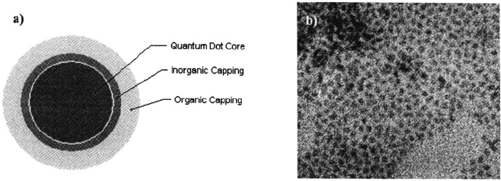
The Bohr exciton radius of prototypical CdSe qdots‚ as illustrated in Fig. 1‚ is ~10 nm.
The unique optical and electronic properties of qdots have spurred a great deal of
research into their potential applications in the design of novel biological probes‚
8‚9
light
emitting diodes‚
10‚11
photovoltaic cells‚
12‚l3
among other devices.
*
A.S., H.C.F., J.W., W. C. W. C., Institute of Biomaterials and Biomedical Engineering, University of Toronto,
Toronto, Ontario, Canada M5S 3G9. H.C.F. and W.C.W.C. are also affiliated with Department of Materials
Science & Engineering, University of Toronto.
37
38
A. SINGHAL ET AL.
Figure 1. a) Schematic representation of quantum dots commonly used for biological labelling‚ b)
Transmission electron micrograph (TEM) of monodisperse CdSe quantum dots (magnification = 200.000X).
14
In this chapter‚ we focus on the biological applications of semiconductor qdots‚
beginning with a brief description of their unique optical and electronic properties. We
then highlight various methods of synthesizing and characterizing qdots‚ as well as
successful biological applications of these fluorophores. Finally‚ we discuss some of the
prospects and challenges associated with future biological applications of qdots.
OPTICAL AND ELECTRONIC PROPERTIES OF SEMICONDUCTOR
QUANTUM DOTS
2.
Quantum dots (Qdots) exhibit unique optical and electronic properties that are only
observed in an intermediate size regime between the size of discrete atoms and that of
bulk solids. In this nanometre-size regime‚ charge carriers (i.e. electrons and holes) are
spatially confined within the dimensions of qdots‚ a phenomenon known as quantum
confinement. Due to this effect‚ the optical properties of qdots are heavily dependent
upon their size‚ shape‚ composition‚ and surface interactions with their local environment.
The use of excitation energy (e.g. via incident UV light) exceeding the bandgap energy of
the qdots leads to the promotion of electrons from the valence band (ground state) to the
conduction band (excited state)‚ creating mobile electrons and holes. The light-induced
excitation (or mobility) of electrons leads to fluorescence light emission in a process
called radiative recombination. In this process‚ the electrons and holes interact to form an
electron-hole pair called an exciton. The excited-state lifetimes of nanocrystals are multi-
exponential with lifetimes of 5 ns‚ 20-30 ns‚ and 80-200 ns‚ with the 20-30 ns dominating.
These processes can be measured using UV-Vis spectrophotometry or spectrofluorimetry.
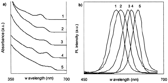
Typical UV-Vis absorbance measurements of qdots produce broad‚ continuous
spectra‚ which are dependent on the physical dimensions and composition of the particles
as illustrated in Fig. 2a. One characteristic feature of the qdot absorbance curves is an
observable peak‚ called the “quantum confinement” peak‚ which represents the lowest
bandgap energy transition. A second characteristic feature of the qdot absorbance curves
is the increasing absorbance at wavelengths shorter than the quantum confinement
BIOLOGICAL APPLICATIONS OF QUANTUM DOTS
39
wavelength. This property is extremely advantageous for biomedical applications since
qdots of all types and peak emission wavelengths can be excited using a single
wavelength.
The fluorescence emission spectra of qdots are narrow and symmetric as exemplified
by Fig. 2b. As with the qdot absorbance spectra‚ the fluorescence peak emission of qdots
is heavily dependent on qdot composition and physical dimensions‚ with an observable
red-shift in the peak fluorescence emission of larger qdots. The excitonic fluorescence
emission for a bulk measurement of capped qdots (e.g.‚ ZnS-capped CdSe) typically
exhibits a full-width at half-maximum (FWHM) of ~30 nm and quantum yield of 20-50%.
Conversely‚ measurements of the fluorescence spectra of single qdots have demonstrated
a FWHM of 13 nm‚ ~ 2.5 times narrower than the typical bulk measurement. Since the
fluorescence emission peak is size-tuneable‚ the broadness of bulk measurements can be
attributed to size distributions of the qdots within the bulk solution. In comparison to
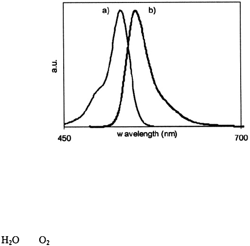
qdots‚ many organic dye molecules (e.g. Rhodamine 6G) have broad‚ asymmetric
emission spectra with FWHM > 45 nm as depicted in Fig. 3.
The fluorescence quality of qdots is often quantified in terms of a fluorescence
quantum yield - the ratio between the number of fluorescence photons emitted when
mobile electrons recombine with holes to the number of photons absorbed upon
excitation. Defect structures both in the internal structure and on the surface of qdots
can produce competing energy states that trap the excited electrons and holes‚ resulting in
lower quantum yields. In a classic study by Alivisatos and coworkers‚
15
the oxidative
decomposition of CdSe qdots was shown to produce a broad-fluorescence peak that was
red-shifted from the excitonic fluorescence peak. In their experiment‚ the oxidation of
qdots produced an excess of unbonded-atoms (dangling bonds) on the qdot surface; this
created low-energy bands to trap the mobile electrons and holes. In correspondence to
the increase in the intensity of the red-shifted defect peak‚ the excitonic fluorescence
intensity decreased.
Figure 2. a) Absorbance spectra and b) Fluorescence Spectra of CdSe/ZnS quantum dots of five different
sizes/emission colours (1 green‚ 2 yellow‚ 3 orange‚ 4 orange-red‚ 5 red-emitting).
14
40
A. SINGHAL ET AL.
Figure 3. a) Absorbance and b) fluorescence spectra of the organic dye molecule Rhodamine 6G.
Approaches to improving the quantum yield of qdots have focused on removal of
internal and surface defect structures. Internal defect structures can be removed by
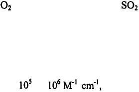
altering the solvent reaction conditions. For instance‚ while the presence of molecules
such as and produces qdots with low quantum yields‚ the use of organic solvents
and a controlled reaction atmosphere in a Schlenk line or glovebox have improved the
quality of synthesized qdots (quantum yields > 5-10 %). Apart from internal defect
structures‚ a major challenge to improving the quantum yield of qdots is to remove
surface defect sites. Qdots have a large surface area-to-volume ratio and therefore‚ a
large population of atoms on their surface. Removal of dangling bonds with organic
stabilizing molecules and/or a second semiconductor layer has shown to dramatically
improve the overall quantum yield of the qdots.
16
Careful selection of surface coating
has produced qdots with quantum yields as high as 85%. In Section 3 - Synthesis and
Characterization of Quantum Dots‚ strategies for coating qdots will be discussed in
greater detail.
Two strategies for tuning the fluorescence emission‚ or bandgap energy‚ of quantum
dots are the alteration of the size and composition of qdots. The bandgap energy is
directly related to the composition of the qdot; for instance‚ CdS qdots exhibit ultraviolet
(UV) fluorescence‚ while InP qdots exhibit near-IR fluorescence. Alloying qdots can
also alter their bandgap energy. Recently‚ Bailey and coworkers demonstrated the shift
in fluorescence emission of CdSe qdots to the near-IR by doping with Te.
17
Apart from
altering qdot composition‚ the fluorescence emission of qdots of a single composition
(e.g.‚ CdSe) can be tuned by changing their size. As described by the quantum
mechanical “particle-in-a-box” model‚ decreasing the size of qdots results in a
corresponding increase in semiconductor bandgap energy (or‚ equivalently‚ shorter
emission wavelengths). For example‚ the fluorescence emission peaks of cadmium
selenide (CdSe) semiconductor qdots can be tuned across the entire visible spectrum from
blue to red by increasing the diameter of the qdots from roughly 2 nm to 6 nm. In the
“particle-in-a-box” model‚ the potential energy is infinite outside of the box and hence
the particle is confined to the dimensions of the box. This particle contains discrete
energy levels and wavefunctions that correspond to the dimensions of the box. This
BIOLOGICAL APPLICATIONS OF QUANTUM DOTS
41
model coincides with the structure of qdots, in which mobile carriers (the particle) are
confined within the dimensions of the qdot (the box) with discrete emission wavelengths
(wavefunctions) and bandgap energy levels. As the physical dimensions of the box
become smaller, the bandgap energy increases.
The fluorescence emission of qdots is extremely stable upon photoexcitation. In
comparative measurements of qdots with small organic fluorophores, qdots have been
shown to be ~ 100 times more stable against photobleaching. Qdots have also been
shown to be more photostable than fluorescent proteins (e.g., phycoerytherin), although
this result has not been quantified.
18
The photobleaching of qdots is believed to arise
from a slow process of photo-induced chemical decomposition. This hypothesis is
supported by our observation of a shift in emission-colour from red to blue when qdot-
aggregates are spread on a glass slide and monitored under an epifluorescence
microscope with high-power UV-excitation. Henglein and coworkers speculated that
CdS decomposition is initiated by the formation of S or SH radicals upon optical
excitation.
19
These radicals can react with from the air to form an complex,
resulting in slow particle degradation. Capping the surface of qdots with a thick second
semiconductor layer has produced qdots with excellent photostability.
Due to the bright luminescence and high photostability of qdots, single qdots can be
easily visualized and imaged under a conventional epifluorescence microscope. In
addition, qdots are brighter than most small organic dye molecules due to their large
molar extinction coefficients. Bawendi and coworkers
20
estimated that the molar
extinction coefficients of CdSe qdots are about to depending on the
particle size and the excitation wavelength. These values are 10-100 times larger than
those of organic dyes, but are similar to the absorption cross sections of phycoerytherin, a
multi-chromophore fluorescent protein. It has been estimated that a single qdot is
approximately equivalent in fluorescence intensity to 10 to 20 small organic dye
molecules.
21
In this section, we have briefly described some of the properties of qdots that make
them appealing for biological applications. For a more in-depth look at the physical
chemistry of qdots, the interested reader can refer to a number of excellent reviews by
Alivisatos
22
, and Murray et al.
23
SYNTHESIS AND CHARACTERIZATION OF QUANTUM DOTS
3.
The most commonly synthesized semiconductor nanocrystals are composed of atoms
from groups II-VI (e.g. CdSe‚ CdTe‚ CdS‚ and ZnSe) and groups III-V (e.g. InP and
InAs) of the periodic table.
24-29
In particular‚ rapid advancements in synthesis and
characterization techniques have led to the development of highly luminescent and
monodisperse CdSe qdots.
16‚30-32
In the following sections‚ we will discuss a number of
different approaches to the synthesis and characterization of high-quality qdots.
Synthesis Techniques
3.1.
Typical techniques for the synthesis of semiconductor qdots involve the growth of
nanocrystals using molecular precursors in either aqueous or organic solutions. In one
approach to qdot synthesis in aqueous media‚ solutions of cadmium and sulphur
precursors are injected into hot aqueous solutions containing stabilizing agents or in
42
A. SINGHAL ET AL.
inverse micelles.
33‚34
The size of the resulting nanocrystals can be controlled through the
use of different solvent additives‚ or varying the solvent pH and temperature. Another
approach to the synthesis of qdots in aqueous media employs yeast cells‚ such as Candida
glabrata or Schizosacharomyces pombe.
35‚36
In the presence of cadmium or zinc ions‚
these yeast cells express proteins with thiol and carboxylic acid residues that bind to the
metal ions and induce nucleation of nanocrystals. Despite the success and simplicity of
both these aqueous methods‚ the resulting nanocrystals have low quantum yields (QY <
10%) and large size distributions (relative standard deviation RSD >15%)‚ resulting in
broad emission spectra (~50 nm full-width at half maximum‚ FWHM).
Improved synthesis schemes in organic media have led to the development of qdots
that are highly luminescent (quantum yield > 50%) and monodisperse (relative size
distributions < 5%).
16‚ 30-32
In one approach‚ qdots are generated by the pyrolysis of
organometallic and chalcogenide precursors injected into a hot coordinating solvent. For
instance‚ CdSe qdots are synthesized through the dissolution of dimethyl cadmium and
selenium shot in either tri-n-butylphosphine (TBP) or tri-n-octylphosphine (TOP) and
subsequent injection into a solution of tri-n-octylphospine oxide (TOPO) at 340-360°C.
Rapid nucleation and growth of the nanocrystals are observed through changes in colour
of the reaction mixture.
As described by La Mer and Dinegar‚
37
nucleation occurs until the temperature and
precursor concentrations drop below the critical “nucleation threshold”. Subsequent re-
heating of the nanocrystals to intermediate temperatures (250-300°C) results in slow
growth of the nanocrystals. Growth of nanocrystals proceeds until the available precursor
material is consumed‚ after which smaller nanocrystals begin to dissolve in order to
supply materials for the growth of larger nanocrystals. This diffusion process‚ known as
Ostwald ripening‚ occurs due to the high surface free energy of smaller nanocrystals‚
which make them more prone to dissolution than larger nanocrystals. This increase in
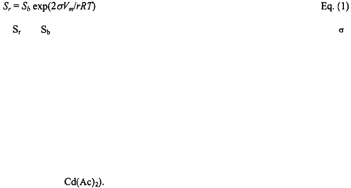
nanocrystal solubility with decreasing nanocrystal size can be described using the Gibbs-
Thomson equation:
38
where and are the solubility of the nanocrystal and the corresponding bulk solid‚ is
the specific surface energy‚
r
is the radius of the nanocrystal‚ Vm is the molar volume of
the materials‚ R is the gas constant‚ and T is the temperature. Since the temperature
required for maintaining steady nanocrystal growth increases with increasing nanocrystal
size‚ careful control of growth temperatures allows for accurate control of the average
size and size distribution of the nanocrystals synthesized in a given reaction. Size and
size distributions are monitored from the peak wavelengths and widths of the absorption
or emission spectra. When the desired properties are attained‚ the temperature is reduced
to prevent further growth or dissolution.
Variations to this organometallic approach have resulted in synthesis schemes that
yield gram-quantities of high-quality qdots. In particular‚ Peng and coworkers
39‚40
have
demonstrated that organometallic precursors (e.g. dimethyl cadmium) can be replaced
with non-pyrophoric and less costly “greener” reagents (e.g. cadmium oxide‚ CdO‚ or
cadmium acetate‚ These “alternative routes” to the synthesis of qdots in
organic media can been used to reproducibly prepare high-quality CdS‚ CdSe‚ and CdTe
nanocrystals.
17‚39‚41
In addition‚ since nanocrystals formed from greener reagents exhibit
slower reaction kinetics (e.g. slower nucleation)‚ extended nucleation periods allow
BIOLOGICAL APPLICATIONS OF QUANTUM DOTS
43
increased quantities of “greener” precursors to be injected at the start of the reaction.
This is a promising approach to scaling up the synthesis of high quality qdots.
3.2. Capping of Quantum Dots
Due to the high surface-to-volume ratio of nanocrystals‚ the structural‚ optical‚ and
electronic features of semiconductor qdots are heavily influenced by their surface
properties. In effect‚ the surface properties of qdots are often manipulated to enhance the
stability‚ processibility‚ and optical properties of these nanocrystals. The following
sections will outline general strategies that are commonly used to manipulate the surface
properties of qdots through the coating (“capping”) of surfaces with organic ligands and
inorganic materials.
3.2.1. Organic Capping
Coating of qdot surfaces with organic ligands typically facilitates the production of
stable‚ high-quality qdots in organic media. A classic example of this effect is observed
in the synthesis of CdSe qdots in hot solutions of amphiphilic tri-n-octylphosphine oxide
(TOPO). In these solutions‚ the hydrophilic phosphine oxide groups coordinate to Cd
sites on the qdot surface while the hydrophobic alkyl chains form a densely packed
surface coating that stabilize nanocrystals against aggregation. The non-polar alkyl chains
at the solid-liquid interface between the nanocrystals and solvent cause the resulting
qdots to be soluble in organic solvents‚ such as chloroform and hexane. As a result‚ these
TOPO-coated qdots can be selectively precipitated out of initial reaction mixtures
containing excess ligands and unreacted precursors through the addition of polar solvents
(e.g. methanol) and subsequent centrifugation. Since increasing the solvent polarity
results in precipitation of smaller nanocrystals‚ repeated precipitation of qdot mixtures
with increasing solvent polarities can be used to separate mixtures of quantum dots with
different sizes; this process is referred to as “size-selective precipitation”. The purified
qdots typically have relative size distributions of less than 5%. Lastly‚ the capping of
organic ligands on the qdot surfaces tends to improve their overall quantum yields.
3.2.2. Inorganic
Capping
The fluorescence efficiency of qdots can be greatly enhanced by the capping of the
nanocrystals inside an inorganic shell. For instance‚ monolayers of zinc sulphide (or
cadmium sulphide) can be grown epitaxially on CdSe qdots by drop-wise injection of
zinc (cadmium) and sulphur precursors into solutions of qdots at moderate temperatures
(150-240°C).
16‚ 31
These reaction conditions favour deposition of the capping precursors
onto the qdot surface over homogeneous ZnS (CdS) nucleation. The resulting core/shell
nanocrystals exhibit luminescence quantum yields up to 85%‚ in contrast to the maximum
quantum yields of ~15% observed for TOPO-capped CdSe qdots.
32
In order for the capping layer to produce increased qdot quantum yields‚ the
inorganic layer must be composed of a semiconductor with larger bandgap energy than
the core qdot. In addition‚ for efficient capping‚ the bond length of the semiconductor
comprising the capping layer must be similar to that of the core. The observed increase in
quantum yield upon growth of an inorganic shell can be attributed to the‘removal of
surface defects (“trap states”) on the nanocrystals‚ a process referred to as electronic
44
A. SINGHAL ET AL.
passivation. For CdSe nanocrystals‚ these surface defects typically result in broad
emission at 700-800 nm‚ which are removed with the growth of an inorganic shell with a
wider bandgap than the CdSe core. The removal of these trap states increases the number
of electrons that undergo radiative recombination as described in Section 2 on Optical
and Electronic Properties of Quantum Dots. Thus‚ the resulting nanocrystals exhibit an
increase in fluorescence efficiency. Furthermore‚ capping with a semiconductor layer
also prevents the photo-oxidation of the core qdot. Thus‚ while uncapped qdots may
degrade within a month or two when stored in air at room temperature‚ long-term storage
of capped qdots under similar conditions can often be achieved with minimal effect on
their optical properties.
BIOLOGICAL APPLICATIONS OF QUANTUM DOTS
4.
Over recent years there has been much excitement surrounding the potential
applications of qdots to biological research‚ including the development of optical probes
for biomedical imaging‚ bioassays and biosensors. For example‚ the novel size-tunable
optical properties of qdots and universal conjugation strategies have generated a great
impetus for the development of biological probes based on qdots.
7
In the coming
sections‚ we will discuss some practical issues in developing qdots suitable for biological
applications‚ and will review some proof-of-concept studies that highlight the unique
advantages of qdots in biological research.
Biocompatible Quantum Dots
4.1.
One of the major challenges to using qdots for developing biological probes and
sensors is their surface chemistry. Since high-quality qdots are synthesized in organic
solvents‚ such as TOPO‚ they are not water-soluble and‚ hence‚ incompatible with
biological systems. In the last six years‚ great efforts have been placed on modifying the
surface chemistry of qdots to render them biocompatible.
8‚ 9‚ 21‚ 42‚ 43
One approach to
achieving qdot biocompatibility has been the use of surface exchange techniques‚ where
TOPO molecules on the qdot surface are displaced by bifunctional molecules‚ such as
mercaptoacetic acid and phospho-alcohols. One end of these bifunctional molecules
contain functional groups (-SH or –P) that interact with metal atoms on the surface of the
qdots and displace non-polar TOPO molecules. The other end of these bifunctional
molecules typically contains polar alcohol or carboxylic acid functional groups‚ thus
rendering the qdots extremely polar and water-soluble. Furthermore‚ the alcohol and
carboxylic groups can react with biomolecules such as proteins‚ peptides‚ and
oligonucleotides through several different synthetic techniques.
In a second approach to achieving qdot biocompatibility‚ molecules can be designed
to interact with the TOPO molecules on the surface of qdots.
21‚ 43
These molecules
typically contain both a hydrophobic and hydrophilic region (e.g.‚ phospholipids). The
hydrophobic end interacts with the TOPO molecule through hydrophobic-hydrophobic
interactions‚ while the hydrophilic end‚ containing carboxylic acid or alcohol functional
groups‚ protrudes from the surface of the quantum dot. The qdots can be locked into the
organic-shell by cross-linking the surface-stabilizing molecule. This prevents the
organic ligands from desorbing from the surface of the qdots and stabilizes the qdots
against flocculation.
BIOLOGICAL APPLICATIONS OF QUANTUM DOTS
45
The functional groups provide a means to conjugate biomolecules (e.g.‚ proteins‚
peptides‚ oligonucleotides) to form an optical probe. Conventional carbodiimide
chemistry has been employed to catalyze this linkage (e.g.‚ qdots containing carboxylic
acids can react to biomolecules containing primary amines to form an amide bond).
Electrostatic interactions can also be used to link biomolecules onto the surface of qdots.
For example‚ Mattoussi and coworkers
44‚ 45
engineered a protein with a positive-charged
leucine zipper and demonstrated the adsorption of such a protein onto the surface of a
negatively-charged qdot.
4.2.
Quantum Dots for Biomedical Imaging Applications
Biocompatible qdots have spurred great interest in their possible use as fluorescent
labels for biological imaging applications. To date‚ qdots have been used to image
cellular components (e.g. DNA‚ nuclear antigens‚
21
and cytoplasmic components)‚ cells‚
9
and tissues
46
to name a few examples. Figure 4 shows a differential interference contrast
(DIC) and fluorescence image of transferrin-coated quantum dots entering HeLa cells
through receptor-mediated endocytosis.
One distinct advantage of using qdots over organic fluorophores for biological
applications is the ability to perform multiplexed colour-coded imaging and detection‚ a
general technique that uses multi-coloured fluorophore labels to simultaneously identify
different biological targets and study the interactions between these targets.
7
Multiplexed imaging of qdots can be used to investigate different biological processes by
tagging different biological targets with qdots of many different emission wavelengths
and simultaneously exciting them with a single excitation wavelength as discussed in
Section 2 on Optical and Electronic Properties of Semiconductor Quantum Dots. For
instance‚ Mattoussi and coworkers used green‚ yellow‚ and red quantum dots to study the
behavioural differences between starved and unstarved AX2 amoebae cells.
45
While the
starved cells were shown to form aggregate centres (e.g. possibly via chemotaxis in
response to signals sent by one another)‚ the unstarved cells did not form such aggregates.
In another study‚ Alivisatos and coworkers labelled mouse fibroblast cells using red and
green coloured silica-coated CdSe-CdS qdots.
8
The red qdots were conjugated to biotin‚
which targets actin filaments‚ while the green qdots were conjugated to tri-
methoxysilylpropyl urea and acetate groups‚ which bind electrostatically to the cell
nucleus. Under both conventional wide-field and laser-scanning confocal fluorescence
microscopes‚ the actin and the nucleus in the fibroblast samples were spatially and
spectrally resolvable to the eye. Multiplexed imaging of qdots was further demonstrated
by Wu and coworkers‚ who performed a series of experiments in which the nuclear
antigens and microtubules in the cytoplasm were simultaneous stained and imaged using
two different-coloured quantum dots.
21
Several in situ studies have also investigated and exploited the resistance of qdots to
photobleaching. In the above-mentioned experiments that probed AX2
