Schlick T. Molecular Modeling and Simulation: An Interdisciplinary Guide
Подождите немного. Документ загружается.

576 Appendix D. Homework Assignments
Summary of Items to Hand in:
(a) Data with the amount of PDB 3D structures for each category of
biomolecules.
(b) Description of the differences in the informational part of the PDB
files for form II and form III of BPTI.
(c) Explanation of the PDB format for storing atomic coordinates.
(d) Description of the structural differences between BPTI (form II) and
the mutated form (7pti).
(e) The table with dihedral angles φ and ψ listed for BPTI.
(f) Scatter plot with points (ψ, φ) for each residue of BPTI.
Background Reading from Coursepack (Appendix B)
• M. Levitt and A. Warshel, “Computer Simulation of Protein Folding”,
Nature 253, 694–698 (1975).
• M. Karplus and J. A. McCammon, “The Dynamics of Proteins”, Sci. Amer.
254, 42–51 (1986).
• Y. Duan and P. A. Kollman, “Pathways to a Protein Folding Intermediate
Observed in a 1-Microsecond Simulation in Aqueous Solution”, Science
282, 740–744 (1998).
• H. J. C. Berendsen, “A Glimpse of the Holy Grail”, Science 282, 642–643
(1998).
• X. Daura, B. Juan, D. Seebach, W. F. Van Gunsteren, and A. Mark, “Re-
versible Peptide Folding in Solution by Molecular Dynamics Simulation”,
J. Mol. Biol. 280, 925–932 (1998).
• R. Bonneau and D. Baker, “Ab Initio Protein Structure Prediction: Progress
and Prospects”, Annu. Rev. Biophys. Struc. 30, 173–189 (2001).
Appendix D. Homework Assignments 577
Assignment 3: Construction and Analysis
of the Pentapeptide Met-enkephalin with the Insight II
Program
1. Building a Pentapeptide. To build the molecule met-enkephalin, whose
amino acid sequence is Tyr–Gly–Gly–Phe–Met, invoke the Biopolymer
module. From
Residue select Append.
Specify the molecule’s name and choose Extended to specify the structural
motif of the backbone. (At this stage, do not worry whether the structure is
correct). Select the first residue, Tyr. Then add each of the remaining four
residues in turn.
You can center the molecule on your screen by clicking on it with the center
mouse button and dragging it to the desired position. You can also rotate the
molecule by pressing the right mouse button and dragging the molecule. By
pressing both (center and right) mouse buttons and dragging the molecule
you can change its position along the z-axis, perpendicular to the screen.
You can rotate the molecule around the z-axis by dragging the mouse while
both left and right mouse buttons are pressed. To change the representa-
tion style of the molecule, select
Molecule / Render and choose any of
the options. Note how the speed of executing commands (e.g., translation,
rotation) is affected by the representation display.
Now you must amend the ends of the structure you built. Switch to
Protein and choose Cap. Change both the N-terminus and C-terminus
moieties to the zwitterionic form (NH
+
3
and COO
−
) to get a proper
oligopeptide.
2. Measurements of Met-enkephalin’s Structural Parameters. Generate a
table listing all the dihedral angles in your met-enkephalin molecule by
using
Protein / List from the Biopolymer module. Select the appropriate
command (Dihedrals)anddirectthedatatoafile(List
to file button on).
This file can be viewed and edited later.
To measure individual bond lengths or distances, bond angles, dihedral an-
gles, etc., use
Measure . Select the atoms for the measurement by clicking
on them with the left mouse button.
3. Rotameric Structures.Byarotamer, Insight II refers to a different confor-
mational arrangement of a side chain of a given amino acid. The rotameric
structures identified in Insight II are correlated with the φ and ψ angles for
a given residue (i.e., are sterically compatible).
Select Manual
Rotamer from Residue of the Biopolymer module.
Press the Evaluate
Energy button. Energy for a given rotameric structure
will be printed in the information window at the bottom of the screen (you
can scroll by pressing on arrows to its right). Keep the Nonbond Cutoff
value — an option which is displayed on the screen once Evaluate
Energy
is chosen — at the default value of 8.0
˚
A.
578 Appendix D. Homework Assignments
Select a residue by clicking on it and sweep through all the rotamers
of that residue (while holding other rotamer conformations fixed). Next
will trigger the execution of the command. In a table, report the ro-
tamer structures for each residue by specifying the dihedral angles χ
1
, χ
2
,
χ
3
,...( Protein / List), along with associated energies.
Now assemble the pentapeptide structure with the lowest rotamer energy
and save it. Thus far we have ignored a global optimization of the structure
and have only built it up from low-energy conformations of its compo-
nents. We will return to optimization later in the course, after studying
minimization techniques.
4. Main-chain Structure. Choose
Protein / Secondary from the Bio-
polymer module. Change the main-chain configuration by selecting
different motifs (Alpha
R Helix, Alpha L Helix, 3–10 Helix, etc.). For
each motif:
(a) prepare a table with the dihedral angles φ and ψ for each of the
residues (
Protein / List),
(b) list all the hydrogen bonds present in the structure.
Measure / HBonds and Molecule / Label will be helpful here.
5. Torsional Rotation. Bring back your Met-enkephalin’s backbone structure
in the extended form. You can use the structure saved in part 3 of this as-
signment (
File / Restore Folder). Change the ψ dihedral angle on the
second residue, Gly,to60
◦
and the φ dihedral angle on the forth residue,
Phe,to−60
◦
.
Torsional motion around a chosen dihedral angle can be performed
with
Transform / Tors ion command or by pressing the To rsion button
on the left of the Insight II screen.
First, click with the left mouse button on the bond which constitutes the axis
of rotation, then press the
Tor sion button. A little cone, defining the direc-
tion of the torsion, will pop up on the screen at one of the bond ends. Now
you can change the torsion angle by horizontally dragging the mouse with
the center button pressed in. To exit the torsion mode press the
Tor sion
button again (the cone will disappear).
Calculate the distance between the N-terminus (N atom) and C-terminus (C
atom) atoms. Use the
Measure / Distance command. Keep the Monitor
button on, and select Monitor Mode/Add. Atom 1 and Atom 2 can be
selected by clicking on them with the left mouse button.
Print a picture of your molecule (keep the distance between the N-terminus
and C-terminus atoms on the screen). To save ink, the background color
should be changed to white every time you print a color or black/white
picture. To do so, go to
Session / Environment,presstheBack-
ground button and change the color to white. Now go to
File and choose
Export
Plot.

Appendix D. Homework Assignments 579
Select postscript, Gray Scale and optionally Ball and Stick.Savethefile
as postscript by using Save
Device File (the file will have the “.ps” ex-
tension). This file can be printed on any postscript printer. Hand in your
printout as part of the homework.
Summary of Items to Hand In:
(a) The table with the dihedral angles for met-enkephalin.
(b) The table with the dihedral angles and energies for the rotameric
structures of met-enkephalin.
(c) The table with the {φ, ψ} dihedral angles for each different backbone
motif of met-enkephalin.
(d) The listing of the hydrogen bonds for each of the backbone motif for
met-enkephalin.
(e) Printout of the met-enkephalin structure with the end-to-end link
marked.
Background Reading from Coursepack
• G. N´emethy and H. A. Scheraga, “Theoretical Determination of Sterically
Allowed Conformations of a Polypeptide Chain by a Computer Method”,
Biopolymers 3, 155–184 (1965).
• P. Y. Chou and G. D. Fasman, “Prediction of Protein Conformation”,
Biochemistry 13, 222–245 (1974).
• M. Levitt and C. Chothia, “Structural Patterns in Globular Proteins”, Nature
261, 552–558 (1976).
580 Appendix D. Homework Assignments
Assignment 4: Creating Ramachandran Plots
from Known Protein Structures and the NDB
1. Ramachandran Plots. Our goal is to generate Ramachandran plots for a
particular amino acid residue or a group of residues. We have assembled
a database of 50 proteins based on the article: M.A. Williams, J.M. Good-
fellow, and J.M. Thornton, “Buried Water and Internal Cavities”, Protein
Science 3, 1224 (1994). These files can be found in the PDB directory of
Insight II prepared for our course.
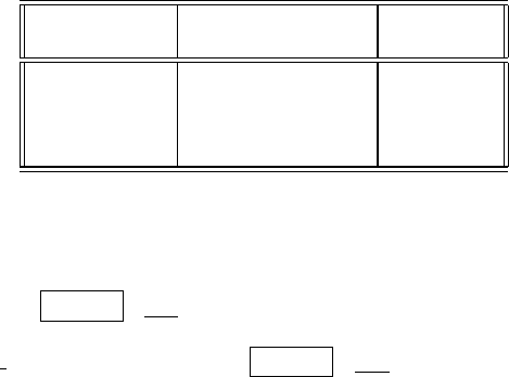
Each of you must generate two Ramachandran plots. Check the chart below
for your particular assignment (on the basis of your last name).
First letter of Subgroup 1 Subgroup 2
your last name
A–N Ala, Val, Leu, Ile Gly
O–R Asp, Asn, Glu, Gln Pro
S–V Lys, Arg, His Ser, Thr
W–Z Trp, Tyr, Phe Cys, Met
Each plot should have the data points for the (φ, ψ) dihedral angles, cor-
responding to the assigned group of residues from all the proteins in the
database. To find the values of the φ and ψ dihedral angles in a protein you
can use Protein / List command from the Biopolymer module. Record
these angles to a file. You can use the Fortran code posted on the website
(aa
select.f) for searching the Protein / List output files (called PDA
files) for the dihedral angles of selected residues. Alternatively, you can
write a suitable program in a different language.
If you use the code from the website, you will need to edit it to replace
all occurrences of the names
ALA, VAL, etc. by the abbreviations of the
residues you are searching for. These abbreviations must be capitalized.
Compile the code and execute it with each PDA file as input. The output
file should contain only two numbers per line, the φ and ψ angles for the
specified residues. Check the numbers for correctness by comparing a few
lines from this output with the numbers in the PDA file.
Note: For plotting, you must use Insight II so that all plots are uniform in
size. We will overlay them in class! Follow the instructions below.
Prepare a file with your data points collected from all the proteins for
each of the assigned groups of residues. These files should have the ex-
tension
tbl (filename.tbl) . In addition, you must format these files
for Insight II by inserting the 12 lines as indicated:

Appendix D. Homework Assignments 581
#
TITLE: Phi (deg)
MEASUREMENT TYPE: Ang
UNITS OF MEASUREMENT: Deg
FUNCTION: dihedral
#
TITLE: Psi (deg)
MEASUREMENT TYPE: Ang
UNITS OF MEASUREMENT: Deg
FUNCTION: dihedral
#
#
−34.5 144.9 This is the first line of your numeric data.
.
.
.
Note that at the top of the file there is space for your own comments and you
can use as many lines as you need. Insight II will start reading the data from
the line without the character # on the first column. That first line should be
as indicated above.
If you have done everything correctly, you are ready for plotting. Press
the
Graph button on the left of the screen and select Get. Specify the
data file name, dihedral1 as X
Function and dihedral2 as Y Function.
Keep the New
Graph on and execute. The zigzag appearance of the plot
now requires fixing. Move the graph box near the bottom-left corner of
the screen. First, connect to the object (your plot) with the command
Transform / Connect. Second, move the plot by clicking on it and drag-
ging to the desired position. The scatter plot will be produced in the next
step. Select Point (only!) from the
Graph / Modify Display dialog box.
Choose Star as the Point Symbol, scale it ten times and execute. Use
Graph / Threshold to change the minimum value to −180.0 and the max-
imum value to 180.0 for both, X Graph Axis and Y Graph Axis. Scale
each axis 4 times with
Graph / Scale Axis command. Change the color
of any white elements in your plot with
Graph / Color command. Change
the background color to white. Print your plot (see instructions below) and
repeat the procedure for the second data file.
Prepare a transparency for each of the two plots and bring to class. (Hand
in the printed version of the plots with your homework.) Also e-mail the
{φ, ψ} files you generated, sending each file separately and specifying your
name and the group of residues in the
Subject line.

582 Appendix D. Homework Assignments
2. The NDB. The next part of the homework will acquaint you to the Nucleic
Acid Database, NDB, on which we will have a guest lecturer.
First, explore the database to discover what is available, and look through
the newsletter archives for current update information. Then, describe the
structures available in the database and the numbers in each class (e.g.,
B-DNA, ribozymes). Explore the different features of NDB.Thereare
many exciting structures and utilities.
3. Sugar Conformations for Canonical A, B, and Z-DNA. To appreciate
the different features of canonical A, B, and Z-DNA forms, choose through
the NDB entries one unmodified form in each DNA class with the largest
number of residues possible. You can view the structure within NDB,orby
porting the PDB file to Insight II and using the
Molecule / Get from the
Viewer module (though the latter may be more difficult).
Learn how to use the Report facility in the NDB Query Interface to gen-
erate a list of all the sugar pseudorotation angles (P ) for each deoxyribose
in each of the three structures you have chosen for your study. (Remember
that there are two sugars per base pair). Print a table for each structure.
Now, on one figure for all the three structures, plot P versus the residue
number. Exclude the two terminal base pairs of each structure for plot-
ting purposes. You may need to connect the points corresponding to each
structure for clarity.
Label the pattern clearly to indicate the A, B, and Z-DNA data. Hand in
this plot, with a description of the patterns you had identified for the sugar
conformation for the three canonical DNA forms, indicating the specific
structures you have chosen.
Summary of Items to Hand In:
(a) Ramachandran plots for the two subgroups of amino acid residues
assigned to you.
(b) Data on the structure class types and the amount of structures in each
class available in NDB.
(c) The figure with P versus residue number for the A, B, and Z-DNA
forms, with complete discussion.
Background Reading from Coursepack
• B. Cipra, “Computer Science Discovers DNA”, in What’s Happening in the
Mathematical Sciences, pp. 26–37 (P. Zorn, Ed.), American Mathematical
Society, Colonial Printing, Cranston, RI (1996).
• M. Gerstein and M. Levitt, “Simulating Water and the Molecules of Life”,
Sci. Amer. 279, 101–105 (1998).
Appendix D. Homework Assignments 583
• J. E. Cohen, “Mathematics is Biology’s Next Microscope, Only Better;
Biology is Mathematics’ Next Physics, Only Better”, PLoS Biology 2
(e439), 2017–2023 (2004).
• A. Hastings et al., “Quantitative Bioscience for the 21st Century”,
Bioscience 55, 511–517 (2005).
Printing Instructions
To save ink, change the background color from black to white at each print-
ing of a color or black/white image. Go to
Session / Environment,pressthe
Background button and change the color to white.
Proceed to
File , choose Export Plot, postscript, Gray Scale and optionally
Ball
and Stick.UseSave Device File to save a postscript file (the file will have
the “.ps” extension). This file can be printed on any postscript printer you can
access. Hand in your printout as part of the homework.
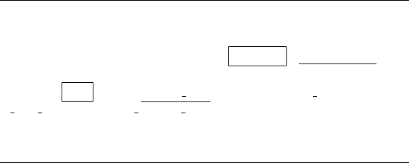
Fortran program for selecting Ala, Ile, Val, and Leu data
(see website)
584 Appendix D. Homework Assignments
Assignment 5: Analysis of Protein/DNA Complexes
with Insight and NDB: Canonical vs. Protein/Bound
DNA, and DNA/Protein Interactions
In this assignment, we will study three nucleic acids structures, two of which
have been crystallized with regulatory proteins: a nucleic acid dodecamer, a DNA
oligomer bound to a prokaryotic protein in the helix-turn-helix (HTH) motif,
and a DNA oligomer (known as the TATA-box sequence) bound to a eukaryotic
transcription factor.
NDB ID code Structure
• BDL078 DNA dodecamer
• PDR010 DNA (20 bp) bound to bacteriophage λ cI repressor
• PDT034 DNA (16 bp) bound to human TATA-Box Binding
protein
1. Structure Downloading. Download each structure from the NDB, already
explored in Assignment 4 (
ndbserver.rutgers.edu:80/). Use the Archives,
Atlas or Search entry points to the NDB; all are useful. In particular, the
Atlas allows you to quickly view the structures. The Search entry point
will be utilized later in this assignment.
Load each structure into Insight II. Separate the two complexes into DNA
and Protein objects using the
Modify / Unmerge command from either
the Biopolymer or Builder modules. Both the DNA and Protein objects
will be used later in this assignment. Use caution with the wildcard char-
acter * in selecting multiple atoms/residues/nucleotides/ proteins in Insight
II. A quick test which you can used to test which object you’ve created
with the
Modify / Unmerge is to blank or blink the object with the
Object / Blank or Object / Blink commands.
In order to create the B-DNA models for specified nucleic acid sequences
(see below) and manipulate the downloaded structures (as in the Unmerge
command), you will need to understand certain general parameters related
to the structure as used in both the PDB file and Insight. These parameters
refer to the DNA sequence, strand and residue labels, and so on. Explore the
text in the downloaded files as well as in the structural displays. Information
about relevant formatted lines in the coordinate files (such as SEQRES and
ATOM) is available from the PDB web site (see Assignment 2).
2. Canonical vs. Protein/Bound DNA Analysis. Create an idealized B-DNA
structure using the
Nucleotide / Append command in the Biopoly-
mer module corresponding to each of the three nucleic acid sequences in
the above structures. The
Nucleotide / Append command both creates
new nucleic acid molecules and appends to existing molecules; when it is
Appendix D. Homework Assignments 585
creating a new molecule, the Append Point is None.
3
You will need to use
the
Nucleic Acid / Cap pulldown menu to replace the phosphate group
with a hydroxyl group at the 5
ends of each strand.
Nucleotide / Append will create your B-DNA model, defining each
strand as a separate object. Your downloaded DNAs, however, are each
composed of a single object in Insight II. To properly superimpose the two
structures (the task in the next part), you will need to
Modify / Merge the
two strands as one object.
Superimpose each B-DNA model upon its respective crystal structure from
the NDB using the
Transform / Superimpose pulldown menu. If your
DNAs are each composed of a single object in Insight II, you will need
to
Modify / Merge the two strands as one object to properly superim-
pose the two structures. Use the Heavy option to avoid superpositioning of
hydrogen atoms. After selecting the B-DNA model and the crystal struc-
ture in the selection boxes, you will need to click the End Definition box
to Execute the Superimpose
command. The root-mean-square deviation
(RMSD) value will be printed at the bottom of your screen.
(a) Record the RMSD values relative to idealized B-DNA for each super-
imposed model/structure by repeating this procedure for each crystal
structure.
(b) Use the Search entry point to the NDB to extract the following pa-
rameters for each base pair of the three structures: P (pseudorotation
sugar pucker); the dihedral angles χ, α, β, γ, δ, , ζ;andthehe-
lical parameters twist, tilt, roll, and propeller twist: Ω, τ, ρ,andω,
respectively.
For each conformational variable (excluding those from the end
residues), calculate the average (μ) and standard deviation (σ) from
the data per structure. Prepare a table in the following form:
Now discuss your results in terms of the differences noted between
the protein-bound DNA and canonical B-DNA (which the BDL078
structure represents):
(c) Which structure is most deformed from B-DNA? Which parame-
ters display the largest changes from B-DNA (consider both μ and
σ)? Based on these parameters, what is similar in the way the two
complexes deform their recognition sites away from B-DNA (look
for similarities in columns 3 and 4 above which are different from
column 2)?
3
The PDR010 structure has an overhanging base at each end, that is a base without a Watson-
Crick partner on the other strand. (This procedure promotes crystal formation.) The recommended
procedure for creating the overhanging base is to use
Nucleotide / Append to create a 21-base-pair
duplex of the correct sequence and then use
Nucleotide / Delete command to delete one base from
each strand prior to using
Nucleic Acid / Cap.
