Research on oil recovery mechanisms in heavy oil reservoirs
Подождите немного. Документ загружается.


E. RANGEL-GERMAN, S. AKIN, L. CASTANIER SPE 54591
22
TABLE 1. RATES AND TIMING FOR THE CORE WITH NARROW FRACTURE
_Fluid Injected_
Time
_ (hrs) _
Flow Rate
_(cm
3
/min)_ _ Ports _
Water 0.0 - 4.0 2.0 Top and bottom closed,
lateral open.
Water 4.0 - 5.0 2.0 Top and bottom open,
lateral closed.
Water 5.0 - 19.8 0.5 Top and bottom open,
lateral closed.
Water 19.8 - 23.0 2.0 Top and bottom closed,
lateral open.
Decane 23.0 - 27.0 2.0 Top and bottom closed,
lateral open.
Decane 27.0 - 28.0 2.0 Top and bottom open,
lateral closed.
Decane 28.0 - 44.7 1.0 Top and bottom open,
lateral closed.
Water 44.7 - 46.0 9.99 Top and bottom closed,
lateral open.
46 Shut down everything
TABLE 2. RATES AND TIMING FOR THE CORE WITH WIDE FRACTURE
_Fluid Injected_
Time
_ (hrs) _
Flow Rate
_(cm
3
/min)_ _ Ports _
Water 0.0 - 5.0 2.0 Top and bottom closed,
lateral open.
5.0 - 21.0 0.0 Top and bottom closed,
lateral closed.
Water 21.0 - 22.5 2.0 Top and bottom open,
lateral closed.
Water 22.5 - 25.6 2.0 Top open, bottom closed,
and lateral closed.
25.6 - 45.0 0.0 Top and bottom closed,
lateral closed.
Decane 45.0 - 48.0 2.0 Top and bottom closed,
lateral open.
Decane 48.0 - 49.2 4.0 Top and bottom closed,
lateral open.
Decane 49.2 - 51.0 4.0 Top and bottom open,
lateral closed.
51.0 - 52.0 0.0 Top and bottom closed,
lateral closed.
Water 52.0 - 54.3 4.0 Top and bottom closed,
lateral open.
Water 54.3 - 68.8 0.5 Top and bottom closed,
lateral open.
Water 68.8 - 71.0 4.0 Top and bottom open,
lateral closed.
71 Shut down everything
SPE 54591 MULTIPHASE-FLOW PROPERTIES OF FRACTURED POROUS MEDIA
23
0.15, 0.02 0.15, 0.010.15, 0.010.15, 0.02 0.15, 0.02 0.15, 0.01 0.14, 0.01
0.15, 0.01 0.14, 0.020.14, 0.020.14, 0.01 0.14, 0.01 0.14, 0.02
0.13, 0.08 0.13, 0.110.14, 0.120.14, 0.11 0.14, 0.12 0.14, 0.12 0.13, 0.11
0.13, 0.09 0.14, 0.080.14, 0.080.12, 0.08 0.13, 0.08 0.13, 0.07
Narrow Fracture
Wide Fracture
0.2
0.0
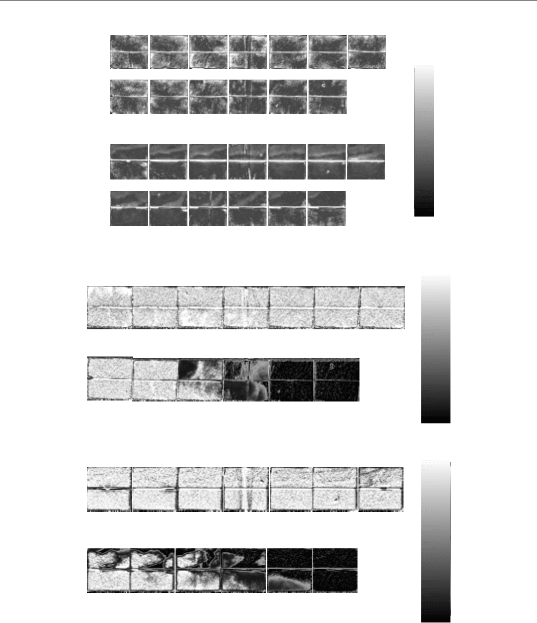
Fig. 5- Porosity distribution for the experimental system.
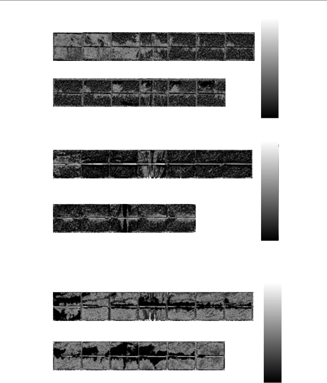
Fig. 6- CT Saturation images for the thin fracture system after 1 hr 30 min of water injection (0.67 PV.)
Fig. 7- CT Saturation images for the wide fracture system after 1 hr 30 min of water injection (0.67 PV.)
0.98, 0.05
0.96, 0.04
0.98, 4.46
0.96, 0.04
0.98, 0.04
1.04, 3.40
0.96, 0.04
0.99, 0.05
1.04, 3.33
0.97, 0.04
0.98, 0.04
1.02, 0.35
0.96, 0.04
0.97, 0.04
1.00, 0.26
0.96, 0.04
0.97, 0.04
1.00, 0.26
0.97, 0.04
0.97, 0.04
1.00, 0.22
0.97, 0.03
0.97, 0.04
0.99, 0.20
0.97, 0.04
0.97, 0.04
0.98, 0.60
0.85, 0.10
0.93, 0.05
0.88, 1.25
0.16, 0.12
0.52, 0.19
0.32, 0.53
-0.01, 0.04
-0.01, 0.04
-0.02, 0.74
-0.01, 0.54
-0.01, 0.04
-0.02, 0.62
after 1 hr. 30 min. press. port
0
0.99, 0.05
0.99, 0.04
0.95, 2.27
0.98, 0.05
0.99, 0.04
0.98, 2.06
0.98, 0.05
0.99, 0.05
1.00, 0.10
0.98, 0.13
0.97, 0.04
0.99, 0.13
0.97, 0.04
0.99, 0.04
0.99, 0.11
0.99, 0.06
0.99, 0.06
0.99, 0.10
0.95, 0.07
0.98, 0.04
0.97, 0.10
0.75, 0.29
0.99, 0.04
0.83, 1.09
0.64, 0.39
0.96, 0.04
0.77, 0.86
0.50, 0.41
0.93, 0.05
0.68, 0.55
0.16, 0.26
0.74, 0.15
0.41, 0.78
-0.01, 0.04
0.20, 0.19
0.09, 0.57
-0.01, 0.03
-0.01, 0.04
0.01, 0.60
after 1 hr. 30 min.
press. port
press. port
1
1
0
E. RANGEL-GERMAN, S. AKIN, L. CASTANIER SPE 54591
24
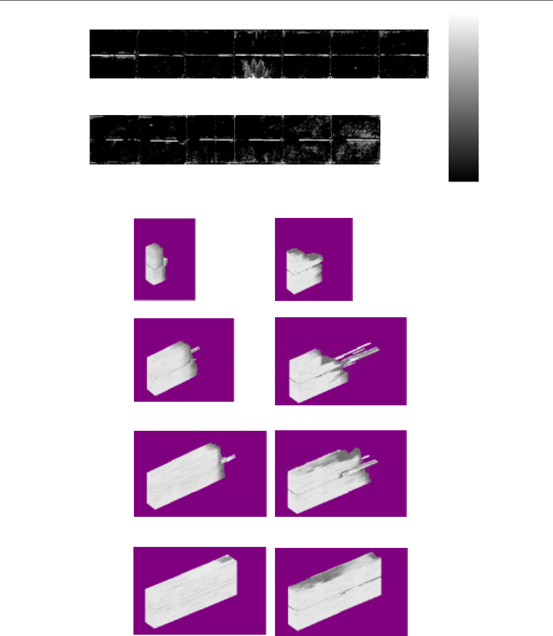
Fig. 8- CT Saturation images for the narrow fracture system after 2 hr 30 min of oil injection (1.13 PV.)
Fig. 9- CT Saturation images for the wide fracture system after 2 hr 30 min of oil injection (1.13 PV.)
Fig. 10- CT Saturation images for the wide fracture system after 16 hours of water injection (7.2 PV.)
0
after 2 hr. 30 min. press. port
0.02, 0.11
0.02, 0.09
0.17,10.15
-0.06, 0.13
-0.05, 0.09
-0.02, 0.92
-0.07, 0.11
-0.05, 0.10
-0.02, 0.45
0.01, 0.23
0.11, 0.23
0.10, 0.43
-0.03, 0.12
-0.01, 0.09
-0.12, 8.15
0.01, 0.12
0.00, 0.10
0.02, 4.61
-0.06, 0.18
-0.05, 0.14
-0.06, 9.22
0.02, 0.12
0.01, 0.12
0.10,14.12
0.02, 0.10
0.01, 0.09
-0.01,12.20
press. port
-0.05, 0.12
-0.03, 0.14
-0.02, 0.26
-0.06, 0.13
-0.03, 0.10
-0.04, 0.25
-0.04, 0.12
-0.03, 0.11
-0.02, 0.25
after 16 hr.
press. port
press. port
0
1
1
0
after 2 hr. 30 min. press. port
0.25, 0.11
0.32, 0.11
0.28, 3.82
0.13, 0.13
0.24, 0.12
0.08, 3.95
0.05, 0.11
0.15, 0.10
0.04, 3.19
0.06, 0.12
0.06, 0.11
0.02, 0.36
0.01, 0.09
0.01, 0.09
-0.02, 0.26
0.00, 0.09
0.00, 0.09
-0.02, 0.24
0.00, 0.09
0.00, 0.10
-0.03, 0.32
-0.01, 0.08
0.00, 0.09
-0.05, 0.81
0.01, 0.09
0.01, 0.10
-0.04, 2.37
0.02, 0.12
0.00, 0.11
0.02, 2.63
0.02, 0.12
0.01, 0.12
-0.03, 6.17
0.01, 0.13
0.00, 0.10
-0.10, 4.14
-0.03, 0.46
0.00, 0.10
-0.01, 0.10
1
SPE 54591 MULTIPHASE-FLOW PROPERTIES OF FRACTURED POROUS MEDIA
25
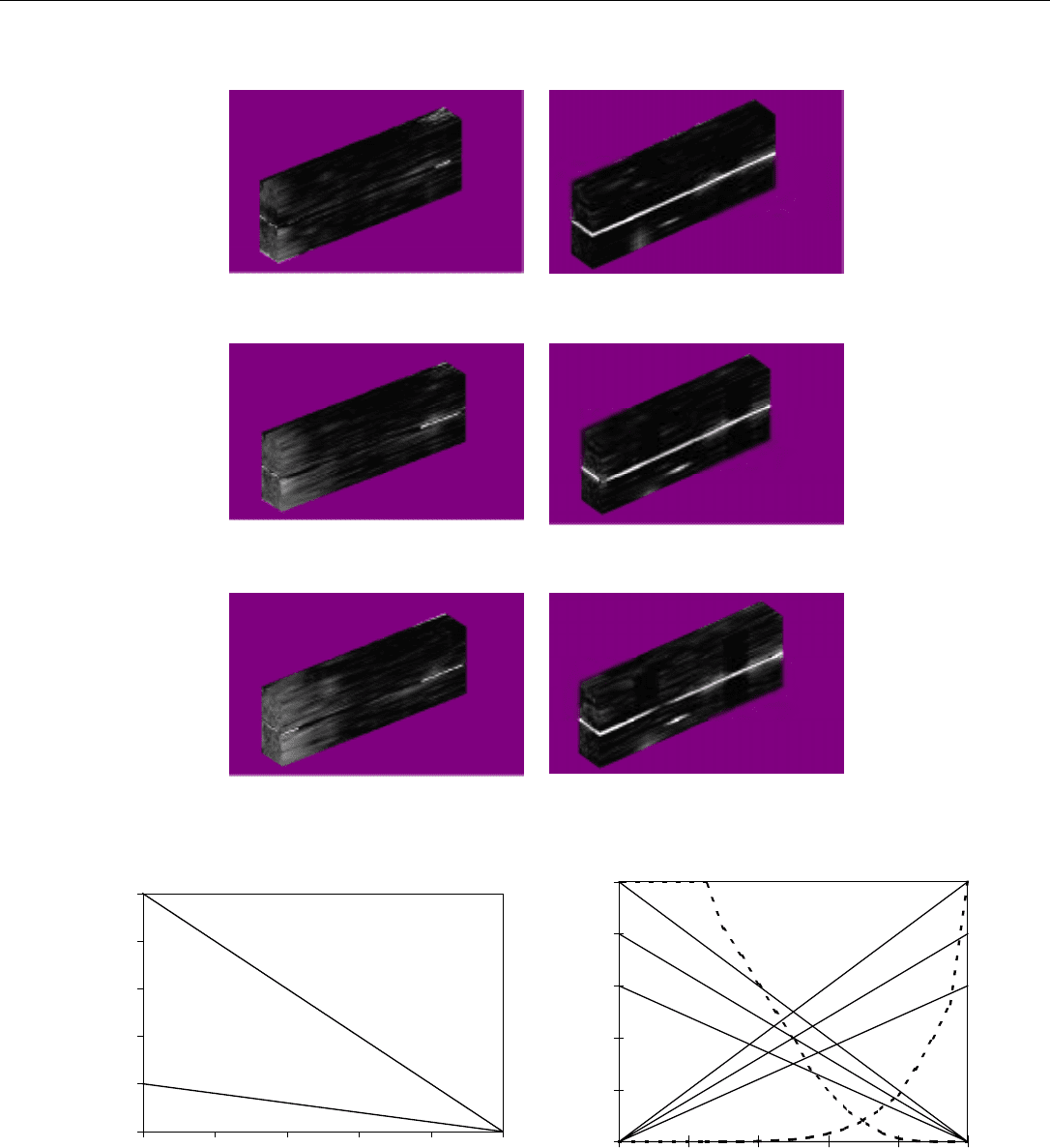
Fig. 11- CT Saturation images for the wide fracture system after 17 hours of water injection (7.7 PV.)
Fig. 12- 3-D Reconstruction for both systems for water injection.
press. port
after 17 hr.
press. port
0.22 PV (30 min)
0.67 PV (1hr. 30 min)
0.89 PV (2 hr.)
0.45 PV (1hr.)
0
1
E. RANGEL-GERMAN, S. AKIN, L. CASTANIER SPE 54591
26
Fig. 13- 3-D Reconstruction for both systems for oil injection.
Fig. 14- Capillary pressure curves and relative permeability curves for the fracture.
2 hr. 30 min
3 hr. 45 min
1hr. 30 min
0
0.2
0.4
0.6
0.8
1
0 0.2 0.4 0.6 0.8 1
Water saturation, fraction
Pcf, atm
Case 1
Case 2
Case 3
0.0
0.2
0.4
0.6
0.8
1.0
0.0 0.2 0.4 0.6 0.8 1.0
Water saturation, fraction
Kr
Case B
Case C
- - - Matrix
___
Fracture
Case A

SPE 54591 MULTIPHASE-FLOW PROPERTIES OF FRACTURED POROUS MEDIA
27
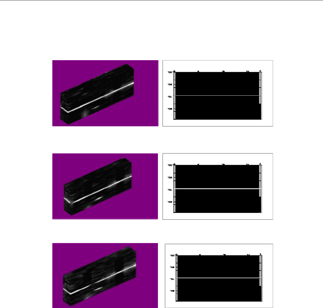
1 hr. 30 min (0.67 PV)
2 hours (0.89 PV)
1 hr. (0.45 PV)
30 min. (0.22 PV)
1 hr. 30 min (0.67 PV)
2 hours (0.89 PV)
1 hr. (0.45 PV)
30 min. (0.22 PV)
Narrow
Wide
Fig. 15- Comparison between experiments and numerical simulations for narrow and wide fracture systems for different PV
of water injection.
E. RANGEL-GERMAN, S. AKIN, L. CASTANIER SPE 54591
28
Fig. 16- Comparison between experiments and numerical simulations for narrow and wide fracture systems for different PV
of water injection.
1.13 PV (2 hr. 30 min)
0.67 PV (1 hr. 30 min)
1.68 PV (3 hr. 45 min)
29
1.3 IMBIBITION STUDIES OF LOW-PERMEABILITY POROUS MEDIA
(S. Akin and A.R. Kovscek)
This paper, SPE 54590, was presented at the 1999 SPE Western Regional Meeting held
in Anchorage, Alaska, May 26-28, 1999, and presented for publication in the Journal of
Petroleum Science and Engineering.
S. AKIN AND A. R. KOVSCEK SPE 54590
30
Copyright 1998, Society of Petroleum Engineers, Inc.
This paper was prepared for presentation at the 1999 Western Regional Meeting held in
Anchorage, Alaska, 26-27 May 1999.
This paper was selected for presentation by an SPE Program Committee following review of
information contained in an abstract submitted by the author(s). Contents of the paper, as
presented, have not been reviewed by the Society of Petroleum Engineers and are subject to
correction by the author(s). The material, as presented, does not necessarily reflect any
position of the Society of Petroleum Engineers, its officers, or members. Papers presented at
SPE meetings are subject to publication review by Editorial Committees of the Society of
Petroleum Engineers. Electronic reproduction, distribution, or storage of any part of this paper
for commercial purposes without the written consent of the Society of Petroleum Engineers is
prohibited. Permission to reproduce in print is restricted to an abstract of not more than 300
words; illustrations may not be copied. The abstract must contain conspicuous
acknowledgment of where and by whom the paper was presented. Write Librarian, SPE, P.O.
Box 833836, Richardson, TX 75083-3836, U.S.A., fax 01-972-952-9435.
Abstract
A systematic investigation of capillary pressure, relative
permeability, and fluid flow characteristics within diatomite (a
high porosity, low permeability, siliceous rock) is reported.
Using an X-ray computerized tomography (CT) scanner, and a
specially constructed imbibition cell, we study spontaneous
cocurrent water imbibition into diatomite samples at various
initial water saturations. Air-water and oil-water systems are
used. Despite a marked difference in rock properties between
diatomite and sandstone, including permeability and porosity,
we find similar trends in saturation profiles and dimensionless
weight gain versus time functions. Diatomite is roughly 100
times less permeable than sandstone, yet it imbibes water at
rates rivaling sandstone. Importantly, the spontaneous
imbibition data when combined with CT-scan images provides
a means to determine dynamic relative permeability and
capillary pressure functions.
Introduction:
Spontaneous imbibition is perhaps the most important
phenomenon in oil recovery from fractured reservoirs. In
imbibition, capillary suction draws wetting liquid into the rock
matrix. In fractured systems, the rate of mass transfer between
the rock matrix and fractures usually determines the oil
production
1,2
. Imbibition is also essential in evaluation of
rock wettability
3
. Because of strong capillary forces, the
smallest pore bodies next to the fracture are usually invaded
first. The rate of imbibition is a function of porous media and
fluid properties such as absolute and relative permeability,
viscosity, interfacial tension, and wettability
4
. Most
experimental work on imbibition behavior has concentrated on
the scaling aspects of the process in order to estimate oil
recovery from reservoir matrix blocks that have shapes, and
sizes different from those of laboratory core samples
4-6
.
Two different approaches are used, typically, to model
imbibition behavior. The first approach employs a sugar-cube
type matrix-fracture system proposed by Warren and Root
1
where the communication occurs at the matrix-fracture
interface
2
. The second approach is based on a representative
elemental volume averaged fracture-matrix system
6-8
. Both
approaches use empirically determined mass transfer
functions.
Kazemi et al.
2
presented numerical and analytical solutions
of oil recovery using empirical, exponential transfer functions
based on the data given by Aronofsky et al.
9
and Mattax and
Kyte
10
. They proposed a shape factor that included the effect
of size, shape, and boundary conditions of the matrix. Later,
this shape factor was generalized by Ma et al.
11
to account for
the effect of viscosity ratio, sample shape, and boundary
conditions. The following equation was proposed:
t
D
= t
k
φ
σ
µ
s
L
c
2
(1)
where
µ
s
=µ
w
µ
nw
(2)
The above scaling equation was used by Zhang et al.
4
who
report that ultimate oil recovery on a pore volume basis by
spontaneous imbibition in Berea sandstone cores is
approximately constant for systems with differing lengths,
viscosity ratios, and boundary conditions.
To date, much of the focus on imbibition has centered on
carbonaceous rocks as a result of the importance of the North
Sea Chalks
12-15
, the West Texas Carbonates, and the Middle
Eastern Limestones. An important, yet relatively unstudied,
low permeability, reservoir rock for which imbibition is
believed to be an important recovery mechanism is diatomite
16
. Imbibition phenomena are also important during steam
injection into diatomite because steam condenses upon
injection into a cool reservoir, and the hot condensate imbibes
into the formation.
SPE 54590
Imbibition Studies of Low-Permeability Porous Media
S. Akin, Middle East Technical University and A. R. Kovscek, Stanford U., SPE Members

SPE 54590 IMBIBITION STUDIES OF LOW PERMEABILITY POROUS MEDIA
31
Diatomites are high porosity (>50%), low permeability
(0.1-10md)
17
rocks of a hydrous form of silica or opal
composed of the remains of microscopic shells of diatoms,
which are single-celled aquatic plankton
18
. Diatomite reservoir
rock is assumed to be moderately to strongly water wet
16
.
Estimates of the original oil in place (OOIP) for diatomite
reservoirs in California range from 10 to 15 billion barrels
19
and primary recovery has been estimated to be about 5% of the
OOIP
20
. Primary recovery is low despite hydraulically
fractured wells that improve injectivity and productivity.
Because of the high porosity, large initial oil saturation (35 to
70%), and large OOIP, the target for potential production is
high.
Multiphase flow in diatomite is assumed to be dominated
by capillary forces, but we lack a good understanding of fluid
flow and capillary pressure behavior in the rock matrix.
Compounding problems, there are only a few reported
capillary pressure curves
16
and little information on the extent
and rate of imbibition
21
. The mechanisms of oil displacement
and trapping are unclear, but assumed to be similar to those in
sandstone. However, rock morphology is very different
22
.
This study presents basic core analysis data and
experimental results of spontaneous water imbibition in
diatomite and sandstone. A state of the art experimental
imbibition cell is employed along with computed tomography
(CT) scanning to quantify the saturation distribution along the
core. It is found that the imbibition data scales according to
the equation proposed by Ma et al.
11
. The major focus of this
article is estimation of relative permeability and capillary
pressure from the experimental saturation data using a
nonlinear history-matching technique.
Experimental Details
Liquids
For water-air imbibition experiments, de-aerated water and air
were used, whereas for water-oil imbibition, n-decane and de-
aerated water were used. Properties of the experimental fluids
are given in Table 1.
Porous Media
Diatomite cores were cut in a direction parallel to the bedding
plane from a block of outcrop diatomite sample (Grefco
Quarry, Lompoc, CA). This sample is fairly homogeneous, but
there are regions of higher density and correspondingly lower
porosity. Figure 1 displays porosity images of the cores used
in this study. The procedure to obtain these images will be
presented, shortly. The average porosity of the samples was
slightly greater than 65% and porosity varied by about 2 to
4%. The liquid permeability varied from 6 to 9 md. Table 2
lists exact values for each core. It should be noted that because
of the brittle nature of the diatomite, the cores were machined
instead of drilled using conventional core bits as described by
Schembre et al.
21
. The lengths (3.45 in. or 8.763 cm) and
diameters (0.95 in. or 2.413 cm) of the cores were all close to
constant. Cores were potted with epoxy in 1 inch (2.54 cm)
Plexiglas tubes. Both ends are left open to enable cocurrent
imbibition.
Experimental Setup
The experimental setup is designed specifically for CT
measurements. It consists of three main parts: (i) a potted
diatomite core inside a (ii) water jacket and (iii) a data
acquisition system based on a personal computer, as shown in
Figure 2. Two end caps hold the imbibition cell in position
within the cylindrical water jacket. The end caps are machined
with spider-web-shaped fluid distribution grooves where the
end cap contacts the core. The entire assembly is placed inside
the CT gantry. Fluid is circulated through the jacket to
maintain a constant temperature up to 90°C (194°F) using a
heating circulator bath. The experiments reported here were
conducted at 20 °C (68 °F). The main function of the water
jacket is to reduce effects of beam hardening. Beam hardening
is the change of overall X-ray attenuation with distance into
the object. It occurs when a polychromatic X-ray beam passes
through a material that preferentially absorbs lower energy
photons. The remaining beam becomes more and more
chromatic at higher energy levels and the beam becomes
“harder”. The effects of this phenomenon are usually more
pronounced at the boundaries where there is a high density
contrast (i.e. air and core).
Three polyethylene lines (input, output, and bypass) allow
the imbibing fluid to enter the imbibition cell and the produced
fluid to exit the cell. The bypass line is used to flush any non-
wetting fluid from the lines, thus allowing the imbibing fluid to
fill the inlet end plate and easily contact the core face. The
amount of water imbibed is found by measuring the weight
loss of the water reservoir using an electronic balance
connected to a personal computer. The CT-Scanner used in
this study is a fourth generation Picker 1200-SX scanner
with 1200 fixed detectors. It allows rapid scanning on a single
vertical volume section in the center of the core as a function
of time. The acquisition time of one CT image is 3 seconds,
whereas the processing time is around 40 seconds. The total
time of measurement is selected such that it is short enough to
capture accurately the position of the imbibition front, yet it is
large enough to provide necessary X-ray energy. Table 3
gives CT scan parameters used in this study.
Imbibition Measurements
For water-air imbibition experiments, the core samples
were dried in a vacuum oven at approximately 70°C (158°F)
for 24 hours before starting the experiments. After pressure
testing for leaks, the imbibition cell is placed into the water
jacket either horizontally or vertically and the jacket is filled
with water. All experiments reported here are in the horizontal
mode. Next, the setup is placed inside the gantry and
positioned such that the main scanning plane is at the center of
the core sample and there is nothing around the setup (i.e. the
patient couch is not in the scanning plane). A reference dry
CT image is obtained using the parameters given in Table 3.
