Peterson D.R., Bronzino J.D. (Eds.) Biomechanics: Principles and Applications
Подождите немного. Документ загружается.


Cardiac Biomechanics 8-5
with age in most species but not in humans. At birth, left and right ventricular weights are similar, but the
left ventricle is substantially more massive than the right by adulthood.
Epicardium
5%
–58°
15%
–43°
25%
–33°
35%
–24°
45%
4°
55%
20°
65%
29°
75%
42°
85%
53°
95%
61°
Endocardium
FIGURE 8.2 Cardiac muscle fiber
orientations vary continuously
throughthe leftventricular wall from
a negative angle at the epicardium
(0%) to near zero (circumferential)
at the midwall (50%) and to increas-
ing positive values toward the
endocardium (100%). (Courtesy
Jyoti Rao, Micrographs of murine
myocardium from the author’s
laboratory.)
8.2.2 Myofiber Architecture
The cardiac ventricles have a complex three-dimensional muscle fiber
architecture (for a comprehensive review see Streeter [7]). Although the
myocytes are relatively short, they are connected such that at any point
in the normal heart wall there is a clear predominant fiber axis that is
approximately tangent with the wall (within 3 to 5
◦
in most regions,
except near the apex and papillary muscle insertions). Each ventricu-
lar myocyte is connected via gap junctions at intercalated disks to an
average of 11.3 neighbors, 5.3 on the sides and 6.0 at the ends [8]. The
classical anatomists dissected discrete bundles of fibrous swirls, though
later investigations showed that the ventricular myocardium could be
unwrapped by blunt dissection into a single continuous muscle “ban”
[9]. However, more modern histological techniques have shown that
in the plane of the wall, the mean muscle fiber angle makes a smooth
transmural transition from epicardium to endocardium (Figure 8.2).
About the mean, myofiber angle dispersion is typically 10 to 15
◦
[10]
except in certain pathologies. Similar patterns have been described for
humans, dogs, baboons, macaques, pigs, guinea pigs, and rats. In the
left ventricle of humans or dogs, the muscle fiber angle typically varies
continuously from about −60
◦
(i.e., 60
◦
clockwise from the circumfer-
ential axis) at the epicardium to about +70
◦
at the endocardium. The
rate of change of fiber angle is usually greatest at the epicardium, so
that circumferential (0
◦
) fibers are found in the outer half of the wall,
and begins to slow approaching the inner third near the trabeculata–
compacta interface. There are also small increases in fiber orientation
from end-diastole to systole (7 to 19
◦
), with greatest changes at the
epicardium and apex [11].
Regional variations in ventricular myofiber orientations are gener-
ally smooth except at the junction between the right ventricular free
wall and septum. A detailed study in the dog that mapped fiber an-
gles throughout the entire right and left ventricles described the same
general transmural pattern in all regions including the septum and
right ventricular free wall, but with definite regional variations [3].
Transmural differences in fiber angle were about 120 to 140
◦
in the left
ventricular free wall, larger in the septum (160 to 180
◦
) and smaller
in the right ventricular free wall (100 to 120
◦
). A similar study of
fiber angle distributions in the rabbit left and right ventricles has re-
cently been reported [12]. For the most part, fiber angles in the rabbit
heart were very similar to those in the dog, except for on the anterior
wall, where average fiber orientations were 20 to 30
◦
counterclock-
wise of those in the dog. While the most reliable reconstructions of
ventricular myofiber architecture have been made using quantitative
histological techniques, diffusion tensor magnetic resonance imaging
(MRI) has proven to be a reliable technique for estimating fiber orien-
tation nondestructively in fixed [13,14] and even intact beating human
hearts [15].
8-6 Biomechanics
The locus of fiber orientations at a given depth in the ventricular wall has a spiral geometry that may
be modeled as a general helix by simple differential geometry. The position vector x of a point on a helix
inscribed on an ellipsoidal surface that is symmetric about the x
1
axis and has major and minor radii,
a and b, is given by the parametric equation,
x = a sin te
1
+ b cos t sin wte
2
+ b cos t cos wte
3
(8.4)
where the parameter is t, and the helix makes w/4 full turns between apex and equator. A positive w
defines a left-handed helix with a positive pitch. The fiber angle or helix pitch angle η, varies along the arc
length:
sin η =
a
2
cos
2
t + b
2
sin
2
t
(a
2
+ b
2
w
2
)cos
2
t + b
2
sin
2
t
(8.5)
If another, deformed configuration
ˆ
x is defined in the same way as Equation 8.4, the fiber-segment-
extension ratio d
ˆ
s/ds associated with a change in the ellipsoid geometry [16] can be derived from
d
ˆ
s/dt
ds/dt
=
|d
ˆ
x/dt|
|dx/dt|
(8.6)
Although the traditional notion of discrete myofiber bundles has been revised in view of the contin-
uous transmural variation of muscle fiber angle in the plane of the wall, there is a transverse laminar
structure in the myocardium that groups fibers together in sheets an average of 4 ± 2 myocytes thick
(48 ±20 μm), separated by histologically distinct cleavage planes [17–19]. LeGrice and colleagues [19] in-
vestigated these structures in a detailed morphometric study of four dog hearts. They describe an ordered
laminar arrangement of myocytes with extensive cleavage planes running approximately radially from
endocardium toward epicardium in transmural section. Like the fibers, the sheets also have a branching
pattern with the number of branches varying considerably through the wall thickness. Recent reports
suggest that, in addition to fiber orientations, diffusion tensor MRI may be able to detect laminar sheet
orientations [20]. The tensor of diffusion coefficients in the myocardium detected by MRI has shown to be
orthotropic, and the principal axis of slowest diffusion was seen to coincide with the direction normal to the
sheet planes.
The fibrous architecture of the myocardium has motivated models of myocardial material symmetry as
transversely isotropic. The transverse laminae are the first structural evidence for material orthotropy
and have motivated the development of models describing the variation of fiber, sheet, and sheet-normal
axes throughout the ventricular wall [21]. This has led to the idea that the laminar architecture of the
ventricular myocardium affects the significant transverse shears [22] and myofiber rearrangement [18]
described in the intact heart during systole. By measuring three-dimensional distributions of strain across
the wall thickness using biplane radiography of radiopaque markers, LeGrice and colleagues [23] found
that the cleavage planes coincide closely with the planes of maximum shearing during ejection, and that the
consequent reorientation of the myocytes may contribute 50% or more of normal systolic wall thickening.
Arts et al. [24] showed that the distributions of sheet orientations measured within the left ventricular
wall of the dog heart coincided closely with those predicted from observed three-dimensional wall strains
using the assumption that laminae are oriented in planes that contain the muscle fibers and maximize
interlaminar shearing. This assumption also leads to the conclusion that two families of sheet orientations
may be expected. Indeed, a retrospective analysis of the histology supported this prediction and more
recent observations confirm the presence of two distinct populations of sheet plane in the inner half of the
ventricular wall.
A detailed description of the morphogenesis of the muscle fiber system in the developing heart is not
available but there is evidence of an organized myofiber pattern by day 12 in the fetal mouse heart that
is similar to that seen at birth (day 20) [25]. Abnormalities of cardiac muscle fiber patterns have been
described in some disease conditions. In hypertrophic cardiomyopathy, which is often familial, there is
substantial myofiber disarray, typically in the interventricular septum [10,26].
Cardiac Biomechanics 8-7
8.2.3 Extracellular Matrix Organization
The cardiac extracellular matrix consists primarily of the fibrillar collagens, type I (85%) and III (11%),
synthesized by the cardiac fibroblasts, the most abundant cell type in the heart. Collagen is the major struc-
tural protein in connective tissues, but only comprises 2 to 5% of the myocardium by weight, compared
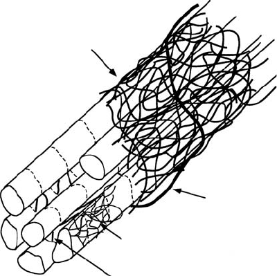
with the myocytes, which make up 90% [27]. The collagen matrix has a hierarchical organization (Fig-
ure 8.3), and has been classified according to conventions established for skeletal muscle into endomysium,
perimysium, and epimysium [28,29]. The endomysium is associated with individual cells and includes
a fine weave surrounding the cell and transverse structural connections 120 to 150 nm long connecting
adjacent myocytes, with attachments localized near the z-line of the sarcomere. The primary purpose of
the endomysium is probably to maintain registration between adjacent cells. The perimysium groups cells
together and includes the collagen fibers that wrap bundles of cells into the laminar sheets described above
as well as large coiled fibers typically 1 to 3μm in diameter composed of smaller collagen fibrils (40 to
50 nm) [30]. The helix period of the coiled perimysial fibers is about 20μm and the convolution index (ra-
tio of fiber arc length to midline length) is approximately 1.3 in the unloaded state of the ventricle [31,32].
These perimysial fibers are most likely to be the major structural elements of the collagen extracellular
matrix though they probably contribute to myocardial strain energy by uncoiling rather than stretching
[31]. Finally, a thick epimysial collagen sheath surrounds the entire myocardium forming the protective
epicardium (visceral pericardium) and endocardium.
Collagen content, organization, cross-linking and ratio of types I to III change with age and in various
disease conditions including myocardial ischemia and infarction, hypertension and hypertrophy (Ta-
ble 8.4). Changes in myocardial collagen content and organization coincide with alterations in diastolic
myocardial stiffness [33]. Collagen intermolecular cross-linking is mediated by two separate mechanisms.
The formation of enzymatic hydroxylysyl pyridinoline cross-links is catalyzed by lysyl oxidase, which re-
quires copper as a cofactor. Nonenzymatic collagen cross-links known as advanced glycation endproducts
can be formed in the presence of reducing sugars. This mechanism has been seen to significantly increase
ventricular wall stiffness independent of changes in tissue collagen content, not only in diabetics, but also
in an animal model of volume overload hypertrophy [34]. Hence the collagen matrix plays an important
role in determining the elastic material properties of the ventricular myocardium.
Woven
perimysial
network
Coiled
perimysial
fiber
Endomysial weave
Transverse
intermyocyte connections
Z-line of sarcomere
Myocytes
FIGURE 8.3 Schematic representation of cardiac tissue structure showing the association of endomysial and peri-
mysial collagen fibers with cardiac myocytes. (Courtesy Dr. Deidre MacKenna.)

8-8 Biomechanics
TABLE 8.4 Changes in Ventricular Collagen Structure and Mechanics with Age and Disease
Types and
Condition Collagen Morphology Crosslinking Passive Stiffness Other
Pressure overload [Hydroxyproline]: Type III: ⇑ [135] Chamber: Perivascular fibrosis
hypertrophy ⇑-⇑⇑⇑ [132,133] ⇑-⇑⇑ [133,134] ⇑⇑[133]
Area fraction: Cross-links: Tissue: ⇑⇑ [137] Focal scarring:
⇑⇑⇑ [133,134] no change [136] [138,139]
Volume overload [Hydroxyproline]: Cross-links: Chamber: ⇓ [143] Parallel changes
hypertrophy no change-⇓: [140,141] ⇑ [136,141]
Area fraction: no Type III/I: ⇑ [141] Tissue: no
change [132,142] change/⇑ [143]
Acute [Hydroxyproline]: ⇓ early [146] Collagenase activity:
ischemia/stunning ⇓ [Charney 1992 #1118] ⇑ late [147] ⇑ [148,149]
Light microscopy: no
change/⇓ [144]
⇓⇓ endomysial fibers [145]
Chronic myocardial [Hydroxyproline]: Type III: ⇑ [153] Chamber: ⇑ Organization:
infarction ⇑⇑⇑ [150,151] early [154] ⇑-⇑⇑⇑ [155,156]
Loss of birefringence [152] Chamber: ⇓
late [154]
Age [Hydroxyproline]: Type III/I: ⇓ [158] Chamber: ⇑ [159] Light microscopy:
⇑-⇑⇑⇑ [148,157] fibril diameter ⇑ [157]
Collagen fiber Cross-links: ⇑ [158] Papillary muscle:
diameter ⇑[157] ⇑ [160]
8.3 Cardiac Pump Function
8.3.1 Ventricular Hemodynamics
The most basic mechanical parameters of the cardiac pump are blood pressure and volume flowrate,
especially in the major pumping chambers, the ventricles. From the point of view of wall mechanics,
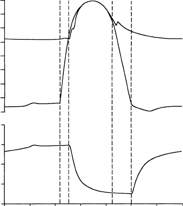
the ventricular pressure is the most important boundary condition. Schematic representations of the
time-courses of pressure and volume in the left ventricle are shown in Figure 8.4. Ventricular filling
immediately following mitral valve opening (MVO) is initially rapid because the ventricle produces a
diastolic suction as the relaxing myocardium recoils elastically from its compressed systolic configuration
below the resting chamber volume. The later slow phase of ventricular filling (diastasis) is followed finally
by atrial contraction. The deceleration of the inflowing blood reverses the pressure gradient across the
valve leaflets and causes them to close mitral valve closure (MVC). Valve closure may not, however, be
completely passive, because the atrial side of the mitral valve leaflets, which unlike the pulmonic and aortic
valves are cardiac in embryological origin, have muscle and nerve cells, and are electrically coupled to atrial
conduction [35].
Ventricular contraction is initiated by excitation, which is almost synchronous (the duration of the QRS
complex of the ECG is only about 60 msec in the normal adult) and begins about 0.1 to 0.2 sec after
atrial depolarization. Pressure rises rapidly during the isovolumic contraction phase (about 50 msec in
adult humans), and the aortic valve opens (AVO) when the developed pressure exceeds the aortic pressure
(afterload). Most of the cardiac output is ejected within the first quarter of the ejection phase before the
pressure has peaked. The aortic valve closes (AVC) 20 to 30 msec after AVO when the ventricular pressure
falls below the aortic pressure owing to the deceleration of the ejecting blood. The dichrotic notch, a
characteristic feature of the aortic pressure waveform and a useful marker of aortic valve closure, is caused
by pulse wave reflections in the aorta. Since the pulmonary artery pressure against which the right ventricle
Cardiac Biomechanics 8-9
16
14
12
10
8
Pressure (kPa)
Pressure (kPa)
6
4
2
0
150
120
90
60
30
0 100 200 300
Time (msec)
400 500 600 700
Aorta
Left ventricle
MVC
AVO
AVC
MVO
FIGURE 8.4 Left ventricular pressure, aortic pressure, and left ventricular volume during a single cardiac cycle
showing the times of mitral valve closure (MVC), aortic valve opening (AVO), aortic valve closure (AVC), and mitral
valve opening (MVO).
pumps is much lower than the aortic pressure, the pulmonic valve opens before and closes after the aortic
valve. The ventricular pressure falls during isovolumic relaxation, and the cycle continues. The rate of
pressure decay from the value P
0
at the time of the peak rate of pressure fall until MVO is commonly
characterized by a single exponential time constant, that is,
P (t) = P
0
e
−t/τ
+ P
∞
(8.7)
where P
∞
is the (negative) baseline pressure to which the ventricle would eventually relax if MVO were
prevented [36]. In dogs and humans, τ is normally about 40 msec, but it is increased by various factors
including elevated afterload, asynchronous contraction associated with abnormal activation sequence or
regional dysfunction, and slowed cytosolic calcium reuptake to the sarcoplasmic reticulum associated with
cardiac hypertrophy and failure. The pressure and volume curves for the right ventricle look essentially
the same; however, the right ventricular and pulmonary artery pressures are only about a fifth of the
corresponding pressures on the left side of the heart. The intraventricular septum separates the right and
left ventricles and can transmit forces from one to the other. An increase in right ventricular volume
may increase the left ventricular pressure by deformation of the septum. This direct interaction is most
significant during filling [37].
The phases of the cardiac cycle are customarily divided into systole and diastole. The end of diastole —
the start of systole — is generally defined as the time of MVC. Mechanical end-systole is usually defined
as the end of ejection, but Brutsaert and colleagues proposed extending systole until the onset of diastasis
(see the review by Brutsaert and Sys [38]), since there remains considerable myofilament interaction and
active tension during relaxation. The distinction is important from the point of view of cardiac muscle
mechanics: the myocardium is still active for much of diastole and may never be fully relaxed at sufficiently
high heart rates (over 150 beats per minute). Here, we will retain the traditional definition of diastole,
but consider the ventricular myocardium to be “passive” or “resting” only in the final slow-filling stage of
diastole.
8-10 Biomechanics
20(a)
16
12
8
4
0
0 50 100 150 200
Pressure (kPa)
20(b)
16
12
8
4
0
0 50 100
LV volume (ml)
150 200
Pressure (kPa)
ESPVR
EDPVR
AVC
AVO
MVC
MVO
Stroke
work
ESPVR
EDPVR
E(200)
=E
max
E(160 msec)
E(120 msec)
E(80 msec)
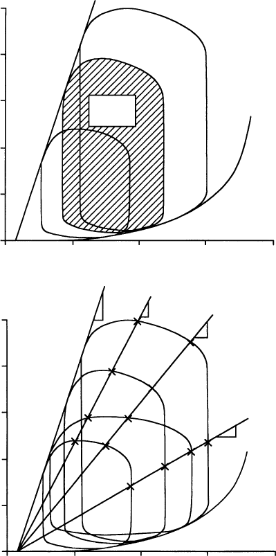
FIGURE 8.5 Schematic diagram of left ventricular pressure–volume loops: (a) End-systolic pressure–volume re-
lation (ESPVR), end-diastolic pressure–volume relation (EDPVR) and stroke work. The three P–V loops show the
effects of changes in preload and afterload. (b) Time-varying elastance approximation of ventricular pump function
(see text).
8.3.2 Ventricular Pressure--Volume Relations and Energetics
A useful alternative to Figure 8.4 for displaying ventricular pressure and volume changes is the pressure–
volume loop shown in Figure 8.5a. During the last 20 years, the ventricular pressure–volume relationship
has been explored extensively, particularly by Sagawa and colleagues [39], who wrote a comprehensive
book on the approach. The isovolumic phases of the cardiac cycle can be recognized as the vertical segments
of the loop, the lower limb represents ventricular filling, and the upper segment is the ejection phase. The
difference on the horizontal axis between the vertical isovolumic segments is the stroke volume, which
expressed as a fraction of the end-diastolic volume is the ejection fraction. The effects of altered loading on
Cardiac Biomechanics 8-11
the ventricular pressure–volume relation have been studied in many preparations, but the best controlled
experiments have used the isolated cross-circulated canine heart in which the ventricle fills and ejects
against a computer-controlled volume servo-pump.
Changes in the filling pressure of the ventricle (preload) move the end-diastolic point along the unique
end-diastolic pressure–volume relation (EDPVR), which represents the passive filling mechanics of the
chamber that are determined primarily by the thick-walled geometry and nonlinear elasticity of the resting
ventricular wall. Alternatively, if the afterload seen by the left ventricle is increased, stroke volume decreases
in a predictable manner. The locus of end-ejection points (AVC) forms the end-systolic pressure–volume
relation (ESPVR), which is approximately linear in a variety of conditions and also largely independent of
the ventricular load history. Hence, the ESPVR is almost the same for isovolumic beats as for ejecting beats,
although consistent effects of ejection history have been well characterized [40]. Connecting pressure–
volume points at corresponding times in the cardiac cycle also results in a relatively linear relationship
throughout systole with the intercept on the volume axis V
0
remaining nearly constant (Figure 8.5b). This
leads to the valuable approximation that the ventricular volume V(t) at any instance during systole is
simply proportional to the instantaneous pressure P (t) through a time-varying elastance E (t):
P (t) = E (t){V(t) − V
0
} (8.8)
The maximum elastance E
max
, the slope of the ESPVR, has acquired considerable significance as an index
of cardiac contractility that is independent of ventricular loading conditions. As the inotropic state of
the myocardium increases, for example with catecholamine infusion, E
max
increases, and with a negative
inotropic effect such as a reduction in coronary artery pressure, it decreases.
The area of the ventricular pressure–volume loop is the external work (EW) performed by the myo-
cardium on the ejecting blood:
EW =
ESV
EDV
P (t)dV (8.9)
Plotting this stroke work against a suitable measure of preload gives a ventricular function curve, which
illustrates the single most important intrinsic mechanical property of the heart pump. In 1914, Patterson
and Starling [41] performed detailed experiments on the canine heart–lung preparation, and Starling
summarized their results with his famous “Law of the Heart,” which states that the work output of the
heart increases with ventricular filling. The so-called Frank–Starling mechanism is now well recognized
to be an intrinsic mechanical property of cardiac muscle (see Section 8.4).
External stroke work is closely related to cardiac energy utilization. Since myocardial contraction is
fueled by ATP, 90 to 95% of which is normally produced by oxidative phosphorylation, cardiac energy
consumption is often studied in terms of myocardial oxygen consumption, VO
2
(ml O
2
.g
−1
.beat
−1
).
Since energy is also expended during nonworking contractions, Suga and colleagues [42] defined the
pressure–volume area (PVA) (J.g
−1
.beat
−1
) as the loop area (external stroke work) plus the end-systolic
potential energy (internal work), which is the area under the ESPVR left of the isovolumic relaxation line
(Figure 8.5a),
PVA = EW + PE
(8.10)
The PVA has strong linear correlation with VO
2
independent of ejection history. Equation 8.11 has typical
values for the dog heart:
VO
2
= 0.12(PVA) + 2.0 ×10
−4
(8.11)
The intercept represents the sum of the oxygen consumption for basal metabolism and the energy
associated with activation of the contractile apparatus, which is primarily used to cycle intracellular Ca
2+
for excitation–contraction coupling [42]. The reciprocal of the slope is the contractile efficiency [43,44].
The VO
2
–PVA relation shifts its elevation but not its slope with increments in E
max
with most positive

8-12 Biomechanics
and negative inotropicinterventions [43,45–48]. However, ischemic-reperfused viable but “stunned” myo-
cardium has a smaller O
2
cost of PVA [49].
Although the PVA approachhas also beenuseful in manysettings, it isfundamentally phenomenological.
Because the time-varying elastance assumptions ignores the well-documented load-history dependence
of cardiac muscle tension [50–52], theoretical analyses that attempt to reconcile PVA with crossbridge
mechanoenergetics [53] are usually based on isometric or isotonic contractions. So that regional oxygen
consumption in the intact heart can be related to myofiber biophysics, regional variations on the pressure–
volume area have been proposed, such as the tension-area area [54], normalization of E
max
[55], and the
fiber stress-strain area [56].
In mammals, there are characteristic variations in cardiac function with heart size. In the power law
relation for heart rate as a function of body mass (analogous to Equation 8.3), the coefficient k is 241
beats.min
−1
and the power α is −0.25 [5]. In the smallest mammals, like soricine shrews that weigh only a
few grams, maximum heart rates exceeding 1000 beats.min
−1
have been measured [57]. Ventricular cavity
volume scales linearly with heart weight, and ejection fraction and blood pressure are reasonably invariant
from rats to horses. Hence, stroke work also scales directly with heart size [58], and thus work rate and
energy consumption would be expected to increase with decreased body size in the same manner as heart
rate. However, careful studies have demonstrated only a twofold increase in myocardial heat production
as body mass decreases in mammals ranging from humans to rats, despite a 4.6-fold increase in heart
rate [59]. This suggests that cardiac energy expenditure does not scale in proportion to heart rate and that
cardiac metabolism is a lower proportion of total body metabolism in the smaller species.
Theprimary determinants of theEDPVRarethematerialproperties of restingmyocardium,the chamber
dimensions and wall thickness, and the boundary conditions at the epicardium, endocardium, and valve
annulus [60]. The EDPVR has been approximated by an exponential function of volume (see e.g., chapter 9
in Gaasch and Lewinter [61]), though a cubic polynomial also works well. Therefore, the passive chamber
stiffness dP /dV is approximately proportional to the filling pressure. Important influences on the EDPVR
include the extent of relaxation, ventricular interaction and pericardial constraints, and coronary vascular
engorgement. The material properties and boundary conditions in the septum are important since they
determine how the septum deforms [62,63]. Through septal interaction, the EDPVR of the left ventricle
may be directly affected by changes in the hemodynamic loading conditions of the right ventricle. The
ventricles also interact indirectly since the output of the right ventricle is returned as the input to the left
ventricle via the pulmonary circulation. Slinker and Glantz [64], using pulmonary artery and venae caval
occlusions to produce direct (immediate) and indirect (delayed) interaction transients, concluded that
the direct interaction is about half as significant as the indirect coupling. The pericardium provides a low
friction mechanical enclosure for the beating heart that constrains ventricular overextension [65]. Since
the pericardium has stiffer elastic properties than the ventricles [66], it contributes to direct ventricular
interactions. The pericardiumalso augments the mechanicalcouplingbetweenthe atria andventricles [67].
Increasing coronary perfusion pressure has been seen to increase the slope of the diastolic pressure–volume
relation (an “erectile” effect) [68,69].
8.4 Myocardial Material Properties
8.4.1 Muscle Contractile Properties
Cardiac muscle mechanics testing is far more difficult than skeletal muscle testing mainly owing to the
lack of ideal test specimens like the long single fiber preparations that have been so valuable for studying
the mechanisms of skeletal muscle mechanics. Moreover, under physiological conditions, cardiac muscle
cannot be stimulated to produce sustained tetanic contractions due to the absolute refractory period of
the myocyte cell membrane. Cardiac muscle also exhibits a mechanical property analogous to the relative
refractory period of excitation. After a single isometric contraction, some recovery time is required before
another contraction of equal amplitude can be activated. The time constant for this mechanical restitution
property of cardiac muscle is about 1 sec [70].
Cardiac Biomechanics 8-13
150
100
Active tension (kPa)
50
0
0 500
Time (msec) Sarcomere length (m)
1000 1.6 1.8 2.0 2.2 2.4
150
100
50
0
Sarcomere length
2.28 m
2.11 m
1.93 m
1.85 m
1.75 m
1.65 m
1.8 M [Ca]i
1.8 M [Ca]i
(a) (b)
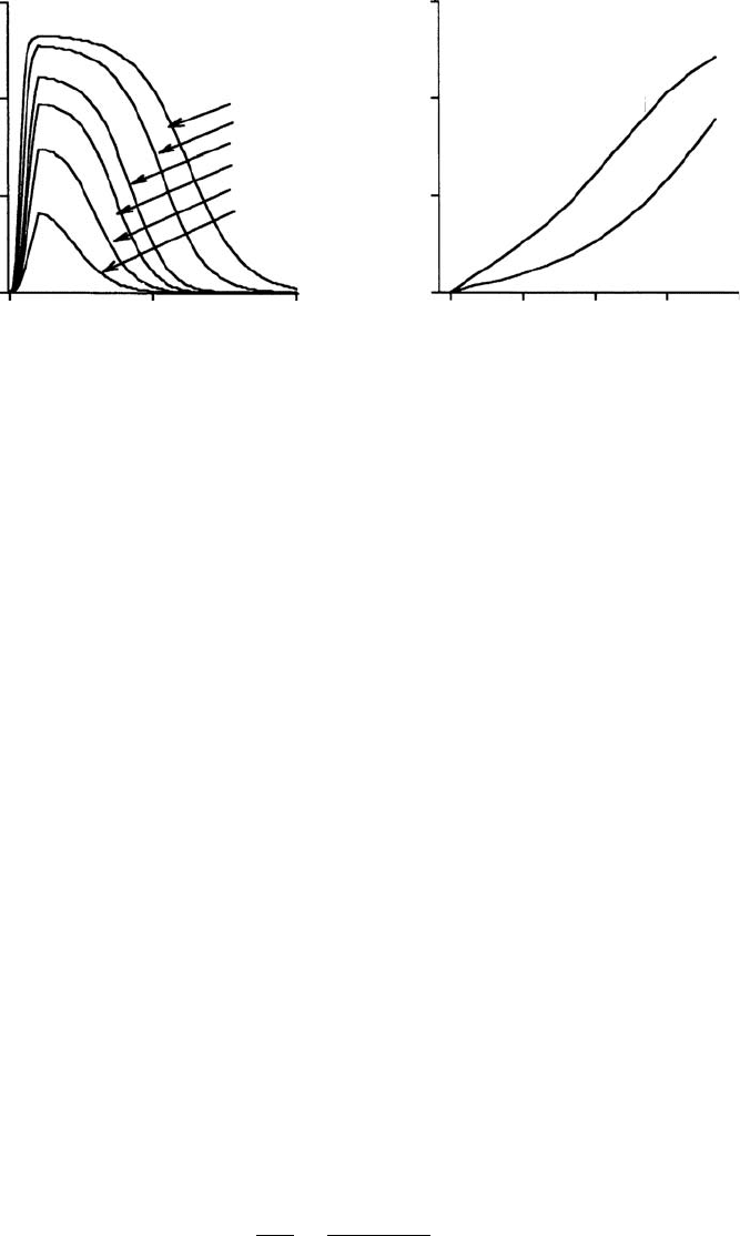
FIGURE 8.6 Cardiac muscle isometric twitch tension generated by a model of rat cardiac contraction (courtesy Dr.
Julius Guccione): (a) Developed twitch tension as a function of time and sarcomere length; (b) Peak isometric twitch
tension vs. sarcomere length for low and high calcium concentration.
Unlike skeletal muscle, in which maximal active force generation occurs at a sarcomere length that
optimizes myofilament overlap (∼2.1 μm), the isometric twitch tension developed by isolated cardiac
muscle continues to rise with increased sarcomere length in the physiological range (1.6 to 2.4 μm)
(Figure 8.6a). Early evidence for a descending limb of the cardiac muscle isometric length–tension curve
was found to be caused by shortening in the central region of the isolated muscle at the expense of stretching
at the damaged ends where specimen was tethered to the test apparatus. If muscle length is controlled so
that sarcomere length in the undamaged part of the muscle is indeed constant, or if the developed tension
is plotted against the instantaneous sarcomere length rather than the muscle length, the descending limb
is eliminated [71]. Thus, the increase with chamber volume of end-systolic pressure and stroke work
is reflected in isolated muscle as a monotonic increase in peak isometric tension with sarcomere length
(Figure 8.6b). Note that the active tension shown in Figure 8.6 is the total tension minus the resting tension,
which, unlike in skeletal muscle, becomes very significant at sarcomere lengths over 2.3 μm. The increase
in slope of the ESPVR associated with increased contractility is mirrored by the effects of increased calcium
concentration in the length–tension relation. The duration as well as the tension developed in the active
cardiac twitch also increases substantially with sarcomere length (Figure 8.6a).
The relationshipbetween cytosolic calcium concentration and isometric muscle tension has mostly been
investigated in muscle preparations in which the sarcolemma has been chemically permeabilized. Because
there is evidence that this chemical “skinning” alters the calcium sensitivity of myofilament interaction,
recent studies have also investigated myofilament calcium sensitivity in intact muscles tetanized by high-
frequencystimulation in the presenceofacompoundsuchasryanodinethatopencalciumreleasesites in the
sarcoplasmic reticulum. Intracellular calcium concentration was estimated using calcium-sensitive optical
indicators such as Fura. The myofilaments are activated in a graded manner by micromolar concentrations
of calcium, which binds to troponin-C according to a sigmoidal relation [72]. Half-maximal tension in
cardiac muscle is developed at intracellular calcium concentrations of 10
−6
to 10
−5
M (the C
50
) depending
on factors such as species and temperature [70]. Hence, relative isometric tension T
0
/T
max
maybemodeled
using [73,74].
T
0
T
max
=
[Ca]
n
[Ca]
n
+C
n
50
(8.12)
The Hill coefficient (n) governs the steepness of the sigmoidal curve. A wide variety of values have
been reported but most have been in the range 3 to 6 [75–78]. The steepness of the isometric length–
tension relation (Figure 8.6b), compared with that of skeletal muscle is due to length-dependent calcium
8-14 Biomechanics
sensitivity. That is, the C
50
(M) and n both change with sarcomere length, L (μm). Hunter et al.
[74] used the following approximations to fit the data of Kentish et al. [76] from rat right ventricular
trabeculae:
n = 4.25{1 + 1.95(L/L
ref
− 1)},pC
50
=−log
10
C
50
= 5.33{1 + 0.31(L/L
ref
− 1)} (8.13)
where the reference sarcomere length L
ref
was taken to be 2.0 μm.
The isotonic force–velocity relation of cardiac muscle is similar to that of skeletal muscle, and A.V.
Hill’s well-known hyperbolic relation is a good approximation except at larger forces greater than about
85% of the isometric value. The maximal (unloaded) velocity of shortening is essentially independent
of preload, but does change with time during the cardiac twitch and is affected by factors that affect
contractile ATPase activity and hence crossbridge cycling rates. deTombe and colleagues [79] using sar-
comere length-controlled isovelocity release experiments found that viscous forces imposes a significant
internal load-opposing sarcomere shortening. If the isotonic shortening response is adjusted for the con-
founding effects of passive viscoelasticity, the underlying crossbridge force–velocity relation is found to
be linear.
Cardiac muscle contraction also exhibits other significant length-history-dependent properties. An im-
portant example is “deactivation” associated with length transients. The isometric twitch tension redevel-
oped following a brief length transient that dissociates crossbridges, reaches the original isometric value
when the transient is imposed early in the twitch before the peak tensionis reached. Butfollowing transients
applied at times after the peak twitch tension has occurred, the fraction of tension redeveloped declines
progressively since the activator calcium has fallen to levels below that necessary for all crossbridges to
reattach [80].
There have been many model formulations of cardiac muscle contractile mechanics, too numerous to
summarize here. In essence they may be grouped into three categories. Time-varying elastance models
include the essential dependence of cardiac active force development on muscle length and time. These
models would seem to be well suited to the continuum analysis of whole heart mechanics [1,81,82] by
virtue of the success of the time-varying elastance concept of ventricular function (see Section 8.3.2).
In “Hill” models the active fiber stress development is modified by shortening or lengthening according
to the force–velocity relation, so that fiber tension is reduced by increased shortening velocity [83,84].
Fully history-dependent models are more complex and are generally based on A.F. Huxley’s crossbridge
theory [52,85–87]. A statistical approach known as the distribution moment model has also been shown
to provide an excellent approximation to crossbridge theory [88]. An alternative more phenomenological
approach is Hunter’s fading memory theory, which captures the complete length-history dependence of
cardiac muscle contraction without requiring all the biophysical complexity of crossbridge models [74].
The appropriate choice of model will depend on the purpose of the analysis. For many models of global
ventricular function, a time-varying elastance model will suffice, but for an analysis of sarcomere dynamics
in isolated muscle or the ejecting heart, a history-dependent analysis is more appropriate.
Although Hill’s basic assumption that resting and active muscle fiber tension are additive is axiomatic
in one-dimensional tests of isolated cardiac mechanics, there remains little experimental information on
how the passive and active material properties of myocardium superpose in two or three dimensions. The
simplest and commonest assumption is that active stress is strictly one-dimensional and adds to the fiber
component of the three-dimensional passive stress. However, even this addition will indirectly affect all the
other components of the stress response, since myocardial elastic deformations are finite, nonlinear, and
approximately isochoric (volume conserving). In an interesting and important new development, biaxial
testing of tetanized and barium-contracted ventricular myocardium has shown that developed systolic
stress also has a large component in directions transverse to the mean myofiber axis that can exceed 50%
of the axial fiber component [89]. The magnitude of this transverse active stress depended significantly on
the biaxial loading conditions. Moreover, evidence from osmotic swelling and other studies suggests that
transverse strain can affect contractile tension development along the fiber axis by altering myofibril lattice
spacing [90,91]. The mechanisms of transverse active stress development remain unclear but two possible
