Nielsen M. Atlas of Human Anatomy
Подождите немного. Документ загружается.

275
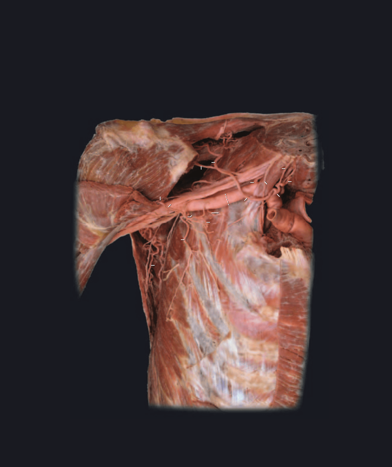
Dissection of subclavian and axillary arteries
Anterosuperior view
1 Brachiocephalic artery
2 Common carotid artery
3 Vertebral artery
4 Subclavian artery
5 Thyrocervical trunk
6 Inferior thyroid artery
7 Ascending cervical artery
8 Suprascapular artery
9 Dorsal scapular artery
10 Axillary artery
11 Superior thoracic artery
12 Thoracoacromial trunk
13 Pectoral artery
14 Acromial artery
15 Clavicular artery
16 Deltoid artery
17 Lateral thoracic artery
18 Subscapular artery
19 Circumflex scapular artery
20 Thoracodorsal artery
21 Posterior circumflex humeral artery
22 Anterior circumflex humeral artery
23 Brachial artery
24 Deep artery of arm
25 Internal thoracic artery
26 Internal thoracic vein
27 Anterior scalene muscle
28 Middle scalene muscle
29 Deltoid muscle
30 Pectoralis minor muscle
31 Pectoralis major muscle
32 Subscapularis muscle
33 Teres major muscle
34 Latissimus dorsi muscle
35 Serratus anterior muscle
36 Phrenic nerve
37 Brachial plexus
38 Clavicle
39 First rib
40 Suprascapular nerve
1
2
3
4
4
6
9
20
5
7
8
8
10
11
12
13
14
15
17
27
28
29
30
31
32
34
35
36
37
38
39
40
1
2
3
4
4
6
9
20
5
7
8
8
10
11
12
13
14
15
17
27
28
29
30
31
32
34
35
36
37
38
39
40
Atlas_Cardio.indd Page 275 15/03/11 7:27 PM user-F391Atlas_Cardio.indd Page 275 15/03/11 7:27 PM user-F391 /Users/user-F391/Desktop/Users/user-F391/Desktop
276
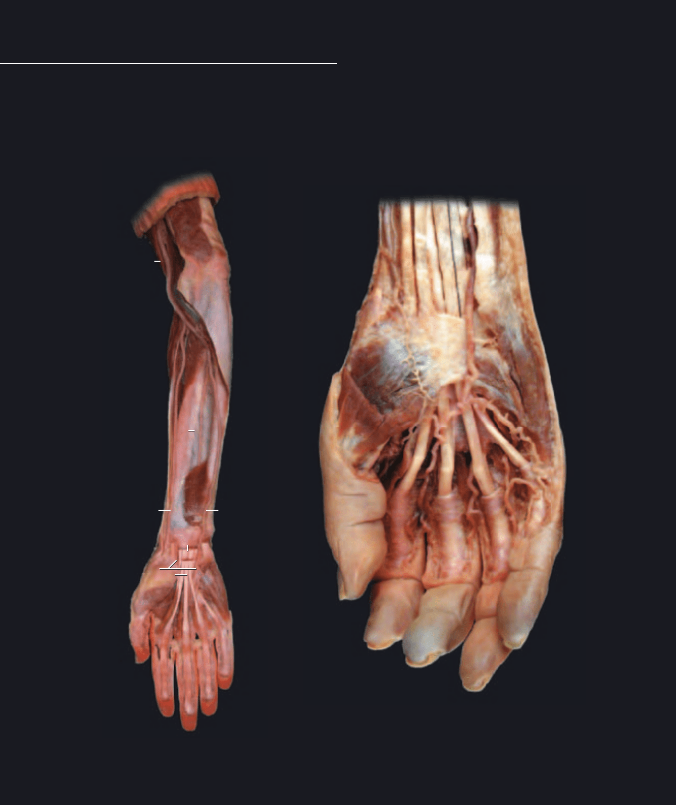
Superior Limb Vessels
Dissection of antebrachial arteries
Anterior view
Dissection of palmar arterial arch and branches to digits
Anterior view
1 Brachial artery
2 Ulnar artery
3 Radial artery
4 Anterior interosseous artery
5 Superficial palmar arch
6 Common digital artery
7 Proper digital artery
8 Deep palmar arch
9 Cephalic vein
10 Median cubital vein
11 Basilic vein
12 Median antebrachial vein
13 Accessory cephalic vein
14 Brachial vein
15 Interosseous membrane
16 Transverse carpal ligament
17 Supinator muscle
18 Pronator quadratus muscle
19 Flexor digitorum superficialis tendons
20 Flexor digitorum profundus tendons
21 Biceps brachii muscle
22 Triceps brachii muscle
23 Pectoralis major muscle
24 Deltoid muscle
25 Deltopectoral groove
26 Serratus anterior muscle
27 Brachioradialis muscle
28 Coracobrachialis muscle
1
2
2
2
3
3
3
4
5
6
7
7
8
15
16
17
18
1818
18
18
19
21
22
1
2
2
2
3
3
3
4
7
7
8
15
17
18
18
19
21
22
Atlas_Cardio.indd Page 276 15/03/11 7:27 PM user-F391Atlas_Cardio.indd Page 276 15/03/11 7:27 PM user-F391 /Users/user-F391/Desktop/Users/user-F391/Desktop
277
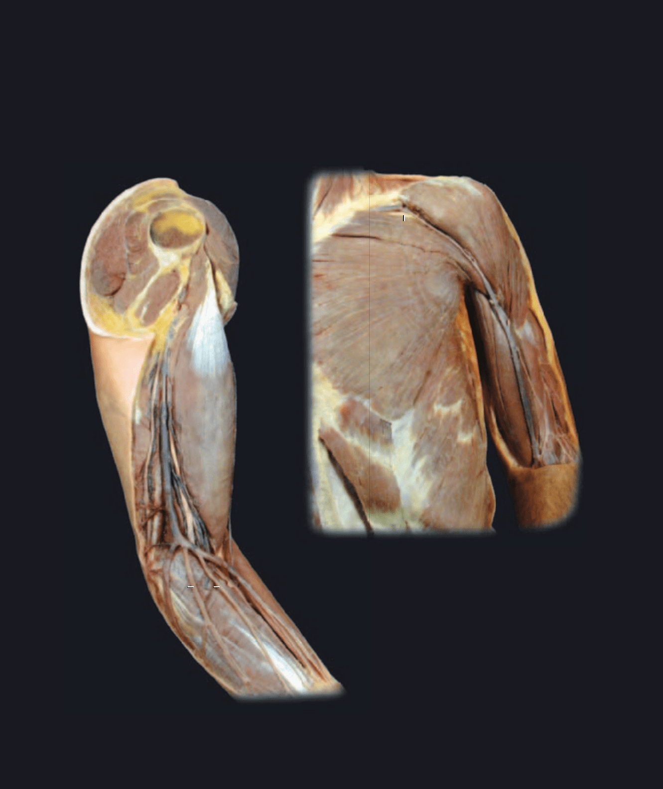
Within the upper limb there are two sets of veins: deep veins that accompany the arteries, and superfi cial veins that course
through the hypodermis without arterial counterparts. The deep veins, running with the arteries of the upper limb, have the
same names as their arterial counterparts. These veins are signifi cantly smaller than the arteries they accompany and form
vena comitans with anastomotic channels around the arteries. The superfi cial veins of the upper limb are large and numerous.
There are three major superfi cial veins into which all the other superfi cial veins fl ow; they are the basilic vein, cephalic vein,
and median cubital vein. The median cubital vein is a connecting vein between the cephalic vein and the basilic vein. The
cephalic and basilic veins eventually pass deep to join the axillary vein at the proximal end of the limb. Most of the venous
return from the upper limb passes through the superfi cial veins.
Dissection of superfi cial vein of upper limb
Medial view of left upper limb
Dissection of cephalic vein
Anterior view
9
9
10
11
11
1312
21
21
22
22
22
23
24
24
24
25
26
27
28
9
9
10
11
11
1312
21
21
22
22
22
23
24
24
24
25
26
27
28
Atlas_Cardio.indd Page 277 15/03/11 7:27 PM user-F391Atlas_Cardio.indd Page 277 15/03/11 7:27 PM user-F391 /Users/user-F391/Desktop/Users/user-F391/Desktop
278
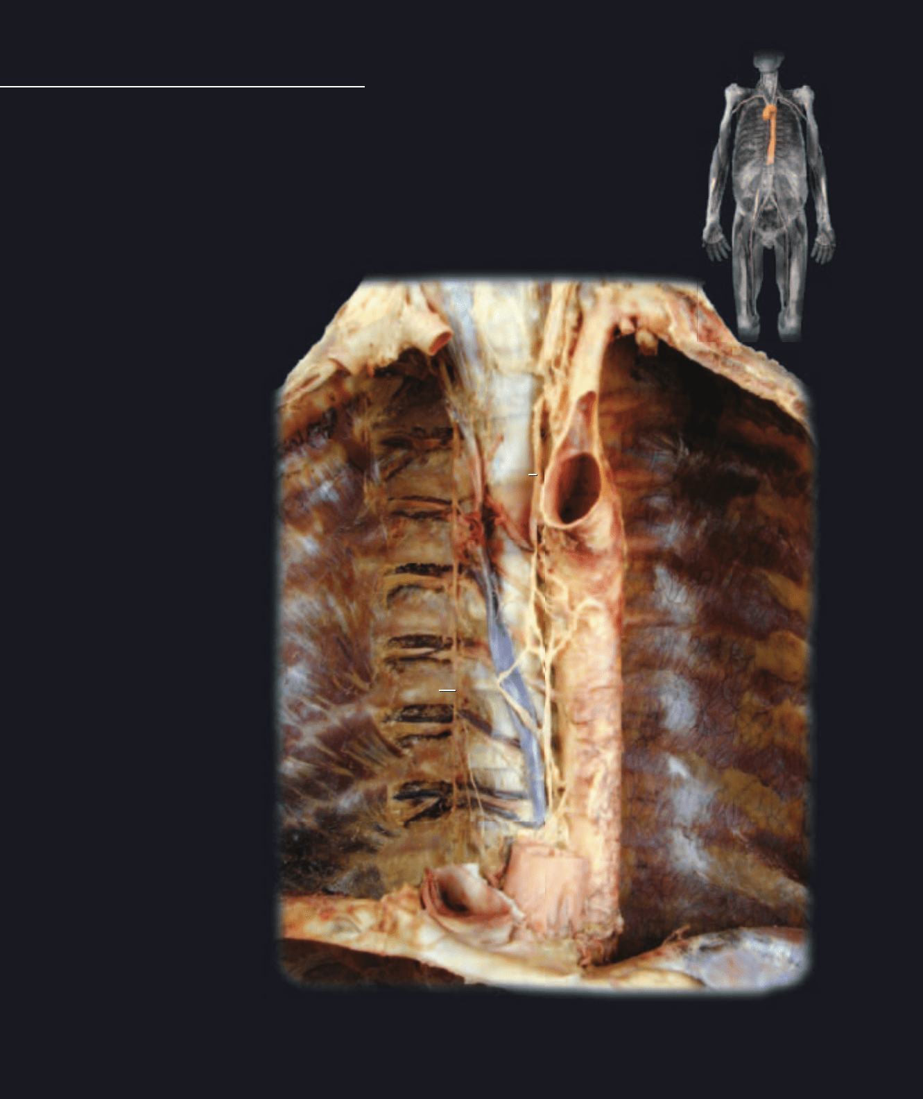
The branches of the aorta that supply the thoracic
region can be divided into two principal groups —
those that supply the thoracic body wall and those
Thoracic Vessels
that supply thoracic viscera. Two arterial supply routes carry blood into the thoracic body wall. Poste-
riorly the aorta courses vertically down the vertebral column, while anteriorly the internal thoracic
arteries arise from the subclavian arteries and course vertically down the inside of the sternum.
Between these anterior and posterior supply arteries are interconnecting collateral arteries. These
collateral vessels are the anterior intercostal arteries and the posterior intercostal arteries, which sup-
ply the tissues of the intercostal spaces and form collateral circuits between the anterior and poste-
rior arterial pathways. All thoracic viscera receive their blood supply from branches of the aorta. The
thoracic viscera include the heart, lungs with their associated bronchial tubes, and the esophagus.
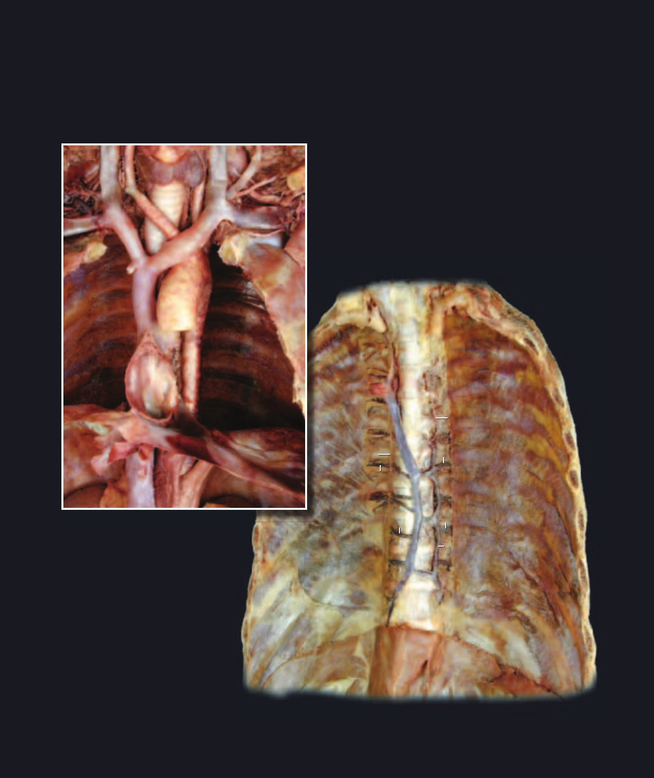
Dissection of vessels of posterior thoracic wall
Anterior view
1 Aorta
2 Posterior intercostal artery
3 Posterior intercostal vein
4 Azygos vein
5 Hemi-azygos vein
6 Accessory hemi-azygos vein
7 Superior vena cava
8 Brachiocephalic vein
9 Subclavian vein
10 Internal jugular vein
11 Inferior vena cava
12 Right atrium (cut)
13 Left subclavian artery
14 Left common carotid artery
15 Right common carotid artery
16 Hepatic vein
17 Trachea
18 Diaphragm
19 Esophageal hiatus
20 Subcostal muscle
21 Innermost intercostal muscle
22 Esophagus
23 Sympathetic trunk nerve
24 Thoracic lymphatic duct
1
2
2
3
4
11
13
18
20
21
22
23
24
1
2
2
3
4
11
13
18
20
21
23
24
Atlas_Cardio.indd Page 278 15/03/11 7:27 PM user-F391Atlas_Cardio.indd Page 278 15/03/11 7:27 PM user-F391 /Users/user-F391/Desktop/Users/user-F391/Desktop
279
Like the arterial supply to the thoracic wall, the venous drainage returns via both anterior-wall and posterior-wall drainage
veins. The veins of the anterior wall have the same names as their arterial counterparts, while the veins of the posterior wall
differ in name and structure. Unlike the aorta, which is the posterior-wall supply artery, the superior vena cava and inferior
vena cava diverge from the posterior thoracic wall to enter the thoracic cavity and return their contents to the heart. In the
absence of vena cavae in the posterior thoracic wall, an azygos system of veins is formed to drain the body wall and the
thoracic viscera. These azygos veins communicate with the superior vena cava to return their contents to the heart. With
the exception of the azygos veins, the veins are similar to the arteries in name and distribution.
Dissection of vena cavae and tributaries
Anterior view
Dissection of azygos veins
Anterior view
1
3
3
3
3
4
5
6
7
8
8
9
9
10
10
11
14
15
16
17
18
19
20
21
23
1
3
3
3
3
4
5
6
7
8
8
9
9
10
10
11
14
15
16
17
18
19
20
21
23
Atlas_Cardio.indd Page 279 15/03/11 7:27 PM user-F391Atlas_Cardio.indd Page 279 15/03/11 7:27 PM user-F391 /Users/user-F391/Desktop/Users/user-F391/Desktop
280
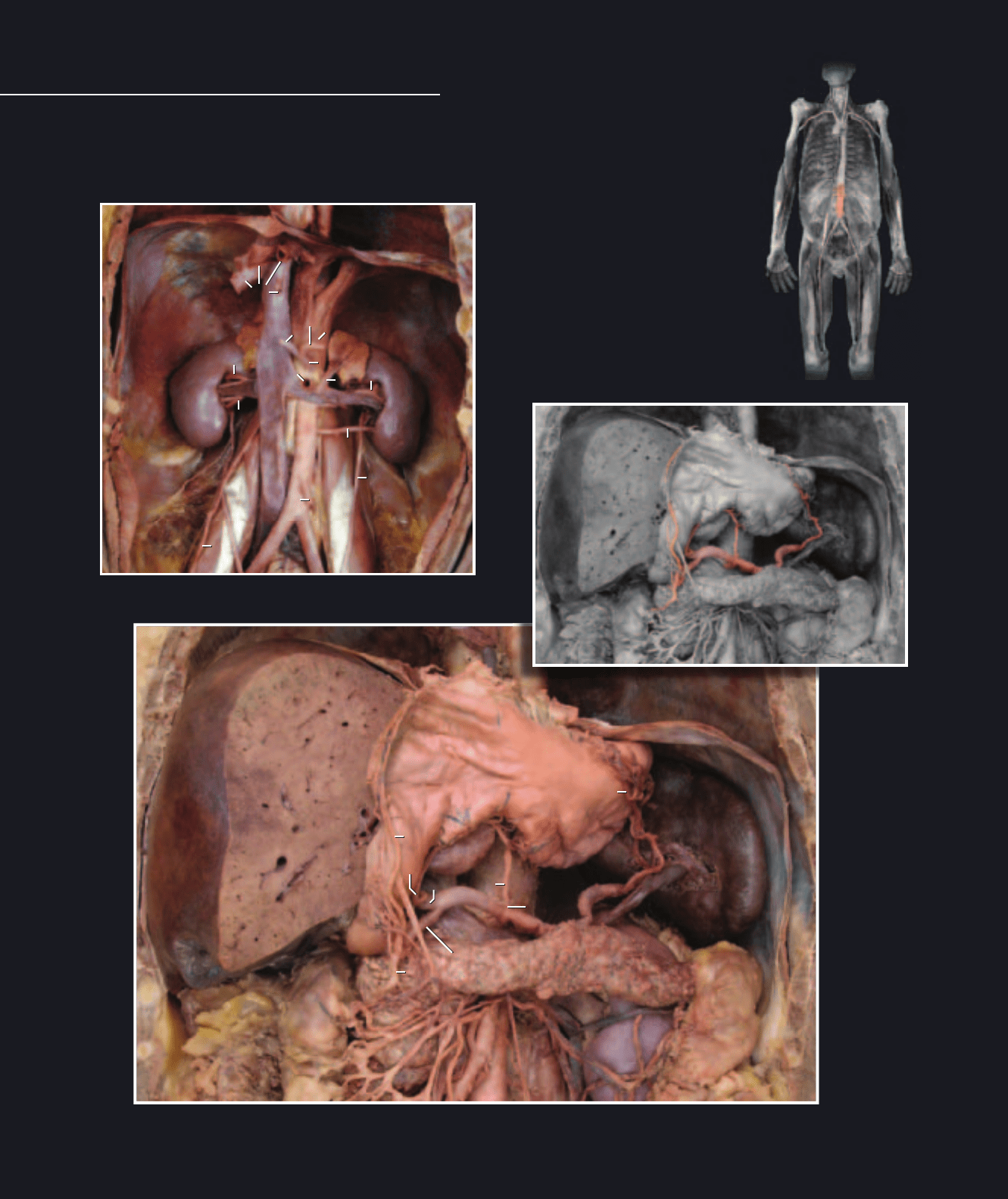
Like the thorax, the abdomen has somatic ar-
teries that supply the abdominal muscle wall
Abdominal Vessels
and visceral arteries that supply the viscera of the abdominal cavity. These vessels follow the same
pattern observed in the thoracic region; that is, the abdominal body wall has both anterior (epigastric
arteries) and posterior (aorta) supply pathways that form interconnecting collateral arteries, while the
viscera receive branches from the aorta — celiac artery to the foregut, superior mesenteric artery to
the midgut, inferior mesenteric artery to the hindgut, and renal arteries to the kidneys.
Deep dissection of abdomen showing renal vessels
Anterior view
Dissection of abdomen showing celiac branches and supply of foregut viscera
Anterior view, stomach reflected upward
Branches of celiac artery
Branches of celiac artery
1
1
2
2
3
3
4
4
5
5
6
7
8
9
10
11
12
12
47
19
23
23
23
23
23
25
26
27
28
29
30
31
32
33
34
35
35
35
36
37
39
39
41
43
48
44
46
46
40
1
1
2
2
3
3
4
4
5
5
6
7
8
9
10
11
12
12
47
19
23
23
23
23
23
25
26
27
28
29
30
31
32
33
34
35
35
35
36
37
39
39
41
43
48
44
46
46
40
Atlas_Cardio.indd Page 280 15/03/11 7:27 PM user-F391Atlas_Cardio.indd Page 280 15/03/11 7:27 PM user-F391 /Users/user-F391/Desktop/Users/user-F391/Desktop
281
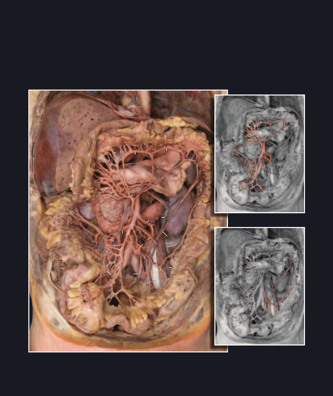
Superior mesenteric artery
Inferior mesenteric artery
Dissection of abdomen showing arterial supply of midgut and hindgut viscera
Anterior view
1 Aorta
2 Celiac artery
3 Splenic artery
4 Common hepatic artery
5 Left gastric artery
6 Right gastric artery
7 Left gastro-omental artery
8 Right gastro-omental artery
9 Proper hepatic artery
10 Gastroduodenal artery
11 Superior pancreaticoduodenal artery
12 Superior mesenteric artery
13 Middle colic artery
14 Marginal artery
15 Right colic artery
16 Ileocolic artery
17 Jejunal arteries
18 Ileal arteries
19 Inferior mesenteric artery
20 Left colic artery
21 Sigmoid artery
22 Superior rectal artery
23 Renal artery
24 Segmental arteries
25 Common iliac artery
26 Inferior vena cava
27 Hepatic vein
28 Renal vein
29 Hepatic portal vein
30 Superior mesenteric vein
31 Inferior mesenteric vein
32 Splenic vein
33 Suprarenal vein
34 Testicular vein
35 Kidney
36 Liver
37 Stomach
38 Transverse colon
39 Suprarenal gland
40 Pancreas
41 Spleen
42 Duodenum
43 Ascending colon
44 Descending colon
45 Ileum
46 Diaphragm
47 Ureter
48 Psoas major muscle
12
13
15
15
14
14
16
17
17
47
18
19
20
20
21
22
24
25
25
30
31
31
34
35
35
36
37
38
41
42
43
44
45
46
40
12
13
15
15
14
14
16
17
17
18
19
20
20
21
22
24
25
25
30
31
31
34
35
35
36
37
38
41
42
43
44
45
46
40
Atlas_Cardio.indd Page 281 15/03/11 7:27 PM user-F391Atlas_Cardio.indd Page 281 15/03/11 7:27 PM user-F391 /Users/user-F391/Desktop/Users/user-F391/Desktop
282
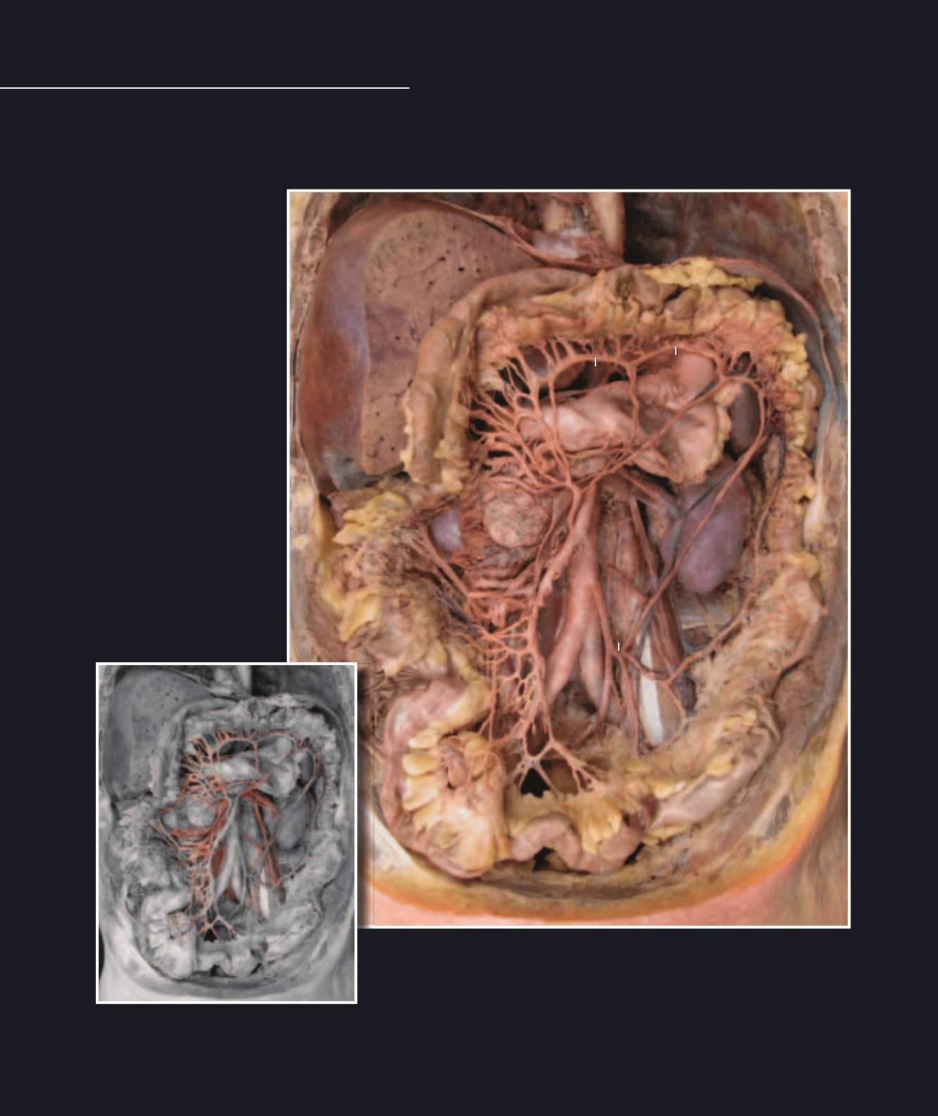
The major difference between the arteries and veins of the abdomen
is the fact that all the visceral venous return from the capillaries of the
digestive system and spleen pass via the hepatic portal system to the
Abdominal Vessels
capillaries of the liver before returning to the heart. Within the liver, both the hepatic artery and hepatic portal vein branch to
form a complex network of specialized capillaries called the hepatic sinusoids. The hepatic sinusoids then drain into the
hepatic veins to return the blood to the inferior vena cava.
Abdominal veins
Dissection of abdomen showing arteries and veins of the intestines
Anterior view
1 Inferior vena cava
2 Hepatic portal vein
3 Superior mesenteric vein
4 Right colic vein
5 Inferior mesenteric vein
6 Renal vein
7 Superior mesenteric artery
8 Inferior mesenteric artery
9 Middle colic artery
10 Marginal artery
11 Left colic artery
12 Common iliac artery
13 External iliac artery
14 Internal iliac artery
15 Superior gluteal artery
16 Inferior gluteal artery
17 Obturator artery
18 Internal pudendal artery
19 Lateral sacral artery
20 Superior vesical artery
21 Vaginal artery
22 Obliterated umbilical artery
23 Uterus
24 Bladder
25 Prostate
26 Rectum
27 Stomach
28 Kidney
29 Upper bands of sacral plexus
30 Sympathetic trunk
31 Inferior vesical artery
32 Middle rectal artery
33 Obturator nerve
34 Uterine artery
1
2
3
4
5
6
7
8
9
10
10
11
12
28
1
2
3
4
5
6
7
8
9
10
10
11
12
28
Atlas_Cardio.indd Page 282 15/03/11 7:27 PM user-F391Atlas_Cardio.indd Page 282 15/03/11 7:27 PM user-F391 /Users/user-F391/Desktop/Users/user-F391/Desktop
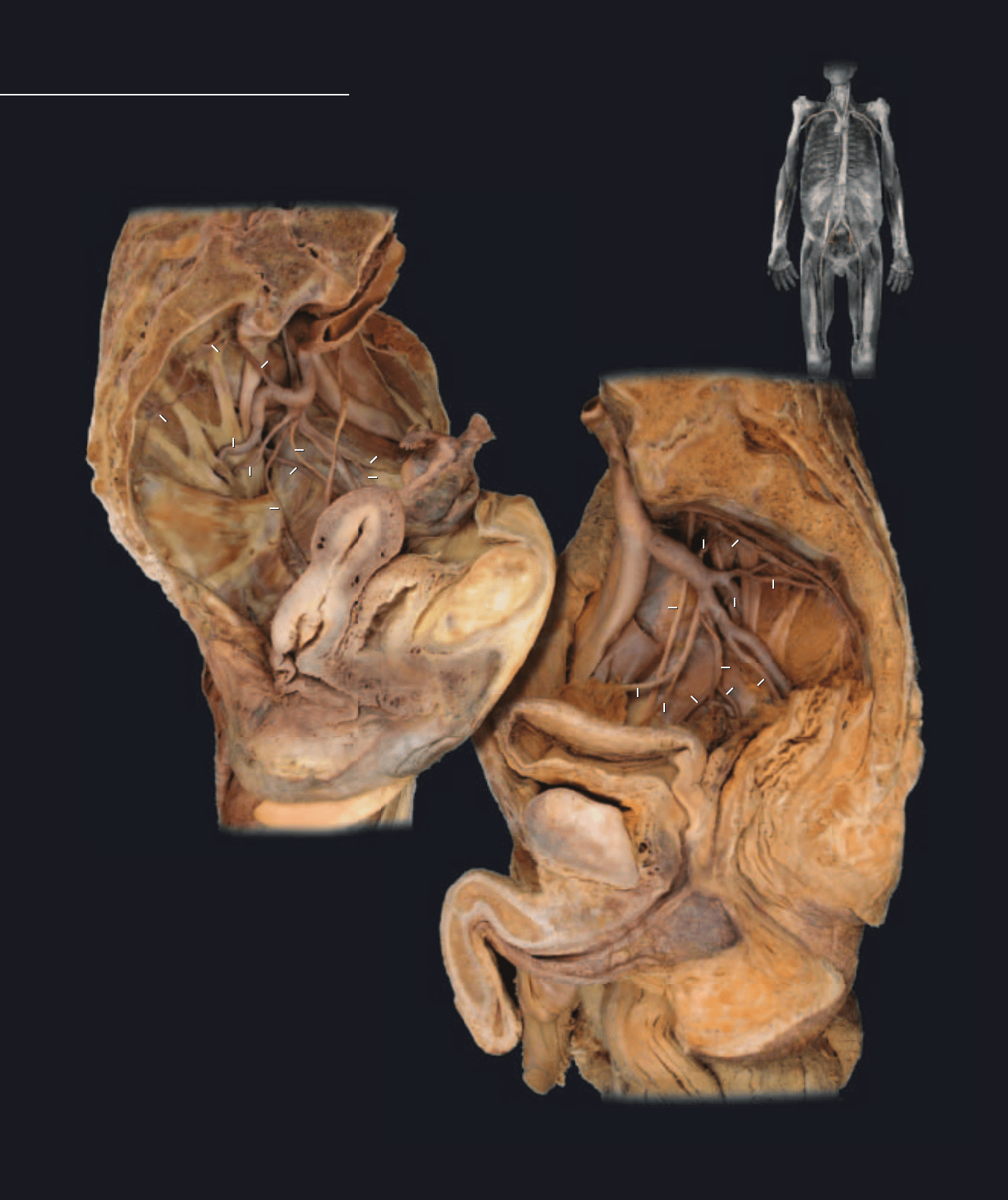
283
The common iliac arteries, the terminal branches of the
aorta, carry all of the blood supply to the lower limbs and
pelvis. All pelvic viscera, along with the body wall anatomy
Pelvic Vessels
of the pelvis and perineal regions, receive their blood supply from the internal iliac artery. Numerous
branches arise from the internal iliac artery to supply the pelvic wall, the perineum, and the gluteal
region. Other branches course into the pelvic cavity to supply the viscera. The veins are similar in
name and course with the corresponding arteries.
Dissection of pelvic arteries of female
Medial view, anterior at left
Dissection of pelvic arteries of male
Medial view, anterior at right
12
12
13
13
14
14
16
16
17
17
18
18
19
19
19
19
20
20
21
22
22
15
23
24
24
25
26
26
29
29
29
29
29
30
30
31
32
33
34
12
12
13
13
14
14
16
16
17
17
18
18
19
19
19
19
20
20
21
22
22
15
23
24
24
25
26
26
29
29
29
29
29
30
30
31
32
33
34
Atlas_Cardio.indd Page 283 15/03/11 7:27 PM user-F391Atlas_Cardio.indd Page 283 15/03/11 7:27 PM user-F391 /Users/user-F391/Desktop/Users/user-F391/Desktop
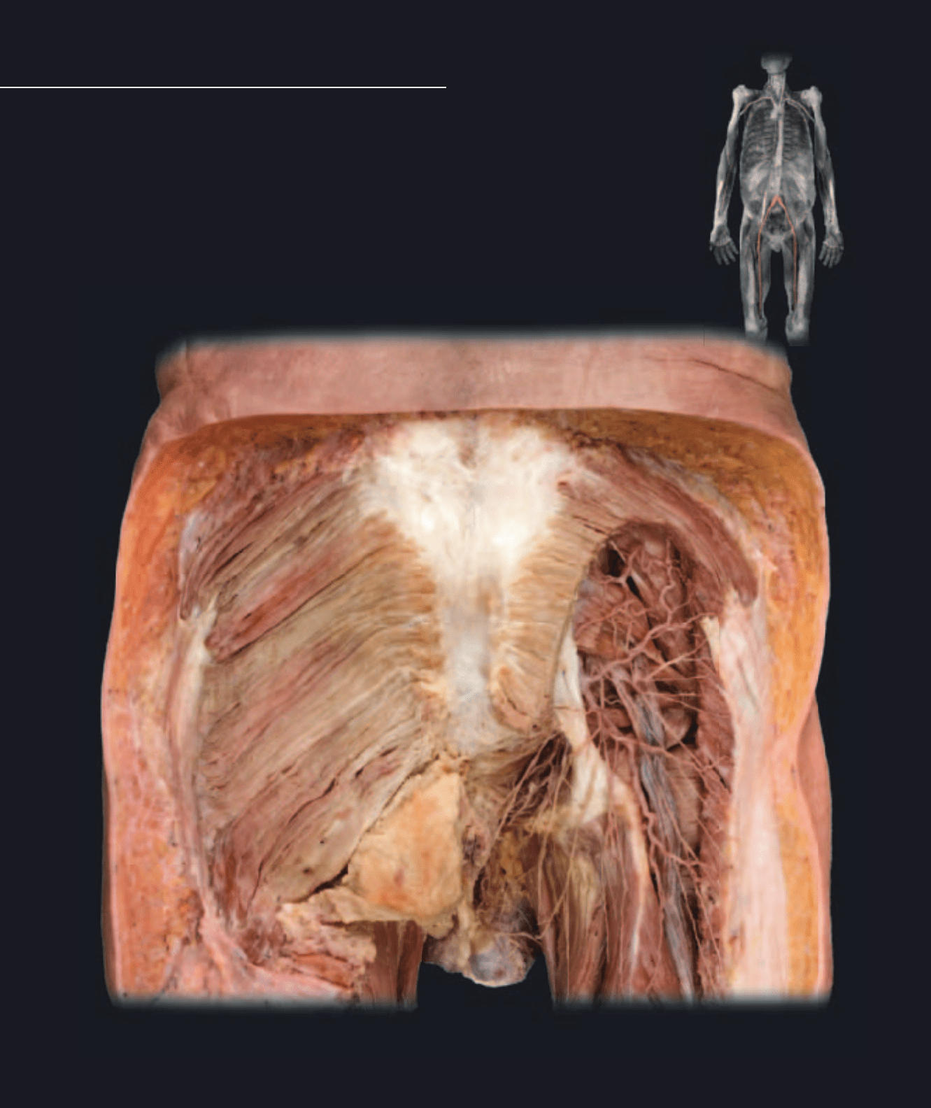
284
As in the upper limb, the main arterial
pathway into the lower limb consists of
a single, major arterial roadway that
Inferior Limb Vessels
gradually tapers as it gives rise to numerous branches on its pathway through the limb. This large
arterial roadway begins as the external iliac artery in the pelvis, passes beneath the inguinal ligament
to enter the thigh as the femoral artery, passes to the back of the knee to become the popliteal artery,
and in the proximal aspect of the leg bifurcates into the anterior tibial and posterior tibial arteries,
which course through the leg and into the foot.
Dissection of gluteal region showing gluteal arteries and nerves
Posterior view
1 Superior gluteal artery
2 Inferior gluteal artery
3 Internal pudendal artery
4 Femoral artery
5 Deep artery of thigh
6 Muscular branches of femoral
7 Femoral vein
8 Great saphenous vein
9 External iliac artery
10 Internal iliac artery
11 External iliac vein
12 Common iliac artery
13 Aorta
14 Gluteus maximus muscle
15 Sacrotuberous ligament
16 Piriformis muscle
17 Spermatic cord (cut)
18 Penis (cut)
19 Adductor longus muscle
20 Rectus femoris muscle
21 Vastus intermedius muscle
22 Gracilis muscle
23 Vastus lateralis muscle
24 Vastus medialis muscle
25 Fascia lata
26 Sartorius muscle
27 Iliacus muscle
1
2
3
14
14
15
16
22
1
2
3
14
14
15
16
22
Atlas_Cardio.indd Page 284 15/03/11 7:28 PM user-F391Atlas_Cardio.indd Page 284 15/03/11 7:28 PM user-F391 /Users/user-F391/Desktop/Users/user-F391/Desktop
