Murray J. Clifford. Angiogenesis Protocols - Methods in Molecular Medicine, Vol. 46
Подождите немного. Документ загружается.

Endothelial Cell Sprouting 151
2. Add 8.5 mL of collagen working solution (2.2 mg/mL ) to the above mixture at
4°C, thus making a total of 10 mL gelling solution (see Notes 12 and 13).
3. Mix by pipetting up and down with a 10 mL pipet, taking care to avoid air
bubbles. The mixing has to be done thoroughly as well as rapidly.
4. Transfer all the gelling solution into the pipette and plate 2 mL onto each of five
35-mm dishes (see Note 13). The tip of the pipet may rest on the base of the dish
while pipeting, to facilitate accurate delivery.
5. Gently tilt and swirl the dish to spread the collagen evenly.
6. Without moving the dishes further, allow the collagen to set in the laminar flow
cabinet. This will take approximately 5 min, but it is better not to move them for
another 5–10 min. Make sure that the surface of the cabinet is level!
7. Repeat steps 1–6 to make the total number of gels required.
8. Once the gels are set, transfer to a humidified tissue culture incubator at 37°C
with 5% CO
2
and incubate for 1–2 hr before adding medium.
9. Add 1 mL appropriate medium per gel (e.g., SF-MEM). Gels may be made 1–3 d
in advance and stored in the incubator.
3.2. Maintenance and Plating of ECs on Collagen Gels
Stock cultures of bovine aortic ECs are maintained on gelatinized dishes in
MEM supplemented with 20% donor calf serum (DCS), 1 mM sodium pyru-
vate, nonessential amino acids (81.4 mg/L), 2 mM glutamine, and 50 µg/mL
ascorbic acid. Glutamine and ascorbic acid are stored at –20°C in aliquots of
convenient volume; these are thawed and added to the volume of medium
required for either 7–10 d (glutamine) or for 1 d (ascorbic acid). Penicillin
(100 U/mL) and streptomycin (100 µg/mL) may also be added. The complete
growth medium is referred to as 20% DCS-MEM (see Note 5). When serum is
omitted, this complete medium is referred to as SF-MEM.
3.2.1. Routine Passage of ECs
Stock cultures of ECs are split at a 1/3 ratio when confluent. This is accom-
plished as follows:
1. Gelatinize the number of new dishes required by adding sufficient gelatin solu-
tion to cover the surface (e.g., 2 mL/90-mm dish) (see Note 14). Incubate for 1 hr
at 37°C, remove the gelatin solution, and wash the dish twice with Hanks’ (see
Note 15).
2. Remove the medium from the stock culture to be passaged and wash the attached
cells twice with 4–5 mL Hanks’.
3. Add a small volume of trypsin EGTA to cover the dish (e.g., 1.5 mL/90-mm
dish). Incubate at 37°C until the cells begin to round and detach from the dish
(about 5 min) (see Note 16).
4. Pipet up and down to detach all cells. Transfer trypsin and floating cells to a
Universal containing 7.5 mL of 40% DCS-MEM (soya bean trypsin inhibitor
152 Schor et al.
may be used if the experiment requires serum-free conditions). Wash the dish
twice with 3 mL SF-MEM, transferring the washes to the same Universal.
5. Distribute the total volume (e.g., 15 mL of 20% DCS-MEM containing the cells
harvested from one 90-mm dish) into 3 gelatinized dishes.
6. Cell attachment occurs rapidly, with 90% of the cells adherent to the dish within
15–30 min. Check microscopically that this is the case. After 1–2 hr remove
medium with nonattached cells and replace with fresh medium. The medium is
normally changed on Mondays, Wednesdays, and Fridays. The volume added
per 90-mm dish is increased from 5 mL when the cells are sparse to 10 mL when
confluent or over the weekend.
3.2.2. Plating ECs on Collagen Gels
Gels of different volumes and surface area may be prepared (see Note 13).
The method described here is for 2 mL gels cast in 30-mm dishes:
1. The gels contain MEM without additives; before plating the cells, they must be
equilibrated so that the interstitial fluid within the gel has the same composition
as the medium in which the cells are to be plated. For example, when the cells are
to be plated and grown in 20% DCS-MEM, the gels are first incubated for 1–2 hr
with 1 mL 60% DCS-MEM and threefold concentration of glutamine, sodium
pyruvate, ascorbic acid, and nonessential amino acids (see Note 17). The equili-
brating medium may be removed before plating the cells if required.
2. Bring the cells to be plated into a stock cell suspension as described in Subhead-
ing 3.2.1., steps 2, 3, and 4.
3. Take a small aliquot (e.g., 0.1–0.5 mL) to count the number of cells present (e.g.,
by Coulter counter or hematocytometer).
4. Take a volume of cell suspension that contains the number of cells required plus
approximately 10–20%. For example to plate 20 gels at 2 × 10
5
cells per gel take
5 × 10
6
cells.
5. Centrifuge the cells at 100g for 5 min.
6. Remove the supernatant and resuspend the cell pellet in the appropriate volume
of 20%DCS-MEM to achieve the desired concentration of cells to be plated per
gel in 1 mL of medium (e.g., 2 × 10
5
cells per mL) (see Note 18).
7. Plate 1 mL of final cell suspension per gel, taking care of distributing it homoge-
neously throughout the gel and not to touch or damage the gel (e.g., by a fast
stream of medium). Shake the dish horizontally in various orientations to achieve
a homogeneous distribution of the cells.
8. Transfer the gels to the tissue culture incubator and incubate for 24 hr.
9. Remove the medium and replace with 1 mL 20%DCS-MEM. Change medium
every 2–3 d (see Note 19).
10. When the cells reach saturation density (Note 20) remove the medium and wash the
gels 4 times with serum-free MEM, incubating the cultures for 1 hr after each wash.
11. Add 1.5 mL of “maintenance medium” (e.g., 1% DCS-MEM ) per gel (see Note
21) and change the medium every 2 d. These cultures are referred to as “resting”.
Endothelial Cell Sprouting 153
3.3. Addition of Test Substances
The required number of resting cell cultures is prepared as described above.
The resting cells may be maintained for several months. These cells remain
metabolically active, and continue to synthesize and deposit a subendothelial
matrix. It is therefore important that care be taken to use cultures of the same
age in order to ensure an optimal level of experimental reproducibility (see
notes 1 and 16).
The factor to be tested is dissolved in maintenance medium and added to
duplicate cultures at a wide range of concentrations. If it is necessary to
dissolve the factor in an organic solvent first (e.g., the phorbol ester PMA is
dissolved in DMSO or acetone), the concentration of the organic solvent should
be kept constant for the different concentrations of the active compound and
for the controls. Alternatively, various solvent concentrations have to be used
for both the active compound and the controls.
In initial experiments, positive controls should be included at different con-
centrations, for example bFGF or vascular endothelial growth factor (VEGF),
at concentrations from 1–200 ng/mL. The various factors can be added in 1–2 mL
volumes per gel and the medium changed every 2 d (see Note 22). Sprouts
normally appear in the positive controls after 24–48 hr and the experiment is
usually terminated in 4–6 d. At this point a small percentage of sprouting cells
have usually migrated measurable distances into the collagen gel. If the cul-
tures are to be maintained for longer periods, it is advisable to change the
medium every day. The morphology of the cells at different stages of the assay
is shown in Fig 2.
3.4. Quantification of Sprouting
The initial assessment of factor activity may be done using duplicate cul-
tures. Once activity has been detected and the active concentration approxi-
mated, it may be necessary to quantify the sprouting-inducing ability of the test
factor (see Note 23). In this case triplicate cultures are used for every concen-
tration and time point tested, as well as positive and negative controls.
The cultures are fixed by removing the medium, washing once with PBS,
and adding 1.5 mL of 10% formaldehyde. These cultures may be left at room
temperature for 1–2 hr or stored at 4°C for 2–3 d. Before quantitation, the
cultures are washed twice with PBS and then 1.5 ml of PBS containing sodium
azide is added. These cultures may be stored for months at 4°C provided that
the PBS is not allowed to dry out (see Note 24).
The extent of sprouting may be quantified by various means. Here we
describe two methods that we have found to be rapid and reproducible and
only require standard phase-contrast microscopy. Both methods may be scored
at the same time, as the information they provide is complementary.
154 Schor et al.
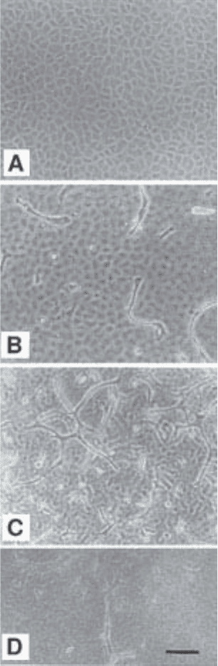
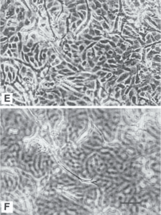
Fig. 2
Endothelial Cell Sprouting 155
Fig. 2. Morphology of the cells at different stages of the assay. (A) Confluent
cobblestone monolayer of resting cells under maintenance conditions. (B–F) Induc-
tion of sprouting cells by addition of angiogenic factors. (B) Early phase. Single sprout-
ing cells directly underneath the cobblestone monolayer, which is seen slightly out of
focus. (C) Late phase. As in A, but showing also sprouting cells connected into com-
plex networks or “aggregates”. (D) Sprouting cells deeper into the 3D gel. (E) Culture
showing extensive network of sprouting cells and cobblestone monolayer of “acti-
vated” appearance (compare with resting cobblestone monolayer in A and B). (F) Same
field as shown in E, but focusing down shows sprouting cells deeper into the gel and
connected to cells underneath to the monolayer. Bar, 100 µm
156 Schor et al.
3.4.1. Method 1: Percentage of Microscopic Fields Containing Sprouts
An arbitrary field is defined by a graticule placed in the eyepiece of the
microscope. The cultures are viewed using phase-contrast optics set at the low-
est magnification that allows a clear distinction between resting cells of the
monolayer (cobblestone) and sprouting cells (e.g., 10× or 20× objective, and
10× eyepiece). Between 10 and 15 fields are randomly selected per culture,
using the same procedure for each culture. The presence of single sprouting
cells and sprouting cell aggregates is recorded as yes or no in each field. The
aggregates are defined by the presence of at least three connected sprouting
cells. This scoring is carried out first focussing on (and directly under) the
surface monolayer and then focussing down into the gel (Fig. 2). Four vari-
ables are therefore recorded per culture, and the results are expressed as per-
centage of fields containing (a) single sprouting cells on the surface, (b) single
sprouting cells in the gel, (c) aggregates on the surface, and (d) aggregates in
the gel.
3.4.2. Method 2: Percentage of Sprouting Cells
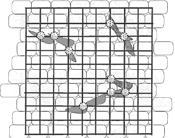
A graticule that contains a fixed number of points (e.g., a Chalkley grid with
100 points) is placed in the eyepiece of the microscope. Using the same proce-
dure as for Method 1 (Subheading 3.4.1.), 10–15 fields are randomly selected
per culture (the same fields and graticule may be used to quantify by both
methods). Each point that coincides with a sprouting cell is recorded, making it
possible that a single sprouting cell be recorded more than once (Fig 3). This is
a classical stereological method that reflects the volume occupied by the sprout-
ing cells. For each culture two variables are recorded: (a) percentage of sprout-
ing cells on the surface, and (b) percentage of sprouting cells in the gel. When
scoring points counted within the 3D gel, a fixed number of focussing planes
are predefined (e.g., four planes at fixed distances under the resting mono-
layer) and only those sprouting cells that coincide with the points of the grid
and are in focus are recorded (note that a single cell may be recorded more than
once).
4. Notes
1. In this assay the 3D extracellular matrix is formed by two components (i) a 3D
substratum (type I collagen matrix or “collagen gel”) on which the endothelial
cells are cultured, and (ii) a cell-produced subendothelial matrix which is
synthesised and deposited by the cells on top of the collagen gel. The basic assay
described here may be adapted in several ways, as follows:
a. The collagen gel used as substratum may be modified by the addition of other
matrix macromolecules in the medium or within the gel (14,15).
Endothelial Cell Sprouting 157
b. The collagen gel covered by a cell-produced matrix may be used as substra-
tum following lysis of the producing cell monolayer (8,10,16).
c. A simpler version of this assay is achieved by using tissue culture dishes
coated with native collagen (or other extracellular matrix component) instead
of the collagen gel. In this case the 3D matrix available for sprouting cell
migration is very thin, being provided exclusively by the cell-produced sub-
endothelial matrix (9,10).
In all cases it is important to bear in mind that the composition of the cell-
produced subendothelial matrix can be modified by the composition and struc-
ture of the substratum in contact with the cells, exogenous factors that may be
added to the culture medium, and by other experimental conditions, such as cell
density of the cells and time in culture. It is therefore essential that experimental
variables are kept constant. We have used this assay with ECs derived from
bovine aorta, retina and brain, human subcutaneous fat, and umbilical vein. The
latter was the only unsatisfactory cell type. Interestingly, new blood vessels may
originate from all types of microvessels and from luminal endothelium of large
vessels (4), but, to our knowledge, it is not known whether umbilical endothe-
lium is angiogenic.
2. Type I collagen preparations (bovine dermal and rat tail) suitable to make gels
for tissue culture are commercially available (Collagen Bio-Materials, Becton &
Dickinson, Sigma). We cannot comment on these, because we find it more effi-
cient to make our own collagen preparation. In spite of carefully controlling the
protocol, not all collagen batches are suitable to use with ECs. Occasionally a
batch appears to prevent proliferation or even be “toxic” for these cells. Interest-
Fig. 3. Quantification of sprouting cells by point counting. Diagram representing
a cobblestone monolayer, sprouting cells and a graticule containing 100 points.
Each point that coincides with a sprouting cell is recorded as positive (marked in the
diagram).
158 Schor et al.
ingly, such toxic batches are only so for ECs and have no detectable deleterious
effect on other cell types, such as fibroblasts. We have also observed that a suit-
able batch of collagen may become toxic upon storage. It is therefore important
to test in a pilot experiment the collagen batch to be used. Ideally, several batches
should be tested before carrying out the assay and the chosen batch retested if it
has been stored for more than three months. The reason for differences amongst
batches prepared in the same way is not clear. It may be due to variations in
collagen crosslinking or even to the presence of active TGF`-1 (see Note 5).
3. To avoid problems due to variations amongst collagen batches it is important to
use the same type of rats. Young rats (e.g., 6 mo-old) are preferable to old ones,
possibly owing to the increase in collagen crosslinking that occurs with aging.
Variations also occur amongst strains. The tails are frozen for convenience and to
facilitate excision of the tendons.
4. Analar or Aristar grade acetic acid is essential for the preparation of collagen; if
inferior grades are used, the collagen may set erratically or precipitate during
dialysis.
5. Either donor or fetal calf sera may be used, but batches should be tested for
growth-promoting properties on the ECs. Great variations occur among batches,
irrespective of their origin. We have found that some serum batches contain
active TGF`-1; these should not be used. However, TGF`-1 becomes activated
by prolonged storage (e.g., 6 mo at –20°C). Therefore, batches should be retested
when used for more than 3–4 mo. Serum-free medium with added growth (and
angiogenic) factors to promote endothelial proliferation is commercially avail-
able (e.g., TCS Biologicals). It should be possible to use this “serum-free endo-
thelial growth medium” instead of serum-supplemented medium (see also Note
19). This, however, has not been tested by us.
6. Cutting the tendons should be done by a second operator. Although this is not
essential, it makes an enormous difference in efficiency. With two operators
working together, the whole procedure takes about 1 min per tail.
7. Following this step, the supernatant may be mixed with an equal volume of 20%
NaCl to precipitate the collagen. The precipitate is recovered by another centrifu-
gation (as for step 3), redissolved in 0.3% acetic acid and dialysed (step 4). We
normally omit this NaCl precipitation, since tests show that it did not appear to
affect the final product or results obtained.
8. The collagen tends to thicken during dialysis and occasionally it may aggregate
and precipitate in the tubing. This is most likely caused by an elevated pH and
can be reversed when the water used for dialysis is brought to pH 4.1 by the
addition of acetic acid.
9. The addition of antibiotics is optional. Products to control fungal contamination
may also be added, but Fungizone should not be used when working with ECs,
for which it appears to be toxic.
10. The collagen solution should only be stored in the refridgerator. Freezing and
thawing alters the physical properties of the resultant gels. The pH of the col-
lagen solution needs to be kept as close to 4.1 as possible (NB: pH 3.9–4.4 is
Endothelial Cell Sprouting 159
acceptable). If it is lower than pH 3.9 the collagen will not set, if higher than pH
4.4, it will set too quickly for proper gel formation. See Note 2 regarding time of
storage.
11. A collagen standard curve is made using a known concentration of collagen
diluted in distilled water acidified to pH 4.1 with acetic acid. In the first instance,
the concentration of collagen is assessed by freeze drying and weighing known
volumes of the stock collagen solution, or using commercially available type I
collagen (such as Vitrogen, 2 mg/mL). At least six dilutions are made so that the
standard curve should include from 0.1–0.6 mg/mL. The absorbance is read at
optical density 230 nm. The concentration of collagen in new batches is deter-
mined by diluting aliquots with distilled water (1/5, 1/10. and 1/15 is usually
sufficient), measuring the optical density at 230 nm and comparing the results
with the standard curve. The values obtained for each dilution that fall within the
curve are used to calculate the concentration of collagen (mean ± SD). The stock
collagen solution is then diluted to a working dilution of 2.2 mg/mL.
12. According to this protocol, the concentration of collagen in the gelling solution is
1.87 mg/mL. The concentration of the collagen working solution may vary (e.g.,
2.0–2.6 mg/mL), as long as the final concentration in the gels used for the assay
is constant and close to 2.0 mg/mL. This is the concentration that allows maximal
migration to occur for various cell types tested (17).
13. The temperature is very important, as the collagen solution is induced to set by
raising the pH and temperature. It is usually convenient to keep the collagen
solution on ice. The total volume of gelling solution (10 mL) may be adjusted to
smaller volumes if required. However, we do not recommend adjusting to vol-
umes larger than 12 mL, because it is crucial to cast the gelling solution as rap-
idly as possible. In practice, we mix 10.5 mL of gelling solution in order to cast
2.0 mL per 5 × 30 mm dishes. Alternatively, one can make 10 mL and cast
1.9 mL per dish. The volume per dish may be reduced, for example 1 mL per
30-mm dish. However, thin gels have a more pronounced meniscus and this pro-
duces problems regarding heterogeneous distribution of cells and their visualiza-
tion. The volume can be adapted for use on smaller dishes (e.g., 24-well trays). It
is convenient to make more gels than required for the experiment, as some may
have to be discarded.
14. To speed up this step, we first wet the surface of the dishes by “washing” them
with the gelatin solution, e.g., 5–10 mL of the solution is transferred from one
dish to the next.
15. Gelatinized dishes may be prepared 1–3 d in advance for convenience and also to
check sterility. In this case, add 3 mL of 20% DCS-MEM per dish and store them
in the tissue culture incubator.
16. If the cells are postconfluent it is difficult to obtain a single cell suspension by
trypsinization. This may be remedied by incubation with a solution of collage-
nase (0.5 mg/mL in MEM) for 15–20 min prior to trypsinization. For optimal
experimental reproducibility, the stock cultures used to set up experiments should
be of similar density, just confluent.
160 Schor et al.
17. It is advisable to make more gels than required for the experiment. The gels may
be made in advance and incubated with equilibrating medium for 1–2 d before
plating the cells. This is for convenience, to recheck sterility, and to discard any
defective gel (e.g., those containing air bubbles).
18. In order to obtain a single cell suspension, the cell pellet is first resuspended in
0.5–1.0 mL of medium and pipeted up and down with a fine bore pipet. More
medium is next added to bring the volume to approximately half of the final
volume required. This intermediate cell suspension is mixed well and a 0.5 mL
aliquot taken to count cell number and estimate viability. Based on these results
the intermediate cell suspension is diluted to the final concentration to be plated.
When the volume of the final cell suspension is more than 25–30 mL, we aliquot
it into plastic universals (15–20 mL per universal). Each is kept closed and mixed
well before plating the cells. The number of cells plated per gel may be sus-
pended in any volume of medium, from 0.2–2.0 mL as long as cell numbers are
accurate and the total volume on the gel is between 1.0 and 2.0 mL (e.g., the gel
may contain 1 mL of equilibrating medium and the cells plated in 0.5 mL). It is
advisable to practice with pilot experiments of varying volumes, and using tech-
niques to plate the cells in order to obtain a homogeneous distribution of the cells
on the gel surface when they attach.
19. At all times take care not to touch the gel. Depending on the type of experiment,
the volume of growth medium added may vary from 1–2 mL/gel, as long as it is
kept constant for every gel in the experiment.
20. The time taken to reach confluence will vary depending on the culture condi-
tions; it is usually 3 d with the conditions given in this example. The cells reach
saturation density 1–2 d after confluence. To gain experience, cell numbers may
be counted and compared with the morphology of the cells. It is, nevertheless,
safe to change from 20% to 1% serum 1–2 d after confluence. Confluence is
defined as the first instance when the cells cover 100% of the surface area. The
cells still divide after that stage, still occupying 100% of the surface area, but less
area per cell.
21. The concentration of serum used at this stage may vary from 0.1–5%, depending
on the growth-promoting properties of the particular batch in use. The aim is to
provide the minimum amount necessary to maintain the cells in a healthy,
confluent, and metabolically active state. As an alternative, a commercially avail-
able serum-free growth medium may be diluted to achieve the same effect (see
Note 5). In either case the right serum (or “serum-free endothelial growth
medium”) concentration to be used for the “maintenance medium” can be tested
in preliminary experiments as follows:
a. Experiment 1: Plate the cells on gelatinized 30-mm dishes at 10
5
cells/dish in
1 mL 20% DCS-MEM. Two days later (cells should be semiconfluent) wash
the cultures three times with SF-MEM, count the number of cells present
(baseline), and add MEM containing from 0.1–10%DCS to replicate dishes.
Change medium every 2 d. Count the number of cells present and viable after
2, 4, and 6 d.