Marulanda J.M. (ed.) Electronic Properties of Carbon Nanotubes
Подождите немного. Документ загружается.

Detection of Carbon Nanotubes Using Tip-Enhanced Raman Spectroscopy
225
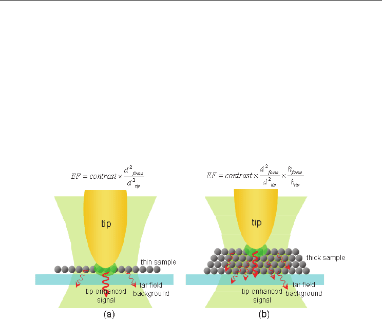
of the tip-enhancement is small the near-field signals may be submerged in the far-field
background noise, and results in low contrast. The phenomenon has been confirmed in
experiments (Mehtani et al., 2005). Strictly speaking, for the nano-dots and nano-wires, the
TERS enhancement factors should also be estimated by using different formulas. Due to the
differences between samples, the calculation of the EF is influenced by subjective factors.
And the contrast, which is calculated just based on the experimentally measured values is of
more practical significance. Nevertheless, the EF is still very necessary for the research on
the electromagnetic enhancement mechanism of TERS. In 2005, Pettinger et al (Pettinger et
al., 2005) ingeniously designed an experiment to avoid the subjective factors mentioned
above. By comparing the photobleaching time of the single-layer Malachite green dye with
and without tip enhancement, the more accurate TERS enhancement factor is obtained as
about 6x10
6
.
Fig. 7. Comparison of enhancement factor calculation (a) thin sample and (b) thick sample.
The lateral spatial resolution of TERS spectral imaging is mainly determined by the radius of
the tip apex. Since the localized electromagnetic field enhancement is highly confined to the
tip apex, the lateral resolution, spectral resolution may even be better than the resolution of
topography. Nowadays, the highest lateral resolution of TERS image reported is less than 15
nm (Anderson et al., 2005). For improving the lateral resolution the topography and spectral
imaging, tips with smaller radius should be used. Moreover, these tips are capable of
contributing more to the tip-enhancement and field enhancement (Downes et al., 2008).
3. Preparation of CNTs specimen
Specimen preparation is an important link of applying the TERS method to the detection of
practical specimens. It will finally determine whether the optimal measurement results can
be obtained as well.
3.1 Requirement of specimen preparation
The in-situ non-destructive spectral detection with high resolution and high sensitivity may
be accomplished with TERS system. Since the Raman spectrum directly reflects the chemical
information of samples, the specimen in TERS detection is dispensed with extra label. In

Electronic Properties of Carbon Nanotubes
226
general, the specimen preparation, especially for the preparation of CNTs samples, needs to
satisfy following requirements:
a. The smooth substrate surface. The fluctuations of the substrate surface should be less
than the sample-size scale.
b. Firmly attaching and immobilizing the specimen to the substrate surface.
c. Transparency. When using transmission-mode TERS system to detect the sample, the
specimen on the substrate should be transmission. The specimen can be highly
dispersed or diluted to obtain good transparency. However, in reflection-mode TERS
system the detected specimen is regardless to its transparency.
d. conductivity (STM-TERS)
According to specific samples and measuring conditions, there might be certain additional
requirements for the specimen preparation.
3.2 Common preparation methods for CNTs specimen
In our transmission-mode TERS system, the laser illuminates the sample from the bottom
and goes through the specimen to focus on the tip apex and locally excited Raman scattering
at the specimen surface. Additionally, the enhanced Raman signal also has to transmit
through the specimen to be collected. Therefore, the detected specimen is optimal to be of
good transparency. The CNTs specimen can be highly dispersed into a thin film.
Meanwhile, the sample is required not to chemically react with the substrate or the tip and
maintaining stabile properties in the whole measuring process. The specimen should also be
firmly attached on the substrate surface to avoid relative movement during the scanning. In
order to obtain high-resolution images, highly dispersed samples are generally used.
Therefore, the transmission-mode TERS system is more suitable for detecting sparse thin
films of CNTs samples with good transparency. For example, the specimen preparation
methods used in our transmission-mode TERS detection are presented here.
3.2.1 Preparation method for single wall carbon nanotube sample (SWNTs)
The SWNTs specimens used in our experiments are bundles of SWNTs. The SWNTs were
synthesized by arc-discharge method. After purification, the high purity (> 95%) SWNTs
powder can be obtained. Then 0.01mg powder was dissolved in 10mL ethanol. The solution
was evenly dispersed by ultrasonic dispersing method. One droplet of the solution was
taken and dripped on a 120μm thickness cover slip. After air-curing, the transparent CNTs
specimen has been prepared as well as for detection (Guan, 2006; Li et al., 2004).
3.2.2 Preparation method for double wall carbon nanotube sample (DWNTs)
The transparent thin DWNTs membrane was prepared following the series of processing
(Wei et al., 2006): firstly, the synthesized DWNTs were soaked in the H
2
O
2
solution with
30% Concentration for 72 hours. Then, HCl solution (37%) was added to remove the
amorphous carbon, catalyst particles and other impurities. The up to 90% (by mass fraction)
purity DWNTs were obtained. By dripping several drops of ethanol or acetone, the purified
macro-membrane of DWNTs were rapidly spreading to ultra-thin film and floating on the
surface of the solution. The film of DWNTs can be easily collected with a cover slip. The flat
and transparent thin film of DWNTs was obtained, and after air-curing the highly dispersed
DWNTs were immobilized on the substrate surface.

Detection of Carbon Nanotubes Using Tip-Enhanced Raman Spectroscopy
227
4. Detection of CNTs
The TERS technology has been widely developed and applied in the fields of physics,
chemistry, material science, and biology in nanometer scale. Especially, the detection of
CNTs will be presented and discussed as an emphasis of this section.
4.1 Applications of TERS
The significant advantage of TERS is that it can provide corresponding topography and
Raman mapping of the nano-material specimen with high spatial resolution and detection
sensitivity. Since 2000, TERS has been experimentally proved practicable (Stöckle et al., 2000;
Hayazawa et al., 2000; Anderson, 2000; Pettingeret al., 2000) and shows the potential of
nanometric spectral detection. Then, the research on TERS’s applications has been being
advanced continually.
Up to now, it has been applied to dye molecule detection (Steidtner & Pettinger, 2008; W. H.
Zhang et al., 2007), semi-conductor material determination(Sun et al., 2003; Lee et al., 2007;
Saito et al., 2008; Sun & Shen, 2001) , biological specimen identification(Anderson et al.,
2003; Watanabe et al., 2004; Bailo & Deckert, 2008), and nanao-material characterization
(Hayazawa et al., 2000; Hartschuh et al., 2003; J. J. Wang et al., 2005; Qian et al., 2006).
It is distinct from the single molecule level or single-molecule state distributed specimen
detection, TERS approach aims on the real single molecule, individual molecule Raman
spectral detection. In 2008, Pettinger et al. directly measured the spatial location and the
corresponding Raman spectra of the single BCB molecule absorbed on Au (111) surface
using STM-TERS system in ultra-high vacuum (Steidtner & Pettinger, 2008). The experiment
shows that under resonance Raman excitation, the enhancement factor is to 10
6
. It is enough
to satisfy the requirement of the single molecule detection sensitivity of the dye molecules in
the TERS characterization.
The in situ spectrum detection of biological samples is another research hotspot in TERS
applications in recent years. Since the Raman spectra directly reflect the molecular structure
information of the sample, the specimen in TERS detection is dispensed with extra label.
Meanwhile, TERS is capable of obtaining high resolution in situ non-destructive detection
with high sensitivity. In 2003, Anderson et al. (Anderson et al., 2003) applied TERS
technique to the detection of drosophila compound eyes, and measured the fine structures
of the eye surface and the near-field Raman spectra at different positions. In 2006,
Hayazawa et al. (Hayazawa et al., 2006) measured the near-field Raman spectroscopy of the
adenine nano-crystal sample. Compared with the standard far-field spectrum, slight
frequency shift of the TERS spectra is reviewed. In 2008, Bailo et al. (Bailo & Deckert, 2008)
detected the topography of single-stranded cytosine RNA and its tip-enhanced Raman
spectra at several different positions. The single-base detection sensitivity of the TERS
system is indirectly proved. The author further noted that although the TERS system is not
yet sufficient to the spectral imaging with the single-base spatial resolution, TERS
technology is still expected to directly sequence the DNA or RNA samples by using certain
detection and data processing method. TERS research on live Biological macromolecules
and virus has been commenced. Recently, Deckert et al. (Cialla et al., 2009) measured the
TERS signals of the single tobacco mosaic virus at different positions, and the characteristic
Raman shifts were identified. Budich et al. (Budich et al., 2008) also reported the TERS
characterization of the Staphylococcus epidermidis cell wall in liquid environment. These
applications all indicate the great potentials of the TERS technique in biological and life
science research.

Electronic Properties of Carbon Nanotubes
228
The demand of nano-material characterization is a major motivation to promote the
development of TERS. CNT is a kind of the typical 1-D nano-materials. Much attention has
been paid to the research and detection of CNT, Due to its outstanding physical, mechanical,
thermal and electrical properties along with application potentials. In 2003, Hayazawa et al.
(Hayazawa et al., 2003) applied TERS method to detect the SWNTs specimen and obtained
the diameter and chirality of the CTNs by analizing the TERS reaults. Harschuh et al.
(Hartschuh et al., 2003) also utilized TERS to characterize SWNTs and simultaneously
acquired the topography and the TERS mapping. Comparing the features in the two images,
the spatial distribution of the CNTs can be accurately recognized. In this detection, the
spatial resolution of TERS mapping reached up to 23 nm, even better than that of
topography, which is 29 nm. With the development of the tip preparation and the TERS
realization, better than 15 nm TERS resolution can be achieved nowadays (Hartschuh et al.,
2005; Anderson et al., 2005; Huihong et al., 2008: 2243-6). In 2005, Hartschuh et al. measured
the TERS and tip-enhanced photoluminescence (PL) mapping of SWNTs with better than 15
nm resolution (Hartschuh et al., 2005). The structural defects and optical properties of CNTs
may be further revealed with the combination of TERS detection and tip-enhanced
photoluminescence (PL). In 2006, Kawata et al. imaged SWNTs bundle within 700 nm×700
nm region. It validated that the radial breathing mode (RBM) feature inducing the Raman
shift corresponding to the vibrational mode of the graphitic layer in the radial direction is
sensitively and directly depended on the diameters of the tube. By imaging a serial of TERS
mappings according to different RBM Raman shifts, the distribution of SWNTs with
different diameters can be obtained separately. Lately, Kawata’s group reported another
way to further improving the resolution of TERS. In this method, the CNTs below the tip are
locally pressed by the tip apex, and the slight Raman frequency shift induced by the
pressure can be accurately detected. The technology is employed in nanometer metrology
and spatial resolution is to 4 nm (Yano et al., 2009).
Carbon nanotubes are categorized as single-walled nanotubes (SWNTs), double-walled
nanotubes (DWNTs) and multi-walled nanotubes (MWNTs). According to the difference in
sizes, structures and features of them, SWNTs and MWNTs will be measured. Also
experimental examples will be provided and discussed respectively in the following passages.
4.2 Detection of SWNTs
Generally speaking, the Raman peaks of SWNTs spectra represent the major vibration
modes in CNTs, the radial breathing mode (RBM), graphite-like mode (G-mode
corresponding to G-band), and defect-related mode (D-mode, D-band)(Dresselhaus et al.,
2005; Rao et al., 1997).
The Raman shifts related to RBM is at the low frequency region of the spectrum. They are
associated with the radial direction vibration modes of the nanotubes and directly reflect the
diameters of the SWNTs. The G-mode vibrations of SWNTs are induced by the planar
vibrations of the carbon atoms. The G-mode vibrations bring in Raman shifts in G-band,
which is generally around 1600 cm
-1
. Also most graphite-like materials all generate the G-
band in Raman spectra. The D-mode of CNTs is related to the out-of-order defects of the
CNTs or the amorphous carbon in the sample. The D-mode vibrations of SWNTs result in
the Raman shifts around 1300 cm
-1
. The ratio of G-band to D-band is an important term for
the evaluation of the purity of the SWNTs specimen (Eklund et al., 1995).
Detection of Carbon Nanotubes Using Tip-Enhanced Raman Spectroscopy
229
4.2.1 Conventional Raman spectrum measurement of SWNTs
For comparison and calibration, the conventional far-field Raman signal of the SWNTs
specimen was measured before the TERS detection. The photon-counter integrated time is
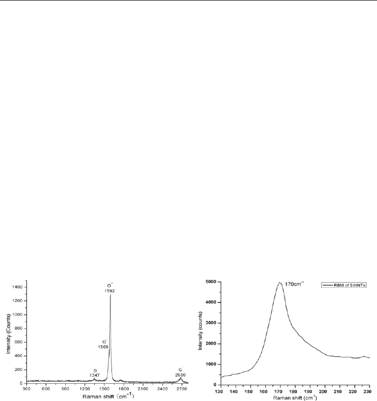
set 100ms in the Raman spectroscope, Renishaw RM2000. The result is shown in Fig. 8(a).
From the far-field Raman spectrum, several Raman shift feature peaks of the SWNTs
specimen can be distinguished. One is the G-band corresponding to the planer vibration. It
breaks up to several characteristic peaks as the fold in Brillouin Zone and mainly represents
as
G
+
band in the high frequency region (1592 cm
-1
) and G-
band in the low frequency region
(1569cm
-1
). Second is the D-band at 1347 cm
-1
, which is resulted from amorphous carbon or
out-of-order defects in SWNTs sample. The purity of SWNTs sample can be evaluated based
on the ratio of G band and D band (Dresselhaus et al., 2005). As the experimental result
indicated, the ratio of G and D band of the specimen is 26.
Another characteristic Raman shift corresponding to RBM was detected in low frequency
region and accumulated 3 times with the integrated time, 10 seconds. Considering that the
sample is bundle of SWNTs and the tube-tube interaction in the bundle, the relationship
between the diameter d
t
(nm) of the nanotube and the RBM band ω
RBM
(cm
-1
) can be
expressed as following (Rao et al.,2001),
ω
RBM
= 224/ d
t
+ 10 (3)
This is a modified formula for a bundle of SWNTs slightly different from isolated SWNTs.
As the detected RBM frequency is 170cm
-1
(Fig. 8 (b)), the diameter of the SWNTs is
estimated to be 1.4 nm.
Fig. 8. (a) Raman spectrum of SWNTs (b) RBM of SWNTs
4.2.2 Topographic and TERS measurement of SWNTs bundle
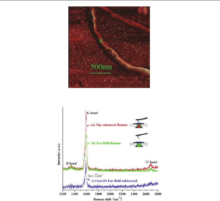
The SWNTs specimen is detected with a transmission-mode TERS system based on an AFM.
Firstly, the topography of the SWNTs specimen is obtained by AFM (Fig.9). It can be
indicated that in the sample most SWNTs exist in the form of bundles due to the action of
Van der Waals force. On the SWNTs bundle one position marked with cross is selected as
the interest point of TERS detection. Then, the position of the specimen is relatively moved
to the near-field region and just below the metallized tip apex. The TERS spectrum of the
SWNTs specimen is shown in Fig.10 (a). Then, the far-field Raman spectrum at the same
position is detected ( Fig.10 (b) ), as the tip is withdrawn from the near-field region of the
specimen (Wu et al, 2009).
Electronic Properties of Carbon Nanotubes
230
Fig. 9. Topographic image of a Single SWNT bundle with diameter 100nm, by TERS.
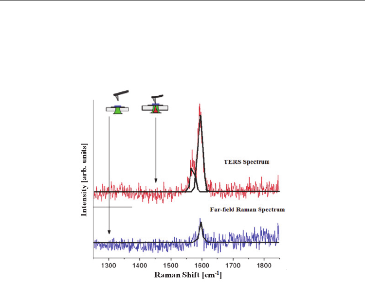
Fig. 10. TERS and far-field Raman spectra of a single SWNT bundle.
Comparing the TERS spectrum with the far-field Raman spectrum, it can be noticed that the
frequency of G-band Raman peak is not obviously shifted and the shape is not evidently
changed. In addition, the characteristic G-band is considerably enhanced as a result of the
tip-enhanced electromagnetic field, while the background noise is not obviously increased.
This can be explained as that the noise is mainly comes from the far-field stray light in the
illuminated area and not Raman scattering localized and enhanced by the tip. While Raman
signal from the localized zone is greatly enhanced by the tip-enhanced electromagnetic field
highly confined to the tip’s apex. It results in the higher SNR detection results. The pure
near-field Raman signal of SWNTs can be obtained as shown in Fig.10(c), by subtracting the
far-field signal from the TERS spectrum. The enhancement factor is calculated to be 230
according to the formula given in Fig. 7 (a) for thin film specimens. The tip enhancement
factor will be much larger than that value, because the practical diameter of far-field laser
spot is always larger than the size of the diffraction-limit used in the estimation. It indicates
adequately that TERS technology is of novel and excellent performances in detection of
weak Raman signal in nanometer localized region.
Detection of Carbon Nanotubes Using Tip-Enhanced Raman Spectroscopy
231
4.3 Detection of MWNTs
Multi-walled nanotubes (MWNTs) are formed with multiple coaxial rolled layers of
graphite. Due to the structures and compositions, they have similar Raman shifts with
SWNTs, but still with several distinct features. Additionally, being the simplest
manifestation of MWNTs, the double-walled carbon nanotubes (DWNTs) are considered to
be an ideal model for researching the interaction between graphite layers, as well as the
connections between the SWNTs and MWNTs.
4.3.1 Detection of DWNTs
A DWNT has the structure of two coaxial hollow cylinders formed by two layers of rolled-
up graphite. The typical interlayer space between the inner and the outer walls, ranges from
0.33 nm to 0.42 nm. DWNTs maintain several outstanding properties of SWNTs, yet have
some distinct ones, such as higher stability and particular electronic and optical properties
(Gennaro et al., 2009). In Raman spectra of DWNTs, the graphite-like band (G-band) region
arose by the graphitic layers is regarded as one of the feature Raman shifts of CNTs. And
the RBM frequency corresponding to the radial direction vibration of the nanotubes is
sensitively and directly depending on the diameters of the inner and the outer walls.
In this practical example, DWNTs specimen was investigated using our home-made
transmission-mode TERS system (Wu et al., 2010). The Raman enhancement factor was
calculated. The tip-enhanced Raman spectra especially in the G-band and the RBM regions
are obtained. A tip-pressure induced RBM band shift of the DWNT is observed, and the
experimental results are discussed.
In this research, a backward-scattering TERS system with a golden AFM tip is used. The
radius of the tip apex is about 30 nm. The DWNTs specimen is prepared using the method
mentioned in section 3.2.2. And the DWNTs used here were synthesized by chemical vapor
deposition (CVD) process. In TERS detection, the excitation laser (532 nm) after expanding
and collimating is focused on the tip by a high-NA objective (NA = 0.95). Also the tip-
enhanced Raman scattering from a confined region of the specimen is efficiently excited.
Then the Raman scattering is collected by the same objective lens and channeled into the
Raman spectroscope to be analyzed.
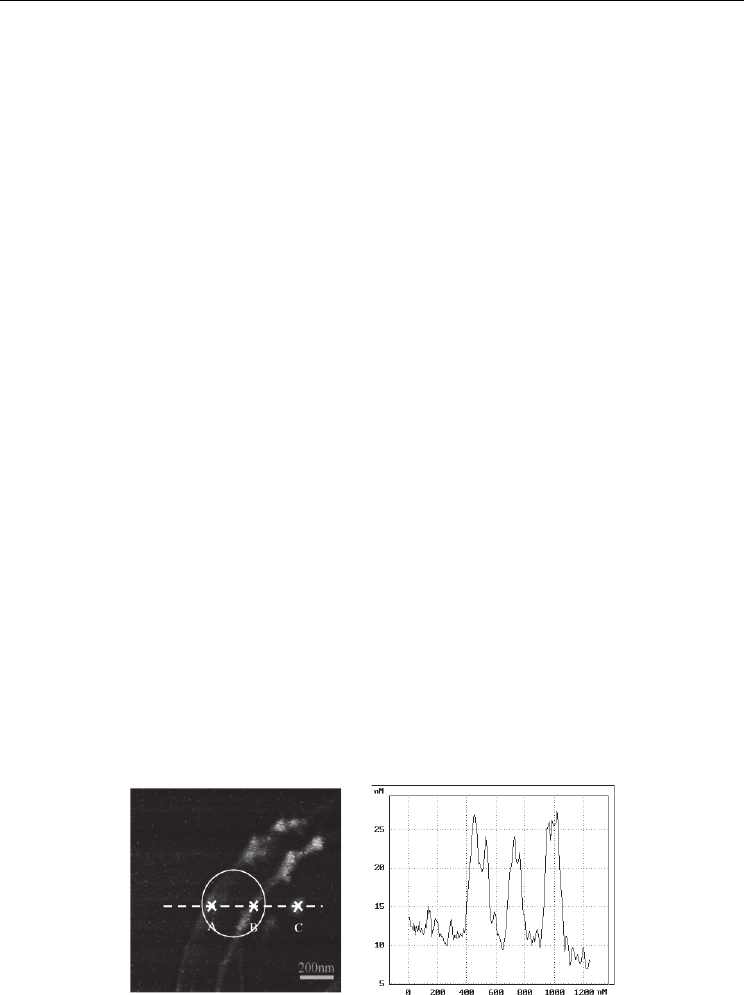
In advance, the topography of the DWNTs specimen was detected (Fig. 11(a)). The height
cross section along the dashed line is shown in Fig. 11(b). It shows that the heights of the 3
figures are all about 10 nm. Judging from the height, the two arc-like bundles each contains
(a) (b)
Fig. 11. (a) Topographic image of the DWNTs specimen (scanning area: 1.2 μm × 1.2 μm).
(b) Height cross section along the dashed line in (a).
Electronic Properties of Carbon Nanotubes
232
estimated about 10 to 15 DWNTs. After acquiring the topography of the specimen, the
region-of-interest could be narrowed down and defined to the three figures. Then, the
single-point TERS detection was carried out at each interested position. The detected TERS
spectrum of position A of the specimen is shown in Fig. 12. For comparison, the far-field
Raman spectrum was detected as the tip was withdrawn from the near-field region. The
separation between the tip and the sample surface was about 1.5 μm then.
Fig. 12. Far-field Raman spectrum and TERS spectrum of DWNTs at position A in Fig. 11(a).
Compared with the far-field spectrum, the TERS spectrum indicates that a strong
enhancement at the G-band is obtained. With Lorentz fitting, it can be observed that the G
band in the TERS spectrum splits mainly into two peaks: 1590 cm
-1
(G
+
) and 1564 cm
-1
(G¯).
However, in the far-field Raman spectrum the G¯ band may be too weak to be distinguished
from the noise. According to the Eq. (2) given in section 2.4, the EF is calculated to be 3.6 ×
10
2
.
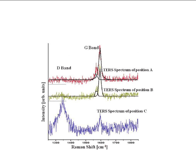
With the tip fixed at the laser focus, the specimen is moved along the dashed line shown in
Fig. 11(a). The TERS spectra of positions A, B, and C are detected in succession (shown in
Fig. 13). TERS spectra of positions A and B both show similar Raman peaks in the G-band
and almost no signals in the D-band. However, at position C the Raman peak in the D-band
is strong and the G band signal is weak. By comparative analysis of the topography and the
TERS spectra it can be ratiocinated that the two arc-like lines are bundles of DWNTs, while
the granule at position C is amorphous carbon respectively. The Raman signal from C
includes the D-band from amorphous carbon and the weak G-band signal might from
DWNTs in the vicinity of C point illuminated area in the far-field.
For further characterization of DWNT specimen, the Raman spectra in the RBM region were
detected and analyzed to estimate the diameters of the DWNTs’ inner and the outer walls,
respectively. The Raman shift in RBM region can be directly determined from the diameter
of the nanotube. Thus, it’s quite useful in estimating the sizes of the inner and the outer
walls of DWNTs and the corresponding deformation of DWNTs under radial pressure.
Detection of Carbon Nanotubes Using Tip-Enhanced Raman Spectroscopy
233
Since the interaction between the inner and the outer walls and the force exerted between
two DWNTs will both induce upshift to the frequencies of the Raman shifts in RBM band
(Bandow et al., 2002), the formula ω
RBM
= 224/ d
t
with about 10% higher RBM frequency
modification is adopted here to calculate the corresponding diameter of the nanotubes, as
Eq. (3) above similar to SWNTs.
Fig. 13. TERS spectra at the 3 positions (A, B, and C) marked correspondingly in Fig. 11(a)
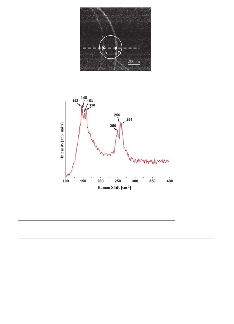
The experimental obtained RBM band consists of two groups of Raman shift peaks, the
range from 140 cm
-1
to 160 cm
-1
and 250 cm
-1
to 270 cm
-1
, which can be observed in Fig.15
(far-field) and Fig.16 (tip-enhanced). Since the RBM frequency depends inversely on the
nanotube diameter, the two groups of peaks correspond to the outer and the inner walls of
DWNTs respectively. The calculated diameters of the nanotubes are shown in Table 2. The
outer walls’ diameters vary between 1.51 nm and 1.69 nm while the inner walls’ diameters
vary between 0.89 nm to 0.93 nm.
In this experiment, the topography of another selected area of the DWNTs sample was
detected by using AFM (shown in Fig. 14) before the Raman spectral investigation. Two arc-
like bundles of DWNTs, whose heights were 3 nm and 6 nm respectively, were observed in
the detected area. With the tip withdrawn from the near-field of the sample, the far-field
Raman spectrum of the DWNTs was obtained (as shown in Fig. 15). Then, for further
detection of the tip-enhanced RBM of the DWNTs, the AFM golden tip was approached to
the near-field of the sample, and the scanning stage was precisely moved to align the
interested DWNT bundle with the tip’s apex. After eliminating the far-field background, the
tip-enhanced RBM of position A and B are shown in Fig. 16(a) and (b) respectively.
Comparing the tip-enhanced RBM with the far-field one, one can draw two conclusions.
First, the excitation area is highly confined. Seven peaks in the RBM region are detected
from the far-field Raman spectrum while 5 and 7 peaks are detected at the position A and B
in TERS, respectively.
Electronic Properties of Carbon Nanotubes
234
Fig. 14. Topographic image of DWNTs.
Fig. 15. Far-field Raman spectrum of a DWNT in the RBM region.
outer wall inner wall
interlayer
space
(nm)
Raman shift
(cm
-1
)
Diameter
(nm)
Raman shift
(cm
-1
)
Diameter
(nm)
142 1.69 250 0.93 0.38
256 0.91 0.39
261 0.89 0.40
148 1.62 250 0.93 0.345
256 0.91 0.355
261 0.89 0.365
153 1.57 261 0.89 0.34
158 1.51 - - -
Table 2. Correspondence of the far-field RBM frequencies and the diameters of the DWNTs.