Lax Alistair J. Bacterial protein toxins: Role in the interference with cell growth regulation (Бактериальные токсины белков: роль в регуляции роста клеток)
Подождите немного. Документ загружается.

P1: IwX/JPJ
052182091Xc03.xml CB786/Lax 0 521 82091 X November 3, 2005 23:51
36
gudula schmidt and klaus aktories
Pak
Erk
Raf
Rac
Cyclin D1
Jun
Cdc42
Rock
Myosin II
RhoA
cell cycle progression
cell division
positioning of
centrosomes
Kip1
SRF
Cyclin A
E2F
Waf / Cip
Cyclin E
mDia
LimK
LimK
Cofilin
?
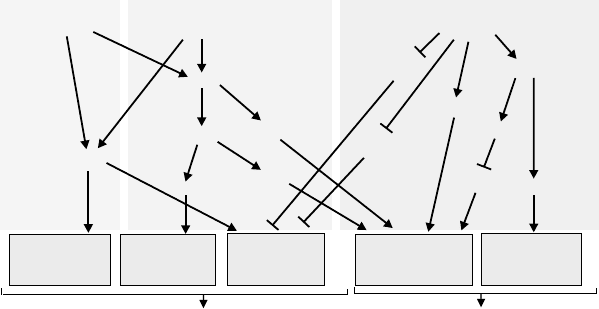
Figure 3.2. Rho GTPases regulate the cell cycle. For regulation of the cell cycle
progression Cdc42, Rac, and RhoA control different members of cell cycle regulatory
proteins of the cyclin family by activating (E2F, PAK) or inhibiting (Kip1, Waf/Cip)
effector molecules. Cyclin A and D are targets for Cdc42 and Rac signalling, whereas
RhoA activity primarily inhibits the CDK inhibitors Kip and Waf/Cip. The GTPases also
control cell division by activating SRF-dependent transcription and regulating cytokinesis.
Rac/Cdc42 is known to activate the Jun kinase signalling cascade with
c-Jun transcriptional activation (Figure 3.2) (Minden et al., 1995). However,
a major role of Rac/Cdc42 is the control of cyclin D1 transcription. Activated
mutants of Rac induce cyclin D1 (Page et al., 1999; Westwick et al., 1997).
Moreover, constitutively activated Rac and Cdc42, but not RhoA, induce E2F
activity and anchorage-independent induction of cyclin A (Philips et al., 2000).
On the other hand, negative Rac mutants inhibit the cyclin D1 induction by
oncogenic Ras (Gille and Downward, 1999). The Rac/Cdc42 effector Pak ki-
nase is involved in integrin-induced Erk activation and stimulates MEK1 and
Raf (Chaudhary et al., 2000). This process additionally involves PI3-kinase.
The major role of Rho in G1 progression appears to be different (Fig-
ure 3.2). Besides an effect of RhoA on cyclin D1 protein accumulation (Danen
et al., 2000), the GTPase was shown to be involved in downregulation of in-
hibitors of cyclin-dependent kinases. Mitogen-induced Erk activation induces
the CDK inhibitor p21
Waf/Cip
(Bottazzi et al., 1999) which is Rho dependent.
Similarly Rho is essential to prevent p21
Waf/Cip
induction by oncogenic Ras
(Olson et al., 1998). Furthermore, Rho appears to be involved in degradation
of the CDK inhibitor p27
kip1
(Hirai et al., 1997). In line with these findings are
studies showing that Rho GTPases are essential for cell transformation and
possess at least some cell transformation activity. The role of Rho GTPases in
P1: IwX/JPJ
052182091Xc03.xml CB786/Lax 0 521 82091 X November 3, 2005 23:51
37
toxins that activate rho
transformation is more obviously appreciated by the observation that many
GEFs are well-established products of oncogenes. This category includes Vav,
(Olson et al., 1996), Lbc (Zheng et al., 1995), Dbl (Hart et al., 1991), TIAM-1
(Michiels et al., 1995), and many others (Jaffe and Hall, 2002). Accordingly, it
was shown that mice, which are deficient in the Rac GEF TIAM, are resistant
to Ras-induced skin tumours (Malliri et al., 2002).
Recently, an important connection between the actin cytoskeleton and
transcriptional activation was described. It was shown that LIM Kinase and
Diaphanous cooperate to regulate serum responsive factor and actin dynam-
ics (Geneste et al., 2002). It has been known for many years that a dynamic
actin cytoskeleton is needed for the cleavage of a dividing cell into two daugh-
ter cells. Moreover, it has been shown in many different cell systems that Rho
GTPases are involved in cell division. Several Rho effectors, including Rho
kinase (ROCK) and citron kinase, are localised at cleavage furrows (Chevrier
et al., 2002). Moreover, Myosin II, which is phosphorylated by Rho kinase, is
an essential motor for cytokinesis (Matsumura et al., 2001). Rho kinase has
been identified as a component of the centrosome. It is required for posi-
tioning of the centrosomes, which play a role in cell division as well as in cell
motility.
RHOPROTEINS AS TARGETS OF BACTERIAL TOXINS
During the last few years, it has been recognised that Rho proteins are ma-
jor eukaryotic targets for various bacterial protein toxins. Some toxins block
the functions of Rho GTPases by covalent modification. For example, C3-
like toxins from C. botulinum, C. limosum, and S. aureus, which share 30 to
70% aminoacid sequence identity, ADP-ribosylate small GTPases of the Rho
family (e.g., at Asn41 of RhoA [Sekine et al., 1989]) and inactivate them. The
prototype of these small toxins (23–30 kDa) is the Clostridium botulinum C3
toxin, which ADP ribosylates RhoA, B, and C (Aktories et al., 1987; Wilde
and Aktories, 2001).
It was assumed that Rho function is blocked due to sterical hinderance
of the GTPase-effector interaction, because the modified residue is located
close to the effector region. However, recent studies indicate that ADP-
ribosylated Rho is still able to interact with at least some effectors (Genth
et al., 2003b). However, the rate of activation of Rho by exchange factors
(e.g., Lbc) is diminished by ADP-ribosylation (Genth et al., 2003b), and it was
suggested that ADP-ribosylation prevents the conformational change, occur-
ring subsequently with GDP/GTP exchange (Genth et al., 2003a). Moreover,
ADP-ribosylated Rho is released from membranes and forms a tight complex
P1: IwX/JPJ
052182091Xc03.xml CB786/Lax 0 521 82091 X November 3, 2005 23:51
38
gudula schmidt and klaus aktories
with GDI, a guanine nucleotide dissociation inhibitor, which keeps Rho in
its inactive GDP-bound form in the cytosol (Genth et al., 2003a).
C3-like exoenzymes consist of only the enzyme domain and lack a specific
cell membrane–binding and translocation unit. The uptake of C3-like toxins
into target cells and their potential roles as virulence factors are not well
understood. Two explanations are possible. First, it was suggested that the
enzymes (at least those produced by S. aureus) are directly released into the
cytosol from bacteria, which are capable of invading eukaryotic target cells
(Wilde et al., 2001). Second, uptake of C3 exoenzymes might depend on the
presence of bacterial pore-forming toxins, which facilitate translocation. C3
toxins are widely used to inactivate RhoA. For the use of the RhoA-specific
C3 toxins as pharmacological tools, toxin chimeras, consisting of C3 and the
binding and translocation domain of “complete” toxins (e.g., C. botulinum C2
toxin), have been constructed (Barth et al., 2002).
Large clostridial cytotoxins comprise a second family of Rho protein inac-
tivating toxins. These toxins modify the GTPases by glucosylation (Busch and
Aktories 2000; Just et al., 1995; Just et al., 2000). Members of this toxin family
are C. difficile toxins A and B, including various isoforms, the lethal and the
haemorrhagic toxins from C. sordellii, and the alpha toxin from C. novyi.
All these toxins are single-chain proteins with molecular masses of 250
to 308 kDa and encompass a catalytic domain and a specific binding and
translocation domain. The substrate specificity of large clostridial toxins is
broader than that of C3-like toxins. For example, C. difficile toxins A and
B glucosylate many GTPases of the Rho family, including Rho A, B and
C, Rac and Cdc42. C. sordellii lethal toxin possesses a different substrate
specificity and modifies Rac but not RhoA. In addition, Ras subfamily pro-
teins (e.g., Ras, Ral, and Rap) are glucosylated (Just et al., 1996). C. novyi
toxin, which shares the substrate specificity of toxin B, is an O-GlcNAc
transferase.
All these transferases modify a highly conserved threonine residue (e.g.,
Thr37 in RhoA) in the switch 1 region of the GTPases, which is involved
in Mg
2+
and nucleotide binding. Modification of this threonine residue
by mono-O-glucosylation has the following effects: (1) causes inhibition of
the interaction of GTPases with their effectors; (2) increases membrane
binding; (3) blocks activation by exchange factors; and (4) inhibits intrin-
sic and GAP-stimulated GTPase activity (Genth et al., 1999; Sehr et al.,
1998). The toxins are taken up from an acidic endosomal compartment
and glucosylate RhoA, Rac, and Cdc42 in the cytosol (Barth et al., 2001).
Glucosylating toxins are also widely used as tools to study the functions of
GTPases.
P1: IwX/JPJ
052182091Xc03.xml CB786/Lax 0 521 82091 X November 3, 2005 23:51
39
toxins that activate rho
Inactivating Rho GTPases is not the only way to influence signal trans-
duction pathways of mammalian host cells. Rho proteins are also activated
due to covalent modification catalysed by bacterial protein toxins like the cyto-
toxic necrotizing factors CNF1 and CNF2 from E. coli and the dermonecrotic
toxin DNT from Bordetella species (described below). Recent studies indicate
that Rho proteins are not exclusively covalently modified by bacterial toxins.
Some bacterial effectors, like the Salmonella SopEs and SptP, modulate the
activity of Rho GTPases by acting as regulatory proteins with GAP (SptP) or
GEF (SopEs) functions (see Chapter 6).
CNFs Activate Rho GTPASES
In 1983, Caprioli and co-workers isolated a toxin from an Escherichia coli
obtained from enteritis-affected children. Because of the necrotising effects
on rabbit skin they called the toxin CNF (cytotoxic necrotizing factor) (Caprioli
et al., 1983). Besides the skin necrotising action, CNF turned out to be lethal
for animals after i.p. injection. The lethal dose (LD
50, mice
) was estimated
to be about 20 ng of purified material (de Rycke et al., 1997). Studies with
cultured cells revealed typical morphological changes, cell body enlargement,
stress fibre formation, and multinucleation. Subsequently, CNF1 and later
the homologue CNF2, first named Vir cytotoxin (Oswald et al., 1989), was
found in various pathogenic E. coli strains isolated from animals (e.g., piglets
and calves) (de Rycke et al., 1987;deRycke et al., 1990) and man.
Structure and Up-Take of CNFs
CNF1 and CNF2 are closely related toxins, sharing more than 90% identity
in their amino acid sequences (Oswald et al., 1994). Whereas CNF1 is chro-
mosomally encoded, CNF2 is encoded by transmissible plasmids (Oswald
and de Rycke, 1990). Both toxins are single-chain proteins with molecular
masses of about 115 kDa. They are constructed like AB toxins, with the cell-
binding and catalytic domains located at the N terminus (amino acids 53 to
190, cell-binding domain) and C terminus (amino acids 720 to 1014, catalytic
domain) of the toxin, respectively (Lemichez et al., 1997). The central part
appears to be involved in membrane translocation. The receptor for the entry
of the toxin into cells is still unknown. Uptake of CNF appears to occur by
clathrin-dependent and -independent endocytosis, which is followed by cell
entry from an acidic endosomal compartment (Contamin et al., 2000). Re-
cently, two hydrophobic helices (aa350–412), which are separated by a short
loop (aa 373–386), have been suggested to be involved in membrane insertion
P1: IwX/JPJ
052182091Xc03.xml CB786/Lax 0 521 82091 X November 3, 2005 23:51
40
gudula schmidt and klaus aktories
CC
ll
63
63
ll
Rho
Rho
OOH
2
ONH
3
NH
2
OH
l
l
l
l
CNF / DNT
Deamidase
Transglutaminase
++
CC
l
l
63
63
ll
Rho
Rho
OONH
3
NH
2
HN—R
H
2
N—R
l
l
l
l
DNT
++
Figure 3.3. Molecular mechanism of Rho activation by CNF and DNT. Glutamine 63 of
RhoA (Gln 61 of Rac and Cdc42) is essential for the hydrolysis of bound GTP. CNF and
DNT deamidate this glutamine residue, creating glutamic acid, and the GTP hydrolysing
activity of the GTPase is blocked. In the presence of primary amines, the toxins can
polyaminate Rho at the same residue, thereby also blocking hydrolysis of GTP.
Polyamination is the preferred activity of DNT whereas CNF is a better deamidase.
in a similar manner to the hairpin helices TH 8–9 of diphtheria toxin (Pei
et al., 2001).
Mode of Action of CNFs
CNFs are cytotoxic for a wide variety of cells, including 3T3 fibroblasts, Chi-
nese hamster ovary cells (CHO), Vero cells, HeLa cells, and cell lines of neu-
ronal origin. The toxins lead to enlargement and flattening of culture cells
in a time- and concentration-dependent manner. These changes are accom-
panied by transient and early formation of filopodia and membrane ruffles
and a dense network of actin stress fibres, indicating that Rho proteins are
involved in the action of these toxins. CNFs change the migration behaviour
of Rho in SDS-PAGE (Oswald and de Rycke, 1990). This finding suggested a
covalent modification of Rho GTPases by CNFs and allowed the elucidation
of their mode of action. To this end, mass spectrometric analysis showed that
CNF1 causes an increase in mass of RhoA by 1 Da. This change in mass is
due to a deamidation at glutamine 63 of RhoA (see reaction scheme in Fig-
ure 3.3). Glutamine 63 in RhoA is essential for the GTP hydrolysing activity
of the GTPase. Thus, GTP hydrolysis is blocked after treatment of Rho with
CNF1.
Moreover, the stimulation of RhoA GTPase activity by GAP is blocked
after CNF1/2 treatment and RhoA is held constitutively active. Similarly,
Rac and Cdc42 are deamidated by CNF (Lerm et al., 1999b). Deamidation
P1: IwX/JPJ
052182091Xc03.xml CB786/Lax 0 521 82091 X November 3, 2005 23:51
41
toxins that activate rho
occurs at the equivalent amino acid residue glutamine 61. Although CNF is
highly specific for Rho GTPases, recent studies show that even a small pep-
tide covering the switch–II region of Rho GTPases is sufficient for substrate
recognition by CNFs.
The crystal structure of the enzyme domain of CNF1 has been solved
(Buetow et al., 2001), showing a novel protein fold. The structure confirmed
the previous suggestions that CNF belongs to the catalytic triad family. In
fact it was shown that a cysteine (Cys 866) and a histidine residue (His 881)
are essential for enzyme activity (Schmidt et al., 1998). As identified from the
crystal structure, the third “catalytic” residue appears to be a valine residue
(Val 833), a finding which is rather unusual among catalytic triad enzymes
(Buetow et al., 2001). Crystallisation of catalytic domains of various bacte-
rial enzyme toxins (e.g., ExoS GAP domain [W
¨
urtele et al., 2001]), which
share regulatory mammalian counterparts, indicates that the overall struc-
ture of the enzyme domain of bacterial toxins is not necessarily similar to
their mammalian counterparts. The same is true for the catalytic domain of
CNF1, which exhibits a different protein fold as compared with mammalian
transglutaminases (Pedersen et al., 1994). A reason for the high specificity
of CNF may be the existence of a deep cleft in the molecule with the cat-
alytic cysteine at the bottom (Buetow et al., 2001). Recently, a potential role in
substrate recognition has been described for three of nine loops located on
the surface of the catalytic domain of CNF1 (Buetow and Ghosh, 2003). The
structure of the CNF1 catalytic region contributes to the idea of a convergent
evolution of the toxins and mammalian enzymes with the same mechanism,
rather than gene transfer.
Cell Biological Effects
As already mentioned, the effects of CNF in cultured cells are characterised
by major changes of actin structure, including stress fibres, lamellipodia,
and filopodia. An increase in the formation of lamellipodia and membrane
ruffles is prototypic for enhanced phagocytic and endocytic activity. Accord-
ingly, activation of Rho GTPases by CNF induces phagocytic behaviour and
macropinocytosis in mammalian cells (e.g., human epithelial cells), which
are non-professional phagocytes (Falzano et al., 1993; Fiorentini et al., 2001).
Quite early, it was found that CNF causes phosphorylation of paxillin and
Fak kinase, which are known to be involved in nuclear signalling. This path-
way is Rho dependent but does not involve the classical Map kinase pathway
(Lacerda et al., 1997). One of the most striking effects observed with CNF is
the formation of multinucleation (Oswald et al., 1989). The effect might be
P1: IwX/JPJ
052182091Xc03.xml CB786/Lax 0 521 82091 X November 3, 2005 23:51
42
gudula schmidt and klaus aktories
caused by blocking cell division without changes in nuclear cycling, or by in-
crease in the rate of nuclear cycles within one cell division cycle (Denko et al.
1997). Recent data show that CNF2 uncouples S-phase from mitosis. Thus,
it affects cytoplasmic division and removes the requirement for a complete
mitosis before starting another S-phase. CNF increases the expression of the
cyclooxygenase-2 gene in fibroblasts (Thomas et al., 2001). This finding is of
importance because an increase in cyclooxygenase expression is observed in
several tumours and it has been suggested that lipid mediators produced by
the enzyme are responsible for tumour progression (Oshima et al., 1996).
Rac, which is also a substrate for CNF-induced deamidation, is involved
in Jun kinase activation and, therefore, CNF1 causes activation of this kinase.
Surprisingly, c-Jun kinase activity is only transiently increased after CNF
treatment of cells, although the GTPases are constitutively activated (Lerm
et al., 1999b). Recently, the reason for the transient activation was identified
as degradation of CNF-activated Rac by a proteasome-dependent pathway
(Lerm et al., 2002). Thus, it appears that the targeted cell is able to block the
persistent activation of Rac induced by deamidation by rapid degradation. So
far, it is not clear whether degradation of activated Rac is part of a general
mechanism in mammalian cells to limit “overactivation” of GTPases or due
to subtle structural changes induced by the deamidation.
DERMONECROTIC TOXIN (DNT) FROM BORDETELLA
A similar mechanism of Rho modification as described for CNF1 was re-
ported for the CNF1-related dermonecrotic toxin (DNT) from Bordetella
species (Horiguchi et al., 1997; Horiguchi 2001; Kashimoto et al., 1999;
Schmidt et al., 1999).
DNT is produced by Bordetella pertussis, B. parapertussis, and B. bronchisep-
tica. Its name is derived from dermonecrotic effects caused by intradermal
injection of the purified toxin (Horiguchi et al., 1989). DNT is a large, 160-kDa
heat-labile protein, which shares significant sequence similarity with CNF
(Figure 3.4). The sequence similarity is restricted to the C terminus of DNT
and CNF, which in part harbours the catalytic activity of the deamidase,
suggesting that both toxins share a similar molecular mechanism. In fact,
DNT induces similar if not identical morphological changes (enlargement
of cells, multinucleation, actin polymerisation) as CNF. Moreover, it was
demonstrated that DNT causes a covalent modification of Rho, resulting
in slightly slower migration of the GTPase in SDS-PAGE (Schmidt et al.,
1999). In line with this notion, it was demonstrated that CNF1 and DNT
induce deamidation of Rho at position Gln63. Similarly, as for CNF, the
P1: IwX/JPJ
052182091Xc03.xml CB786/Lax 0 521 82091 X November 3, 2005 23:51
43
toxins that activate rho
N
NC
C
C
CN
N
DNT (1-1451)
CNF1 (1-1014)
1250
824
1314
888
∆DNT
1136 1451
709
1014
∆CNF
V C H
CH
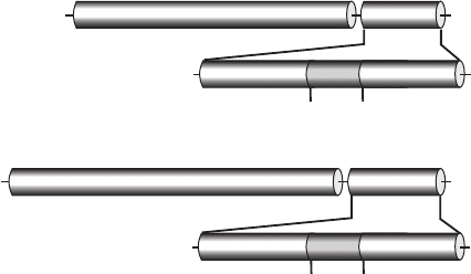
Figure 3.4. Homology between CNF1 and DNT. CNF and DNT consist of 1014 and 1451
amino acids, respectively. The toxins share homology within their catalytic domains
(CNF, aa 709 to 1014 and DNT, aa 1136–1451) that are located at the C termini of the
proteins. Highest sequence similarity is observed in a stretch of 64 amino acid residues,
which covers residues 1250 through 1314 of DNT, showing ∼45% sequence identity,
whereas the amino acid sequence of the whole catalytic domain of DNT is only ∼13%
identical with the sequence of the catalytic domain of CNF1.
DNT targets are not only Rho but also Cdc42 and Rac. Both toxins were
shown to catalyse the polyamination of glutamine 63/61 of Rho GTPases in
the presence of rather high concentrations of amines. However, it appears
that DNT prefers transglutamination, whereas CNF is primarily a deamidase
(Figure 3.3) (Schmidt et al., 1999). Recently, putrescine, spermidine, and
spermine have been identified as in vivo substrates for the transglutamina-
tion (Masuda et al. 2000; Schmidt et al. 2001). Lysine is a very good substrate
for transglutamination by DNT at least in vitro (Schmidt et al., 2001).
STRUCTURE–FUNCTION RELATIONSHIP BETWEEN
CNFs AND DNT
CNFs and DNT are both AB toxins, with a cell-binding domain located at
the N-terminus and a C-terminal catalytic domain. Amino acids 1 to 531 of
DNT blocked the intoxication of cells by full-length DNT, suggesting that this
fragment retains the cell-binding domain of DNT (Kashimoto et al., 1999).
More recently, the receptor-binding domain of DNT was mapped to amino
acids 1 to 54 (Matsuzawa et al., 2002). Moreover, a Furin cleavage site within
this binding domain was identified. Proteolytic processing of DNT by Furin
seems to be necessary for translocation of the toxin across cellular membranes
(Matsuzawa et al., 2004). It is of interest that the Pasteurella multocida toxin
P1: IwX/JPJ
052182091Xc03.xml CB786/Lax 0 521 82091 X November 3, 2005 23:51
44
gudula schmidt and klaus aktories
shares significant sequence similarity with the N terminus of DNT and with
the transmembrane domain of CNF (see Chapter 2). Thus, it is suggested that
both DNT and PMT share the same or the same type of membrane receptors,
however, not the same molecular mechanism (Lemichez et al., 1997; Walker
and Weiss, 1994).
As the catalytic domain of DNT is located at the C-terminus, a fragment
of DNT was constructed covering residues 1136 through to 1451, which was
fully active to cause transglutamination and deamidation of RhoGTPases
in vitro. The highest sequence similarity with CNF is observed in a stretch
of 64 amino acid residues, which covers residue 1250 through to 1314 of
DNT, showing ∼45% sequence identity, whereas the amino acid sequence
of the minimal active fragment of DNT (residues 1136–1451) is only ∼13%
identical with the sequence of the minimal active fragment of CNF1 (residues
709–1014).
As mentioned above, the catalytic triad of CNF shares a typical cys-
teine and histidine residue with the catalytic centre of transglutaminases.
DNT also possesses these conserved cysteine and histidine residues, which
are Cys 1292 and His 1307. These catalytic residues share the same spac-
ing as in CNF. However, biochemical studies also indicate differences in
the enzyme activities of DNT and CNFs. For example, the minimal Rho
sequence allowing deamidation or transglutamination by CNF1 is a pep-
tide covering mainly the switch-II region (D59–D78) of RhoA. By contrast,
DNT appears to need further interaction sites, and modifies exclusively the
GDP-bound form of Rho GTPases (Lerm et al., 1999a). Therefore the en-
zyme substrate interaction for DNT appears to be more complex than for
CNFs.
CNFs AND DNT AS VIRULENCE FACTORS
Initially, the role of CNF as a virulence factor was debated. However, recently,
several studies have suggested that CNFs are important for E. coli–caused dis-
eases. It has been shown that colonisation and tissue damage of the urinary
tract of mice induced by CNF-producing E. coli strains are more severe than
with CNF1-deficient isogenic strains (Rippere-Lampe et al., 2001b). The same
group has reported that tissue damage of rat prostates with CNF1-producing
uropathogenic E. coli strains is more extensive than after infection with iso-
genic CNF1-negative mutants (Rippere-Lampe et al., 2001a). Furthermore,
it was reported that the toxin is involved in the in vitro invasion of brain
microvascular endothelial cells by E. coli and contributes to the traversal of
the blood–brain barrier in a meningitis animal model (Khan et al., 2002).
P1: IwX/JPJ
052182091Xc03.xml CB786/Lax 0 521 82091 X November 3, 2005 23:51
45
toxins that activate rho
In contrast, no difference between CNF-producing and -deficient strains has
been found when studying lung and serosal inflammation (Fournout et al.,
2000). Nevertheless, taken together, one can say that CNF1 is involved in
E. coli virulence and pathogenicity.
Another topic is of interest and concern. Rho-GTPases are increasingly
recognised to be essential for proliferation, development of cancer, and
metastasis. Because CNFs cause activation of Rho GTPases and mediate
signalling leading to cell transformation, it is feasible that chronic carriers
of CNF-producing E. coli might be challenged by a tumourigenic potential
of the toxin (Lax and Thomas, 2002). This might be especially important for
prostate carcinoma and colon tumours. In this respect, it appears important
that CNFs also affect apoptotic processes. Although an increase in apoptosis
by CNF was observed in uroepithelial 5637 cells (Mills et al., 2000), major
anti-apoptotic effects of CNF were also reported (Fiorentini et al., 1998). The
differences in outcome might be due to differences in cell types or toxins
concentration.
The role of DNT as a virulence factor of Bordetella bronchiseptica, B. per-
tussis, and B. parapertussis is also not precisely defined. In fact, the toxin was
described quite early as a virulence factor for whooping cough (Horiguchi,
2001). However, it is now accepted that DNT is not a major factor involved in
this disease. By contrast, DNT is considered to be one of the major virulence
factors in turbinate atrophy in pigs (Magyar et al., 1988). Its role in the patho-
genesis of respiratory diseases has been shown by comparing isogenic DNT
mutants with the corresponding wild-type strains in the efficiency of colo-
nization of the respiratory tract of pigs. These studies showed that production
of DNT by B. bronchiseptica is essential to induce lesions of turbinate atrophy
and bronchopneumonia in pigs (Brockmeier et al., 2002). Accordingly, DNT
was reported to affect osteoblastic MC3T3-E1 cells in vitro and to impair bone
formation in neonatal rats (Horiguchi et al., 1995).
CONCLUSIONS
The deamidating and transglutaminating toxins CNFs and DNT, which ac-
tivate Rho GTPases, have multiple effects on morphology, motility, prolif-
eration, differentiation, and apoptosis of cells. Recent studies have shown
that CNFs and DNT are major virulence factors in various infection models.
Moreover, because Rho GTPases are crucial switches in signalling pathways
responsible for cell transformation and metastasis, it is plausible (although
still speculative) that they play a potential role in the pathogenesis of certain
types of cancer.