Koster G., Rijnders G. (Eds.) In situ Characterization of Thin Film Growth
Подождите немного. Документ загружается.

50 In situ characterization of thin film growth
© Woodhead Publishing Limited, 2011
P.J. Dobson. An introduction to reection high energy electron diffraction. In A. Howie
and U. Valdre, editors, Surface and Interface Characterization by Electron Optical
Methods, volume 191 of NATO ASI Series B, page 159, Plenum Press, 1988.
D.P. Field. Recent advances in the application of orientation imaging. Ultramicroscopy,
67: 1, 1997.
M. Gajdardziska-Josifovska and J.M. Cowley. Brillouin zones and Kikuchi lines for
crystals under electron channeling conditions. Acta Crystallographica Section a, 47:
74–82, 1991.
T. gareth. Transmission Electron Microscopy of Materials, Wiley, 1979.
C.J. Harland, P. Akhter, and J.A. Venables. Accurate microcrystallography at high
spatial-resolution using electron backscattering patterns in a eld-emission gun
scanning electron-microscope. Journal of Physics E–Scientic Instruments, 14 (2):
175–182, 1981.
Z. Hiroi, M. Azuma, M. Takano, and Y. Bando. A new homologous series Sr
n–1
Cu
n+1
O
2n
found in the SrO-CuO system treated under high-pressure. Journal of Solid State
Chemistry, 95 (1): 230, 1991.
Y. Horio. Kikuchi patterns observed by new astrodome RHEED. e-Journal of Surface
Science and Nanotechnology, 4: 118, 2006.
Y. Horio, Y. Hashimoto, and A. Ichimiya. New type of RHeeD apparatus equipped with
an energy lter. Applied Surface Science, 100: 292–296, 1996.
A. Ichimiya and P.I. Cohen. Reection High Energy Electron Diffraction. Cambridge
University Press, 2004.
N.J.C. Ingle, R.H. Hammond, and M.R. Beasley. Molecular beam epitaxial growth of
SrCuO
2
: Metastable structures and the role of epitaxy. Journal of Applied Physics,
91 (10): 6371–6378, 2002. DOI 10.1063/1.1466876.
N.J.C. Ingle, A. Yuskauskas, A. Wicks, M. Paul, and S. Leung. Topical review: the
structural analysis possibilities of reection high energy electron diffraction. Journal
of Physics D – Applied Physics, 43: 133001, 2010.
J.D. Jorgensen, P.G. Radaelli, D.G. Hinks, J.L. Wagner, S. Kikkawa, G. Er, and F.
Kanamaru. Structure of superconducting Sr
0.9
la
0.1
CuO
2
(T = 42K) from neutron
powder diffraction. Physical Review B, 47 (21): 14654, 1993.
S. Kikuchi. Diffraction of cathode rays by mica. Japanese Journal of Physics, 5: 83,
1928.
H.J. Kohl. In J.M. Sturgess, editor, Proceedings of the 9th International Congress on
Electron Microscopy (Toronto), page 198, 1978.
N.C.K. Lassen, D.J. Jensen, and K. Conradsen. Image-processing procedures for analysis
of electron back scattering patterns. Scanning Microscopy, 6 (1): 115–121, 1992.
J.R. Michael and J.A. eades. Use of reciprocal lattice layer spacing in electron backscatter
diffraction pattern analysis. Ultramicroscopy, 81 (2): 67–81, 2000.
H. Nakahara, T. Hishida, and A. Ichimiya. Inelastic electron analysis in reection high-
energy electron diffraction condition. Applied Surface Science, 212: 157–161, 2003.
DOI 10.1016/S0169-4332(03)00057-6.
K. Omoto, K. Tsuda, and M. Tanaka. Simulations of Kikuchi patterns due to thermal
diffuse scattering on MgO crystals. Journal of Electron Microscopy, 51 (1): 67–78,
2002.
V. Randle. Microtexture Determination and its Application. Bourne Press, 1992.
l. Reimer. Scanning Electron Microscopy. Springer-Verlag, 1985.
R.A. Schwarzer. Automated crystal lattice orientation mapping using a computer-controlled
SeM. Micron, 28 (3): 249–265, 1997.
51Inelastic scattering techniques for in situ characterization
© Woodhead Publishing Limited, 2011
J.A. Small, J.R. Michael, and D.S. Bright. Improving the quality of electron backscatter
diffraction (eBSD) patterns from nanoparticles. Journal of Microscopy – Oxford,
206: 170–178, 2002.
P. Staib, W. Tappe, and J.P. Contour. Imaging energy analyzer for RHEED: energy
ltered diffraction patterns and in situ electron energy loss spectroscopy. Journal of
Crystal Growth, 201: 45–49, 1999.
K.Z. Troost, P. Vandersluis, and D.J. Gravesteijn. Microscale elastic-strain determination
by backscatter Kikuchi diffraction in the scanning electron-microscope. Applied Physics
Letters, 62 (10): 1110–1112, 1993.
A. Vailionis, A. Brazdeikis, and A.S. Flodstrom. Observation of local oxygen displacements
in CuO
2
planes induced by a mist strain in heteroepitaxially grown innite-layer-
structure Ca
1–x
Sr
x
CuO
2
lms. Physical Review B, 55 (10): R6152, 1997.
J.A. Venables and R. Binjaya. Accurate micro-crystallography using electron backscattering
patterns. Philosophical Magazine, 35 (5): 1317–1332, 1977.
J.A. Venables and C.J. Harland. Electron backscattering patterns – new technique for
obtaining crystallographic information in scanning electron-microscope. Philosophical
Magazine, 27 (5): 1193–1200, 1973.
J.A. Venables, C.J. Harland, and Bin-Jaya. Developments in Electron Microscopy and
Analysis, page 101. Academic Press, 1976.
R. von Meibom and E. Rupp. Wide-angle electron diffraction. Zeitschrift für Physik,
82: 690, 1933.
Z.L. Wang. Reection Electron Microscopy and Spectroscopy for Surface Analysis.
Cambridge University Press, 1996.
O.C. Wells. Comparison of different models for the generation of electron backscattering
patterns in the scanning electron microscope. Scanning, 21 (6): 368–371, 1999.
M. R. Went, A. Winkelmann, and M. Vos. Quantitative measurements of Kikuchi bands
in diffraction patterns of backscattered electrons using an electrostatic analyzer.
Ultramicroscopy, 109 (10): 1211–1216, 2009. DOI 10.1016/j.ultramic.2009.05.004.
A.J. Wilkinson. Measurement of elastic strains and small lattice rotations using electron
back scatter diffraction. Ultramicroscopy, 62 (4): 237–247, 1996.
A.J. Wilkinson and P.B. Hirsch. Electron diffraction based techniques in scanning electron
microscopy of bulk materials. Micron, 28 (4): 279–308, 1997.
A.J. Wilkinson, G. Meaden, and D.J. Dingley. Mapping strains at the nanoscale using
electron back scatter diffraction. Superlattices and Microstructures, 45 (4–5): 285–294,
2009. DOI 10.1016/j.spmi.2008.10.046.
A. Winkelmann. Principles of depth-resolved Kikuchi pattern simulation for electron
backscatter diffraction. Journal of Microscopy – Oxford, 239 (1): 32–45, 2010. DOI
10.1111/j.1365-2818.2009.03353.x.
A. Winkelmann, C. Trager-Cowan, F. Sweeney, A. P. Day, and P. Parbrook. Many-beam
dynamical simulation of electron backscatter diffraction patterns. Ultramicroscopy,
107 (4–5): 414, 2007. DOI 10.1016/j.ultramic.2006.10.006.
C. Yasuda, S. Todo, K. Hukushima, F. Alet, M. Keller, M. Troyer, and H. Takayama.
Neel temperature of quasi-low-dimensional Heisenberg antiferromagnets. Physical
Review Letters, 94 (21): 217201, 3 2005. 217201.
S. Zaefferer. On the formation mechanisms, spatial resolution and intensity of
backscatter Kikuchi patterns. Ultramicroscopy, 107 (2–3): 254, 2007. DOI 10.1016/j.
ultramic.2006.08.007.
52 In situ characterization of thin film growth
© Woodhead Publishing Limited, 2011

© Woodhead Publishing Limited, 2011
55
3
Ultraviolet photoemission spectroscopy
(UPS) for in situ characterization of
thin film growth
K. M. S hen, Cornell University, USA
Abstract: This chapter discusses the application of ultraviolet photoemission
spectroscopy (UPS) and angle-resolved photoemission spectroscopy
(ARPES) to study the electronic structure of thin lms synthesized in situ
using lm growth techniques such as molecular beam epitaxy (MBE) and
pulsed laser deposition (PLD). The chapter rst describes the principles
of photoemission spectroscopy and the current experimental state of this
technique. Then, particular examples such as UPS and ARPES studies of in
situ thin lms of SrRuO
3
and La
1–x
Sr
x
MnO
3
are detailed.
Key words: photoemission spectroscopy, angle-resolved photoemission
spectroscopy (ArPeS), correlated electronic materials, in situ thin lm
spectroscopy.
3.1 Introduction
One of the prime motivators for the growth of materials in thin lm form is
the potential for manipulating their electronic and magnetic properties. One
of the forefronts of modern condensed matter physics is the search for new
states of quantum matter with exotic physical properties, and thin lms and
superlattices present a unique opportunity for designing and realizing such
novel materials. One way of achieving this control is to create atomically
perfect interfaces between two different materials. Modern technologies such
as the transistor or laser are dependent on interfacial phenomena. Remarkable
scientic discoveries such as the integer and fractional quantum Hall effects
(Nobel Prizes 1985, 1998) were discovered at semiconductor interfaces
engineered using molecular beam epitaxy (MBE). Since 2005, interfacial
phenomena at the atomically abrupt interface between dissimilar materials
have yielded a urry of breakthroughs. To fully benet from the tailoring of
these novel thin lm interfaces and heterostructures, one needs to be able to
characterize the state of electronic matter created in each system. In order to
do so, it will be desirable to utilize sophisticated 21st century tools to achieve
this aim, such as high-resolution angle-resolved photoemission spectroscopy
(ARPES). The ARPES technique follows from the development of electron
spectroscopy for chemical analysis (ESCA) pioneered by Kai Siegbahn (Nobel
56 In situ characterization of thin film growth
© Woodhead Publishing Limited, 2011
Prize 1981) by probing not only the energy but also momentum distribution
of the photoelectrons to directly observe band structure and Fermi surface
of materials. Without the detailed information about the electronic structure
that ultraviolet photoemission spectroscopy (UPS) and ArPeS can provide,
a deep understanding of how and why each thin lm material exhibits the
properties it does is lacking. In turn this greatly diminishes the capabilities
for rational design of new electronic materials in thin lm form.
By extracting electrons from within a material and measuring their
momentum and energy states, ARPES can measure a quantity known as the
electrons’ ‘Green’s function’, a quantity which encodes all the information
about the collective motion of electrons, how they are entangled, and provides
full knowledge of the electronic structure. Despite its power, ARPES has
one major limitation: the requirement of atomically pristine surfaces which
demands large, bulk single crystals which yield at, well-dened surfaces
when cleaved under vacuum. This is where photoemission spectroscopy on
thin lms grown in situ can provide an entirely new avenue into research
into the electronic properties of thin lms. Growth techniques such as MBE
have the distinct advantage of occuring at very low partial pressures (10
–7
or 10
–8
torr) and yielding atomically at surfaces. These points are key to
achieving the atomically pristine, at well-dened surfaces that are essential
for ArPeS.
One important aspect of correlated electronic systems, of which transition
metal oxides are a major constituent, is that the electrons are strongly
entangled, and it is their intricate collective motion that gives rise to their
remarkable attributes. This is in contrast to conventional materials where
the individual electrons essentially move as independent particles and thus
cannot produce these effects. However, understanding this entanglement
which is so essential to these properties is an extremely difcult scientic
problem because our theoretical physical understanding is based largely on
the non-interacting paradigm of materials such as silicon or copper.
3.2 Principles of ultraviolet photoemission
spectroscopy (UPS)
Photoemission spectroscopy is a technique which has rather illustrious
roots. The explanation of the photoelectric effect by Albert Einstein in 1905
has been widely heralded as one of the greatest scientic breakthroughs in
human history. While his work on relativity captured more of the spotlight
and the public’s imagination, it is ‘especially for his discovery of the law
of the photoelectric effect’ for which he was awarded the 1921 Nobel Prize.
Moreover, the elucidation of the photoelectric effect opened the door to
the quantum world, which has formed our basis for understanding all of
modern science. It was not until the late 1950s and early 1960s that it was
57Ultraviolet photoemission spectroscopy (UPS)
© Woodhead Publishing Limited, 2011
observed that the photoelectric effect could yield interesting information on
the nature of the electronic states of the illuminated cathode material. Owing
to the conservation of energy, it was noticed that the energy distribution of
photoemitted electrons could provide information on the density of electronic
states in the cathode material. In particular, the photoemission work of
Berglund and Spicer (1964) on Cu and Ag showed the edges of the d bands
at 2 and 4 eV below the Fermi energy, in agreement with the predictions
of the non-interacting band theory. Later, as previously mentioned Kai
Siegbahn would share the 1981 Nobel Prize in Physics for his development
of electron spectroscopy.
The basic principles of UPS using photons in the energy range between
10 and 100 eV are the same as those of X-ray photoelectron spectroscopy
(XPS). However, XPS is primarily used to study atomic core levels which
are typically hundreds or even thousands of electron volts below the Fermi
energy (E
F
) which can provide crucial information about chemical composition,
oxidation states, and chemical bonding. UPS is mainly employed to study
the states near E
F
(within 0–20 eV). The photoelectron cross-sections at these
lower photon energies is much more favorable to states near E
F
than X-rays
and much higher energy resolutions (DE ~ meV) are achievable in UPS than
in XPS, which is critical for studying low-lying states near E
F
.
3.2.1 ARPES
As early as 1964, Kane argued that the momentum-dependent band structure
could be mapped from the angle and energy dependence of the photoemission
spectra. However, during the formative years of photoemission, all experiments
were exclusively angle-integrated. In the early 1970s, work began in earnest
on measuring the angular distribution of the photoelectrons. It was not until
1974 that the rst angular dependent band-mapping was performed using
photoemission by Traum, Smith, and DiSalvo (1974), which also used
two-dimensional (2D) layered compounds TaS
2
and TaSe
2
to avoid the
complications of the uncertainty in the transverse momentum, k
z
, and were
able to show good agreement with band structure calculations.
The energy resolution of photoemission experiments in the late 1970s and
1980s was in the order of 100 meV. In contrast, the thermodynamic properties
of solids are determined by the electronic states within a thin strip of energy
about k
B
T wide where k
B
is Boltzmann’s constant, around the Fermi energy.
At room temperature, this corresponds to a thermal energy of 30 meV, but
at low temperatures where phenomena such as superconductivity occur (~
10 K), this corresponds to an energy of roughly 1 meV. Therefore, an energy
resolution of 100 meV (corresponding to roughly 1000 K, in the order of
the melting temperature of most solids) was clearly insufcient to address
anything beyond the gross electronic structure of solids. However, substantial

58 In situ characterization of thin film growth
© Woodhead Publishing Limited, 2011
advances in detector technology in the late 1990s and early 2000s resulted
in order-of-magnitude improvements in the energy and angular resolutions,
along with the data acquisition efciency. At present, multichannel angular
and energy detection in normal ARPES experiments typically occur using
DE ~ 5 meV, Dq ~ 0.2°, and with over 100 angular channels acquired
simultaneously. Using the latest generation of analyzers and laser-based
light sources, sub-meV energy resolutions have recently been demonstrated
(Kiss et al. 2008).
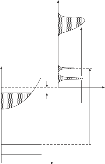
Figure 3.1 shows the energetics of the photoemission process in a simplied
density-of-states picture. Within the solid, there is an electronic density of
states governed by the intrinsic band structure and interaction effects, where
states are lled up to the Fermi energy. There is a nite potential energy
barrier between the rst occupied electronic state (at T = 0) at E
F
, and the
vacuum level (the potential energy zero innitely far away from the sample),
which is the work function of the material, F, and this is what holds the
E
kin
E
F
Spectrum
hn
F
hn
Valence band
Core levels
Sample
N(E)
N(E)
E
B
E
F
E
vac
E
3.1 The left side of the figure shows energy levels in the crystal
in the initial state. The photoemission process occurs with the
absorption of a photon with energy hn. The right side of the figure
shows the measured photoemission spectrum, starting from the
vacuum level. From Damascelli et al. (2003).

59Ultraviolet photoemission spectroscopy (UPS)
© Woodhead Publishing Limited, 2011
electrons inside the crystal. Therefore, when an electron absorbs a photon
(and does not experience any inelastic losses), the binding energy of the
electron in the initial state relative to the Fermi energy, E
B
, can be related
to the measured kinetic energy, E
kin
, by hn – F – E
F
. The work function in
question is that of the detector, since all materials have slightly different
work functions depending on the characteristics of the surface. If the work
function of the sample and detector are different, then a potential will be
set up between the sample and detector.
To relate the relationship of the photoelectron momentum to the momentum
of the photohole left in the crystal, we consider an electron in a crystal with
a well-dened quasimomentum K, and energy E
B
. If the incident photon
has enough energy to promote the electron above E
vac
, then the electron
can be photoemitted into free space. Because the electron has to propagate
a macroscopic distance (~ 1 m) to the detector, it can essentially be treated
as a free plane wave with E = hk
2
/2m. Therefore, by knowing the takeoff
angles of the electron relative to the crystal axes and the detector, we can
determine the momentum wavevector of the outcoming photoelectron. The
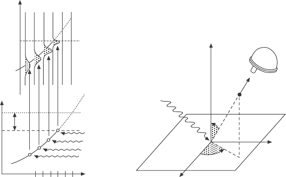
geometrical considerations for the photoemission process are illustrated in
Fig. 3.2. From this we can use momentum conservation to relate this to the
quasimomentum of the electron in the initial state. We can roughly generalize
Kinetic energy, E
kin
Energy
E
vac
E
F
k
1
k
3
k
5
k
2
k
4
k
6
Momentum
(a) (b)
f
hn
hn
q
f
hn
hn
hn
ARPES spectra
k
6
k
5
k
4
k
3
k
2
k
1
E
n
kin
= –E
B
+ hn
E
kin
= E + hn
z
e
–
Electron
analyzer
y
x
Sample
3.2 (a) Illustration of optical (direct) transitions from electrons in a
band in the crystal, to photoelectrons in free space. (b) ARPES and
measured observables, being photoelectron counts, photoelectron
kinetic energy, and polar angles of photoelectron relative to sample
crystal axes. From Damascelli et al. (2003).
60 In situ characterization of thin film growth
© Woodhead Publishing Limited, 2011
the real situation to a semi-innite crystal (innite in the lateral directions,
but with a discontinuous step – the crystal surface – in the z direction). In
this case, the momentum in the lateral direction, k
parallel
is conserved due
to translational symmetry, but the momentum in the z direction is clearly
not conserved, owing to the work function step potential at the surface (the
‘lost’ k
z
momentum of the photoelectron across the barrier is then taken
up by a recoil of the crystal). Therefore, we can obtain an exact measure
of the in-plane wavevectors of the electron in the initial state, k
parallel
, but
extracting k
z
is more challenging. To determine k
z
, one should determine the
so-called ‘inner potential’ of the crystal, which is essentially determining
which photoelectron kinetic energy corresponds to k
z
= 0 (the (0,0,0) point)
(Hufner 1995). This is typically rather involved and requires spanning a
wide range of incident photon energies, hn, in real measurements.
It is generally most appropriate to view the experimentally measured
photocurrent as a photoinduced transition between electron initial and nal
states. In UPS, the incoming photon carries negligible momentum (for hn
= 10 eV, Q ~ 5 ¥ 10
–3
A
–1
), so all transitions are essentially direct, but this
should clearly not be the case for XPS. To reach an available nal state, the
photoelectron should then be translated by a reciprocal lattice vector, G, so
that the wavevector of the outgoing photoelectron K = k + G, demonstrating
that the photoemission process requires the presence of a lattice potential to
conserve momentum. The above analysis allows us to relate the photoelectrons
measured at some kinetic energy and momentum to electrons with some
different binding energy and momentum in the initial state, inside the
crystal before the photoemission process. However, it does not tell us the
direct relationship between the electronic states of the N and N–1 system.
For this, one must make further assumptions beyond simple kinematics and
conservation relations to obtain meaningful information about the N–1 system
of the photohole from the experimentally measured photocurrent.
Rigorously speaking, the best theoretical description of the photoemission
process is the so-called ‘one-step model’. In the one-step model, the
photoemission process is treated as a single, entirely quantum-mechanical
process. However, the phenomenological model which is nearly exclusively
used in the experimental photoemission community is the ‘three-step’ model,
popularized by Berglund and Spicer in 1964. The steps in the three-step
model are as follows:
1. Optical excitation of the electron inside the solid.
2. Transport of the photoelectron to the surface.
3. escape of the photoelectron into the vacuum.
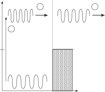
The independent steps in the three-step model are shown in Fig. 3.3. In this
semiclassical picture, only the rst step is treated quantum mechanically,
and the three steps are treated as independent of one another, in contrast

61Ultraviolet photoemission spectroscopy (UPS)
© Woodhead Publishing Limited, 2011
to the correct quantum-mechanical one-step approach. The beauty of the
three-step model is that it actually removes the complicated photoemission
process itself from photoemission. The three-step model effectively reduces
the photoemission process to an optical excitation. Steps (2) and (3) in the
three-step model are simply attenuation factors and kinematic considerations,
respectively, leaving (1) as the only nontrivial process. Given the dramatic
simplications of the three-step model, it works remarkably well and provides
surprisingly accurate results.
Here we will discuss a few additional aspects of the photoemission
process. First, photoemission is a highly surface-sensitive process because
of the strong Coulomb interactions between the photoelectron and the rest of
the electrons left in the solid, with a typical mean free path on the order of
1 nm for photon energies in the vacuum ultraviolet (VUV). Because the mean
free path depends on the cross-section of the photoelectrons to scatter with
plasmons, the mean free path should be even lower for ‘bad metals’ with low
plasmon energies such as many strongly correlated transition metal oxides.
This high degree of surface sensitivity is the reason for extremely stringent
ultra-high vacuum conditions necessary for reliable, high-resolution ARPES
experiments, and experiments are typically performed at pressures of better
than 5 ¥ 10
–11
torr. Samples are usually cleaved in situ at low temperatures
and base pressure to ensure an atomically clean surface layer. Even at these
pressures, we can occasionally observe surface degradation, although this
varies signicantly on the particular compound and contaminants in the
vacuum chamber.
z (Å)
E
initial
E
final
E
vac
1
2 3
w
3.3 Illustration of the three-step model, with (1) optical excitation
of an electron in the bulk, (2) transport of the photoelectron to the
surface, and (3) escape of the photoelectron into vacuum free space.
