Hoque. Advanced Applications of Rapid Prototyping Technology in Modern Engineering
Подождите немного. Документ загружается.

2
The Use of Rapid Prototyping
in Clinical Applications
Giovanni Biglino, Silvia Schievano and Andrew M. Taylor
Centre for Cardiovascular Imaging, UCL Institute of Cardiovascular Sciences, London,
United Kingdom
1. Introduction
This chapter will present a brief overview of the possible applications of rapid prototyping
in the medical context. Different options of clinical inputs will be discussed as well as five
detailed case studies which will demonstrate the flexibility and clinical usefulness of this
technique.
Rapid prototyping broadly indicates the fabrication of a three-dimensional (3D) model from
a computer-aided design (CAD), traditionally built layer by layer according to the 3D input
(Laoui & Shaik, 2003). Rapid prototyping has also been indicated as solid free-form,
computer-automated or layer manufacturing (Rengier et al., 2008). The development of this
technique in the clinical world has been rendered possible by the concomitant advances in
all its three fundamental steps:
1. Medical imaging (data acquisition),
2. Image processing (image segmentation and reconstruction by means of appropriate
software) and
3. Rapid prototyping itself (3D printing).
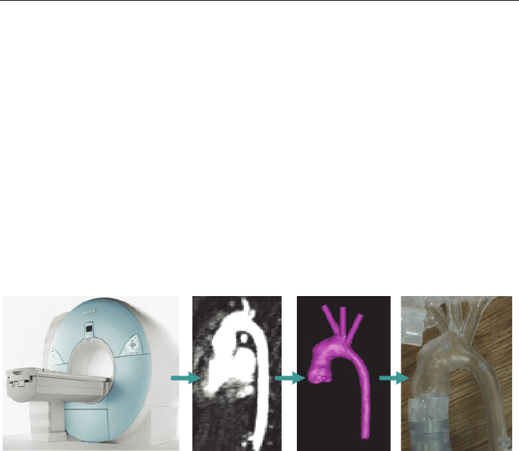
These steps are visually summarised in Figure 1.
In clinical terms, the possibility of observing, manipulating or manufacturing an anatomical
model can serve a range of significant functions (Kim et al., 2008). For instance, it can
address visualisation issues that virtual examination cannot always resolve. Also, it can be
adopted as a simulation tool or a teaching device. Moreover, it allows medical practitioners
and researchers to fully make use of the “patient-specific” concept, in terms of prosthesis
design and implant fitting but also in terms of ad hoc simulations. Finally, it can facilitate the
communication between the clinician and the patient.
The functions of rapid prototyping in the current clinical world are several (Adler &
Vickman, 1999):
Pre-surgical planning: A 3D model not only can be useful in surgical practice (i.e. a
better fitting, purposefully designed implant), but it can also help a surgical team in
visually analysing the location, size and shape of the problem. In the event of a long
operation, the model can also be used to plan and customise the surgery. This can be
especially valuable when the surgery is performed on anatomical abnormalities.
Mechanical replicas: A 3D model can be tailored to specific material properties,
including non-homogenous variations within a region. Specifically, mechanically
Advanced Applications of Rapid Prototyping Technology in Modern Engineering
22
correct bone replicas are useful in evaluating the behaviour of the bone under different
testing conditions.
Teaching aids: Offering both visualisation of anatomical details and the possibility of
practicing directly on a specimen without involving a patient, 3D models can be a
valuable tool for training nurses and doctors.
Customised implants: Instead of using a standard implant and adapting it to the
implantation site during the surgical procedure, rapid prototyping enables the
fabrication of patient-specific implants, ensuring better fitting and reduced operation
time.
Microelectromechanical systems (MEMS): These are micro-sized objects that are
fabricated by the same technique as integrated circuits. MEMS can have different
applications, including diagnostics (used in catheters, ultrasound intravascular
diagnostics, angioplasty, ECG), pumping systems, drug delivery systems, monitoring,
artificial organs, minimally invasive surgery.
Forensics: Reconstruction of crime scene and wound are also benefiting from rapid
prototyping. In particular, in the case of a surviving victim where a wound is of difficult
access, e.g. the skull, a model can be used for detailed analysis.
Fig. 1. Stages of rapid prototyping in a clinical setting. From left to right: data acquisition (in
this case with magnetic resonance (MR) imaging), image processing, 3D volume
reconstruction with appropriate software (in this case, Mimics®, Materialise, Leuven,
Belgium) and final 3D model printed in a transparent resin. The above example (aortic arch
of a paediatric patient) is discussed further in paragraph 4.2.
Despite its clinical use to the present day is still somewhat limited, considering the potential
and flexibility of this technique, it is likely that applications of rapid prototyping such as
individual patient care and academic research will be increasingly utilised (Rengier et al.,
2010).
2. Anatomical data and image acquisition
The clinical input for rapid prototyping is represented by all the information contained in
imaging data. Most commonly, MR and computerized tomography (CT) imaging are used
for this purpose. Other sources include laser surface digitizing, ultrasound and
mammography. The output of the imaging acquisition process and input of the rapid
prototyping following appropriate processing is a DICOM image (Digital Imaging and
Communications in Medicine), which is the outcome of virtually all medical professions

The Use of Rapid Prototypingin Clinical Applications
23
utilising images, including endoscopy, mammography, ophthalmology, orthopaedics,
pathology and even veterinary imaging (Lim & Zein, 2006).
2.1 Magnetic resonance imaging
MR imaging is an imaging technique based on detecting different tissue characteristics by
varying the number and sequence of pulsed radio frequency fields, taking advantage of the
magnetic relaxation properties of different tissues (Liu et al., 2006). MR imaging has the
crucial advantage of not emitting X-ray radiations. Instead, the MR scanner provides a
strong magnetic field, which causes protons to align parallel or anti-parallel to it. MR
measures the density of a specific nucleus, normally hydrogen, which is magnetic and
largely present in the human body, approximately 63% (Hornak, 1996), except for bone
structures. The speed at which protons lose their magnetic energy varies in different tissues
allowing detailed representation of the region of interest. This measurement system is
volumetric, producing isometric 3D images (i.e. the same resolution in all directions).
2.2 Computerized tomography
Hard tissues and bony structures, which are assessed less well by MR imaging, can be
captured by means of CT. This is a radiographic technique that uses a narrow fan X-ray
beam to scan a slice of tissue from multiple directions. The absorption of different tissues is
calculated and displayed according to gray-scale values. The resolution of CT data can be
increased by decreasing the slice thickness, producing more slices along the same scanned
region. However, the resulting longer scanning time has to be weighed by the clinician
against the consequence of increased radiation dose (Liu et al., 2006). The technology known
as spiral CT allows for shorter scanning time and small slice intervals with respect to
previous scanners. In this case the patient is translated continuously through the gantry
while the X-ray tube and detector system are continuously rotating, the focus of the X-ray
tube essentially describing a spiral.
2.3 Other methods
Laser surface digitizing is a technique that permits acquisition only of external data, while
MR and CT comprise both internal and external data, thus reducing scanning time and file
size (Liu et al., 2006). This technology is based on a laser probe emitting a diode-based laser
beam which forms profiles on the surface of the anatomy being imaged. Each profile is
collected as a polyline entity and the combination of profiles yields a 3D volume. Apart from
the speed of acquisition, this method has the advantage of not emitting any radiation. An
early proposed application of laser surface digitizing regarded the case study of an ear
prosthesis model (Ching et al., 1998).
3D ultrasound has also been used as input for rapid prototyping applications, as in the case
of foetal modelling (Werner et al., 2010)
3. Model fabrication
The methods used for manufacturing a physical model by rapid prototyping can be
generally divided into two major categories: “additive” and “subtractive”. Additive
manufacturing indicates the fabrication of a part by adding materials to a substrate. On the
other hand, a subtractive process involves machining using high-speed spindles and fairly

Advanced Applications of Rapid Prototyping Technology in Modern Engineering
24
machinable aluminium alloys in order to provide fast turnarounds for tooling and
functional parts (Destefani, 2005). The choice between additive and subtractive rapid
prototyping requires the evaluation of parameters such as speed of manufacturing, desired
accuracy and budget (Mishek, 2009). In the clinical context, since subtractive techniques
have the limitation of reduced ability in printing complex geometries and of requiring hard
materials, additive techniques are more commonly employed.
Stereolithography: A stereolithographic system includes a bath of photosensitive resin, a
model-building platform and an ultraviolet laser for curing the resin (Winder & Bibb,
2005). The input image is divided into slices and such data is fed to the
stereolithography machine. Layers are cured in sequence, the laser guided onto the
surface of the resin by means of a PC-controlled mirror. The support platform is
lowered following the completion of each layer. Further curing occurs in an apposite
cabinet once the model is removed from the resin bath. Support structures are added to
the model in order to aid layers adhesion and then removed once the model is printed.
It is regarded that stereolithography provides the most accurate 3D models with best
surface finishing.
Fused deposition modelling: Similarly to stereolithography, this is a layer-by-layer process,
the main difference between the two being that the layers are deposited as a
thermoplastic that is extruded from a fine moving tip (Laoui & Shaik, 2003). As for
stereolithography, support structures are necessary and are extruded with a second
nozzle. The supporting elements are often printed in a different colour or using soluble
material (Winder & Bibb, 2005).
Selective laser siltering: In this case an infrared laser is used to cure a thermoplastic
powder. This technique does not require supporting structures, facilitating the cleaning
process of the models (Berry et al., 1997).
Laminated object manufacturing: Models produced with this technique are formed by
layers of paper, cut using a laser and bonded by a heating process. By nature this is an
inexpensive printing method, thus advantageous for large volumes. In clinical terms,
however, hollow structures cannot be properly modelled by this technique, so its
clinical application is limited. It has been used to produce bioceramic bone implants
and prostheses for craniofacial defects (Laoui & Shaik, 2003).
Alongside these additive-printing methods, a subtractive rapid prototyping technology can
be employed for clinical applications:
Computerised numerically controlled milling: In this case the printing process consists in
removing a layer at a time from a block of material. Albeit the complexity of the
surfaces and the detail of internal finishing are limited, this subtractive technology has
been applied to medical modelling. One example is the construction of custom titanium
implants for cranioplasty (Joffe et al., 1999).
4. Clinical case studies
The following case studies present a range of different, specific applications of rapid
prototyping in the clinical context.
4.1 Cardiac I: Refining the process of patient selection
In the past two decades, great advances in transcatheter treatment of several cardiovascular
disorders have been reported. In September 2000, Bonhoeffer et al. reported the first successful

The Use of Rapid Prototypingin Clinical Applications
25
case of a minimally-invasive procedure known as percutaneous pulmonary valve
implantation, or PPVI (Bonhoeffer et al., 2000). PPVI combines the replacement of a functional
valve with relief of stenosis of the right ventricular outflow tract (RVOT) in patients with
repaired congenital heart disease who require pulmonary valve replacement (Hijazi et al.,
2006; Lurz et al., 2008). This technique potentially offers a major alternative to surgical valve
replacement, but the success of PPVI is greatly dependent on patient selection based on
assessment of implantation site morphology and dimensions (Schievano et al., 2007).
Rapid prototyping can be a valuable instrument in assessing patient-specific characteristics
determining PPVI suitability, as demonstrated by a recent study (Schievano et al., 2007). A
population of twelve patients was retrospectively investigated, including a range of
different anatomical configurations. All patients had been referred for possible PPVI
treatment. All patients also underwent MR examinations and the MR angiogram data was
used as input for rapid prototyping development. Image processing was carried out using
Mimics® software (Materialise, Leuven, Belgium). Imaging data was viewed in 2D (sagittal,
coronal and transverse planes) and in 3D following segmentation. Segmentation masks were
appropriately modified in order to highlight the area of interest, i.e. the RVOT. Following
operations of thresholding and region-growing, a 3D volume was obtained by means of
pattern recognition and interpolation algorithms. Such volume corresponds to the blood
volume of the RVOT and, if further modifications are necessary, it is possible to operate on a
pixel-by-pixel basis on the corresponding segmentation mask and render an “updated” 3D
volume. The outer surface of the resulting volume essentially corresponds to the inner
surface of the RVOT walls. The final step of RVOT model creation is hence to create an
additional layer of fixed thickness (in this case 2 mm) around the blood volume and delete
the latter. The final volume is saved as a standard stereolithography solid-to-layer format
(STL file) and is ready to be exported into a rapid prototyping machine. The 3D printer
employed in this study was a drop-on-demand machine using thermoplastic resin (Stratasys
Genisys, Eden Prairie, MN, USA). The printer operates by means of a nozzle driven by an x-
y stage to create outlines of each layer, whose thickness was 0.33 mm. The software
controlling the machine is able to determine optimal orientation for printing the object and
the supports necessary during the printing phase. Total time for printing one model was 3-4
hours. The thin-layer finishing of the printer ensures fine definition of the physical model.
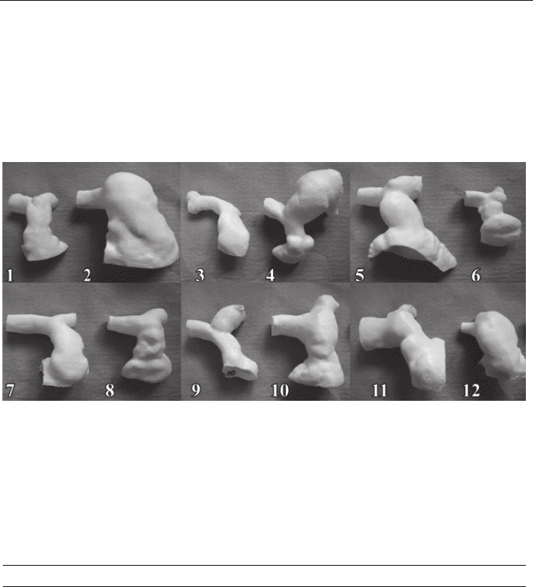
All twelve anatomical models are shown in Figure 2.
For all patients, clinical decision regarding PPVI suitability was agreed by cardiologists,
image experts and cardiac surgeons. The result was that four patients were judged as
unsuitable for PPVI, while for the remaining eight cases, where PPVI was attempted, the
procedure was successful only in four patients.
The utility of the 3D models was then evaluated retrospectively. 3D MR images alone or 3D
physical models alone were given randomly to two cardiologists who were unaware of the
clinical outcomes and who blindly re-evaluated each case solely on the base of the data
provided. For the four cases previously clinically rejected for PPVI, both cardiologists
confirmed that PPVI should have not been performed. Regarding the remaining eight
patients, the two observers correctly determined PPVI suitability in four and two cases
respectively based on MR images alone. However, when assessing the 3D models, PPVI
assessment was correct in five cases each (Table 1).
In the present application, some advantages of rapid prototyping were clearly shown, such
as facilitating clinical assessment, enabling measurements and providing a quick and
instinctive appreciation of different morphologies. One limitation of the aforementioned
Advanced Applications of Rapid Prototyping Technology in Modern Engineering
26
RVOT models is represented by the rigidity of the surface and its lack of transparency. A
compliant surface could mimic more closely, and to varying degrees of accuracy, the
mechanical properties of blood vessels. In the case of a valved stent-graft positioned in the
RVOT, such as PPVI procedure, this additional element would allow the model’s wall to
deform and accommodate the device, thus simulating the in vivo case more correctly. In
addition, wall transparency could facilitate assessment of the position of the device and also
render the model suitable for visualisation experiments. Both these points will be further
discussed in the following paragraphs.
Fig. 2. Rapid prototyping 3D models of right-ventricular outflow tract, printed by means of
stereolithography, for assessment of percutaneous pulmonary valve implantation in twelve
patients. Note, these are 12 patients with the same congenital heart disease (tetralogy of
Fallot), who have undergone the same surgical repair as neonates (complete repair), but
present with a wide variety of anatomies 10-15 years later. This demonstrates the patient-
specific nature and importance of understanding these individual anatomies. Image from
Schievano et al., 2007.
Diagnosis with MRI Diagnosis with rapid prototyping
Observer 1 Observer 2 Observer 1 Observer 2
Correct
Incorrect
Correct
Incorrect
Correct Incorrect Correct Incorrect
PPVI 4 4 2 6 5 3 5 3
no PPVI 4 0 4 0 4 0 4 0
Correct
diagnosis
14/24 18/24
Table 1. Two operators evaluating patient suitability for percutaneous pulmonary valve
implantation (PPVI): in comparison to assessment based on MR images alone, assessment
based on rapid prototyping 3D models alone increased the number of cases evaluated
correctly.
The Use of Rapid Prototypingin Clinical Applications
27
4.2 Cardiac II: Planning first-in-man device implantation
Following directly from paragraph 4.1, the wide range of RVOT anatomies impinges on the
suitability of PPVI in up to 85% of patients (Shievano et al., 2007). For this reason a second-
generation device for PPVI was conceived in order to suit a larger proportion of patients.
While the first-generation device (Melody
TM
, Medtronic Inc., Minneapolis, MN, USA) is a
cylindrical platinum-iridium stent, the new device is an hourglass-shaped nitinol covered
stent (Schievano et al., 2010). At the time of first-in-man implantation, following bench and
animal testing, rapid prototyping proved to be a precious tool for refining the procedure.
The patient-specific anatomy of RVOT, pulmonary trunk and proximal pulmonary arteries
was reconstructed from 4D CT data. The model was printed in transparent rigid resin and
the interventional cardiologists involved in this case of a novel PPVI device implantation
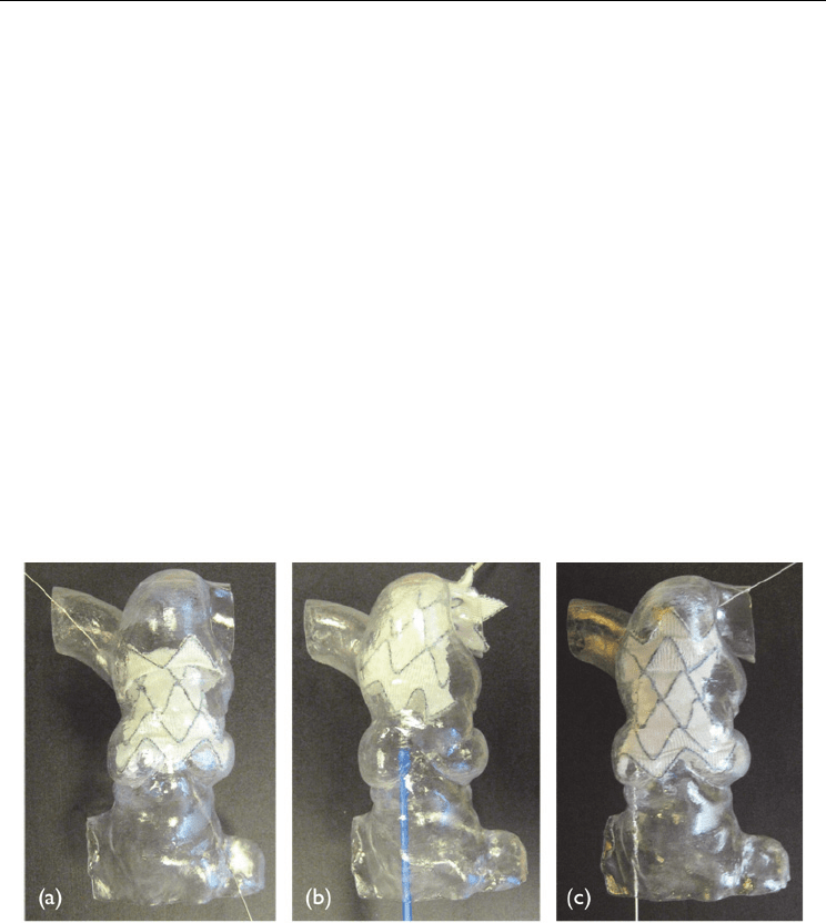
could study access route and placement on the 3D phantom. As a result, the implanters
could identify an optimal approach: guide wire in the left pulmonary artery, device
deployment with the distal portion just within the left pulmonary artery, pullback of the
device from the delivery system until correct positioning in the pulmonary trunk is
achieved. This approach, together with the alternative and unsuccessful approach via the
right pulmonary artery, is shown in Figure 3.
In this case, rapid prototyping enabled the interventional cardiologists with a visualisation
tool that they cannot normally rely on, as opposed to a surgeon who has direct visual access
to the area of interest. Testing correct positioning of the guide wire and practicing the
implantation were important steps in ensuring procedural success.
Fig. 3. Implantation of a new PPVI device into the same, patient-specific rapid prototyping
model (a) via the right pulmonary artery (RPA) and (b & c) the left pulmonary artery (LPA).
It was impossible to place the device accurately via the RPA, but implantation into the LPA
(b) with pullback into the pulmonary trunk (c) was successful. This trial implantation
directed the implantation used in the actual first-in-man procedure, which was performed
via the LPA.
4.3 Cardiac III: Bench-top experiments to integrate clinical knowledge
The first stage of Fontan palliation for neonates with hypoplastic left heart syndrome
(HLHS), namely the Norwood procedure, aims to increase the flow of oxygenated blood to

Advanced Applications of Rapid Prototyping Technology in Modern Engineering
28
the systemic circulation while, simultaneously, provide a source of pulmonary blood flow in
these single-ventricle patients (Norwood, 1991). This operation involves enlargement of the
hypoplastic aorta by means of a patch, reconstruction of aortic coarctation and increase of
pulmonary flow, the latter by means of an arterio-pulmonary (Norwood, 1991) or
ventriculo-pulmonary (Sano et al., 2003) shunt or stenting of the ductus arteriosus
(Galantowicz & Cheatham, 2005). It is thus evident that Norwood patients present a very
specific and complex arrangement of their circulatory system.
A computational model of the Norwood circulation has been already introduced
(Migliavacca et al., 2001). On the experimental side, mock circulatory systems are
acknowledged as a tool for addressing fluid mechanics questions in a systematic and
rigorous way, allowing to isolate a variable of interest in a reproducible environment.
Recent work from our group has shown the development of an in vitro setup suitable for
studying features of the circulation following the Norwood procedure and focusing initially
on the presence of aortic coarctation (Biglino et al., 2011). The setup is broadly based on the
“multiscale” concept, as it includes an anatomically accurate 3D element (the region of
interest, in this case the aortic arch) attached to a lumped parameter network (Quarteroni &
Veneziani, 2003). Rapid prototyping technology was thus employed to manufacture the 3D
elements for this first – to our knowledge – Norwood mock circulatory system.
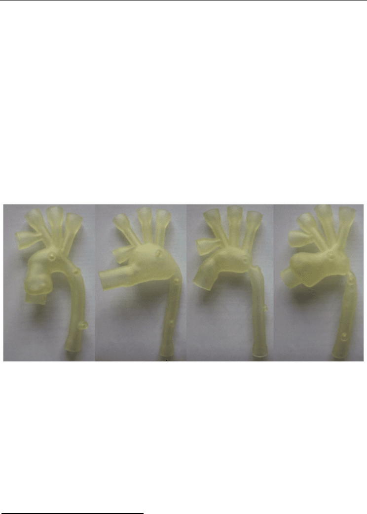
Initially, four distinct aortic arch geometries were selected: (a) “control” morphology, with
straight unreconstructed arch, (b) enlarged arch, (c) aortic coarctation (coarctation index
1
=
0.5) and (d) severe aortic coarctation (coarctation index = 0.3). Retrospective MR
angiographic data were used as input for the rapid prototyping process. Images were
analysed in Mimics® (Materialise, Leuven, Belgium) as described in paragraph 4.1. Once a
first volume rendering was available, each 3D model was modified considering the purpose
of the study. In fact, since the aim was to comment on the effect of aortic coarctation in vitro,
the brachiocephalic vessels were modified so that the variations in their dimensions from
one case to the other would not influence flow distribution, thus rendering more difficult to
discern the effect of varying arch geometry alone and nullifying one of the main benefits of
bench experiments, i.e. the ability of varying one variable at a time. Instead, CAD cylindrical
elements of equal, physiologically reasonable diameter and length were placed in the
position of the brachiocephalic vessels. Also, another element was added on all models on
one of the brachiocephalic branches (corresponding to the innominate artery) providing an
attachment for an arterio-pulmonary (or modified Blalock-Taussig) shunt-equivalent
conduit. Furthermore, conical elements were merged at all endings (shunt, upper body
vessels, and descending aorta) in order to facilitate the insertion of the model into the mock
circuit. Finally, in order to take pressure measurements at different locations, three small
cylinders the size of a 4F catheter were placed at different locations on the models (arch, just
after the coarctation – if present – and descending aorta) in order to create three ports for
pressure catheters insertion. All these volumes were merged in a unique volume, extruded
with a thickness of 1.5 mm and exported as a STL file for printing.
Each model was printed twice, employing a rigid transparent resin and a compliant opaque
composite, each offering different advantages. On the one hand, rigid models are suitable
for visualisation experiments (such as particle image velocimetry) and, albeit non-
1
The coarctation index (CI) defines the severity of a coarctation as the ratio of the narrowest diameter at
the isthmus (D
1
) and the distal diameter in the descending thoracic aorta (D
2
), CI = D
1
/D
2
(Lemler et al.,
2000).
The Use of Rapid Prototypingin Clinical Applications
29
physiological, they allow observing flow distribution without the additional variable of
compliant walls. On the other hand, flexible models are more physiological and can
replicate the arterial Windkessel.
Both versions of the models were printed with a PolyJet machine (Object Geometries Inc,
Billerica, MA, USA). The transparent resin is a commercially available material (Watershed®
XC11122). Until recently, printing a compliant phantom proved to be more difficult,
involving processes such as dripping or dipping and using rigid stereolithographic models
as scaffolds (Armillotta et al., 2007). PolyJet technology, however, allows printing flexible
models and the material of choice appears to be TangoPlus FullCure 930® or 980®, the only
difference between the two being that the first one is opaque while the latter has a black
finishing. Preliminary work from our group reveals that this material can implement
physiological compliance, depending on the wall thickness and the part of the vasculature
being modelled (Biglino et al., 2011). Both rigid and compliant models could be printed
within 24 hours
2
. The four printed geometries are shown in Figure 4.
This study showed the usefulness of PolyJet technology in producing accurate patient-
specific vascular models that can be inserted into mock circulatory systems.
Fig. 4. Four neonate aortic arches, modified for in vitro experiments, printed in TangoPlus
FullCure 930® material. Four patient-specific morphologies were selected: (a) control, (b)
enlarged aortic arch, (c) aortic coarctation and (d) severe aortic coarctation.
4.4 Dental: The use of stereolithography in maxillofacial surgery
“Can rapid prototyping ever become a routine feature in general dental practice?” asked
Harris & Rimell less than ten years ago (Harris & Rimell, 2002). Certainly, since they
highlighted the potential of this technique in their study, experience has shown that rapid
prototyping can play a role in this field. The importance of modelling in this context is
further confirmed by publications of the late 1980s and early 1990s already exploring the
potential of stereolithographic technology for maxillofacial surgery (Arvier et al., 1994; Bill
et al., 1995; Karcher et al., 1992; Lambrecht et al., 1989).
2
All models described in this paragraph were printed by Rapidform, Royal College of Art, London, UK.
(a) (b) (c) (d)
Advanced Applications of Rapid Prototyping Technology in Modern Engineering
30
Robiony et al. recently showed an integrated process involving maxillofacial surgeons,
radiologists and engineers for dental virtual surgical planning (Robiony et al., 2008). In this
case, the input data for the printing process is represented by CT images. Once the images
are imported in the dedicated software (Mimics®), the anatomical region of interest is
contoured by segmentation algorithms and the 3D structure is described by a triangle mesh
which is exported as STL file for rapid prototyping. The printing process is a standard
stereolithographic technique using liquid resin and polymerisation by a UV laser beam.
While acknowledging the importance of the physical 3D model per se, this study also
stressed the importance of being able to simulate a surgical procedure on the digital model.
Manipulation of the STL file, rather than other formats such as IGES, appeared to be the best
solution. Surgeons and engineers were thus able to import the skull model in the digital
environment and replicate a surgical procedure. This study reports that 11 patients have
been treated using this method: 3 cases of mandibular reconstruction, 5 cases of elongation
of the vertical ramus and 3 cases of sagittal elongation of the mandible.
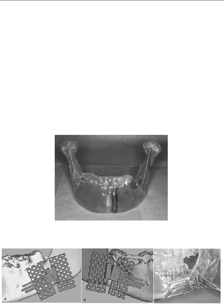
More specifically, one of the reported cases (surgical planning of emimandibular resection in
oral cancer) shows how the rapid prototyping model can highlight cancerous tissues, enable
the surgeon to make hypotheses of intervention for tumour resection and plan accurate
postoperative reconstruction (Figure 5).
Fig. 5. Stereolithographic model of the mandible printed with rigid resin. Image from
Robiony et al., 2008.
Fig. 6. Virtual simulation of mandibular elongation (A and B) and transfer of the surgical
solution to the stereolithographic model (C). Image modified from Robiony et al., 2008.
