Heitjans P., Karger J. (Eds.). Diffusion in Condensed Matter: Methods, Materials, Models
Подождите немного. Документ загружается.

374 Paul Heitjans, Andreas Schirmer, and Sylvio Indris
Then all nuclei have the same resonance frequency and a narrowed NMR line
is observed. This phenomenon is called motional narrowing. In this temper-
ature regime the spin-spin relaxation rate is approximately given by
T
−1
2
∝ J(0) ∝ τ
c
. (9.10)
The functional dependence of the linewidth on temperature allows one to
determine τ
c0
and E
A
.
The situation when at even higher temperatures the correlation rate ex-
ceeds the Larmor frequency,
τ
−1
c
>ω
L
, (9.11)
is called extreme motional narrowing. The full expression for the spin-spin re-
laxation rate in the motional narrowing and the extreme motional narrowing
regime for 3D diffusion is given in (9.28) in the Appendix (Sect. 9.9). Then
the spin-spin relaxation rate T
−1
2
and the spin-lattice relaxation rates T
−1
1
and T
−1
1ρ
should all have the same value, and the residual NMR linewidth is
determined by the inhomogeneity of the magnetic field.
Additional contributions to the lineshape can occur for nuclei with spin
I ≥ 1. These have a quadrupolar moment which interacts with electric field
gradients, when present due to low crystal symmetry at the site of the nuclei.
The general features of the relaxation rates T
−1
1
, T
−1
1ρ
and T
−1
2
in the
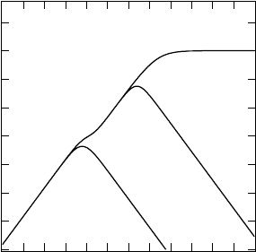
standard case of 3D diffusion via random jumps are summarized in Fig. 9.5.
The rates are plotted here as a function of ω
L
τ
c
so that it can readily be
seen that the T
−1
1
maximum occurs for ω
L
τ
c
≈ 1. As mentioned above, the
maximum of T
−1
1ρ
shows up for ω
1
τ
c
≈ 0.5 and the constant value for T
−1
2
is
reached when 2π∆ν
R
≈ 1. In the plot we used ω
L
: ω
1
:2π∆ν
R
=10
4
:20:1.
10
-3
10
-1
10
1
10
3
10
5
relaxation rate [a.u.]
10
-3
10
0
10
3
10
6
ω
L
τ
c
T
1
-1
T
2
-1
T
1
ρ
-1
Fig. 9.5. Schematic representa-
tion of the relaxation rates T
−1
1
,
T
−1
1ρ
and T
−1
2
vs. ω
L
τ
c
for 3D
diffusion via random jumps.

9NMRandβ-NMR Studies of Diffusion 375
∆ν
R
is typically some tens kHz. The shoulder in the curves of T
−1
1ρ
and T
−1
2
is due to contributions by the spectral densities at ω
L
and 2ω
L
in addition
to those at 2ω
1
and ω = 0, respectively (see (9.30) and (9.28)).
Concluding this section, we note that besides the description of diffusion
induced NMR relaxation in the perturbation approach by spectral density
functions, sketched here in a qualitative way, there are stochastic models.
These yield a more general access to spin relaxation being not confined to
the motional narrowing regimes (see, e. g., [31, 32] and references therein).
9.3 Basics of NMR Relaxation Techniques
Solid-state NMR, being based on the Boltzmann polarization of stable nuclei,
is a very broad field and for a comprehensive treatment the reader is referred
to various monographs [13, 14, 33–35]. Furthermore, detailed descriptions of
NMR relaxation techniques are available (e. g. [36, 37]), and corresponding
pulse programs are nowadays implemented in standard NMR spectrometers.
We here confine ourselves to a brief outline of the basic principles of the
measurements of longitudinal and transverse, i. e. spin-lattice and spin-spin
relaxation times, respectively. A T
1
or T
2
measurement proceeds in two steps
in the time domain:
(i) tilting the nuclear magnetization
M
0
= N ·
γ
2
2
I(I +1)
3k
B
T
· B
0
(9.12)
of an ensemble of nuclei (number density N ,spinI) at equilibrium in a static
magnetic field B
0
at temperature T by a pulsed alternating field.
(ii) detection of the magnetization M (t)asitrelaxesbacktoM
0
.
The experimental set-up consists of the sample placed in a strong mag-
netic field B
0
(of the order of some Tesla) in the z-direction and a coil wound
around the sample for application of a perpendicular alternating field B
1
(t)
with frequency ω/2π in the radio-frequency (rf) regime. In this arrangement
an equilibrium magnetization M
0
is built up in the z-direction due to the
Boltzmann population of the Zeeman energy levels (Fig. 9.6), which is given
by (9.12).
The effect of simple rf-pulse sequences is discussed in the classical picture
of a magnetization M moving in an external magnetic field. The equation of
motion is [14]
dM
dt
= M × γ [B
0
+ B
1
(t)] . (9.13)
For discussion of the motion in an alternating magnetic field applied in the x-
direction (B
1
(t)=B
x0
cos(ωt)e
x
), time dependences are eliminated by using
a coordinate system rotating about the B
0
direction with angular velocity ω.
Its axes are denoted in the following with x
, y
and z
, see Fig. 9.7. In such

376 Paul Heitjans, Andreas Schirmer, and Sylvio Indris
E
B 0¹B =0
m = -1/2
m = +1/2
DE
m = -3/2
m = +3/2
0
0
I
I
I
I
L
L
DnwEh==h
DE
Fig. 9.6. Zeeman splitting of the
nuclear energy level due to inter-
action of the nuclear spins with
a static external field B
0
,here
shown exemplarily for spin I =
3/2, γ>0.
a system the effective magnetic field B
eff
is composed of the static field B
0
,
the alternating field B
1
and a term ω/γ which results from the transition to
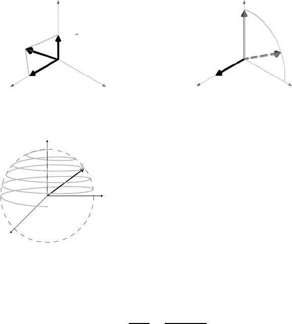
an accelerated reference frame (see Fig 9.8 (a)). When the x
-axisischosen
along B
1
the equation of motion can be rewritten as
dM
dt
= M × γ
B
0
−
ω
γ
e
z
+ B
1
e
x
= M × γB
eff
(9.14)
which means that the magnetization behaves as if it experienced a stationary
magnetic field B
eff
. If the resonance condition ω = ω
L
= γB
0
is fulfilled for
the alternating B
1
field, the z
component of B
eff
vanishes and the effective
field is B
1
e
x
and M will rotate in the z
−y
plane with a frequency ω
1
= γB
1
(see Fig. 9.8). The application of an alternating B
1
with the duration t
p
will
result in a flipping of M to the angle θ
p
= γB
1
t
p
. In the laboratory reference
frame this results in a nutational motion as shown in Fig. 9.9. By proper
choice of t
p
, M can be inverted (θ
p
= π) or tilted into the x − y plane
(θ
p
= π/2), where it will precess (in the laboratory reference frame) and
induce an observable voltage in the coil. This is the free induction decay
(FID).
z,z
x
y
x
,
y
,
,
Fig. 9.7. The rotating reference frame rotat-
ing about the direction of the external magnetic
field (z axis) with angular velocity ω.
9NMRandβ-NMR Studies of Diffusion 377
(a) (b)
x
,
y
,
z
,
B
B
B
0
1
eff
/
x
,
y
,
z
,
B
M
0
1
Fig. 9.8. (a) The effective magnetic field in the rotating reference frame. (b) The
motion of the magnetization in the rotating reference frame for ω = ω
L
.
z
x
y
M
Fig. 9.9. Nutational motion of the magnetization
in the laboratory reference frame due to an alter-
nating magnetic field perpendicular to the static
external field which is applied along the z axis.
After the π or π/2 pulse M will relax towards its equilibrium value M
0
along the +z-direction in the laboratory reference frame. The instantaneous
M
z
value may be described by
dM
z
dt
=
M
0
− M
z
T
1
(9.15)
with T
1
being the longitudinal or spin-lattice relaxation time. Equation (9.15)
yields
M
z
(t)=M
0
[1 − A
p
exp(−t/T
1
)] (9.16)
with A
p
= 2 (1) for an initial π pulse (π/2 pulse). M
z
(t) is monitored by
the amplitude of the FID after a π/2 reading pulse at the evolution time τ
e
which is varied.
In case that the magnetization M is not aligned parallel to the external
magnetic field B
0
the magnetization will precess around the magnetic field.
Furthermore, the transverse components M
x
and M
y
will decay due to a
dephasing of the nuclear spins which are in phase only directly after the rf
pulse. The equilibrium values of M
x
and M
y
are zero. The magnitudes of
M
x
and M
y
obey the equations
378 Paul Heitjans, Andreas Schirmer, and Sylvio Indris
/2
(b)
t=0 t=
B -locking field
1
/2
(a)
t=0 t=
M (t)
FID
FID
/2
(c)
t=0
t=
spin echo
t=2
FID
M (t)
M (t)
y
z
ee ee
,
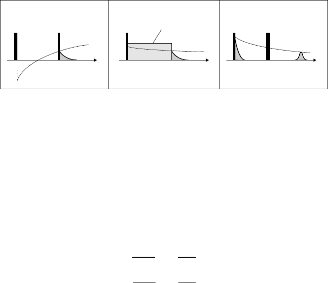
Fig. 9.10. (a) Principle of a T
1
measurement with the inversion-recovery pulse
sequence (π − τ
e
− π/2): After inversion and evolution time τ
e
the magnetization
M(τ
e
)isprobedwiththeπ/2 pulse. (b) Principle of a T
1ρ
measurement: After
a π/2 pulse M
ρ
(t)islockedbyaπ/2 phase shifted B
1
-locking field and decays
according to M
ρ
(t)=M
ρ
(0) exp(−t/T
1ρ
) which is probed at t = τ
e
after switching
off the locking field. (c) Principle of a T
2
measurement with a spin echo pulse
sequence (π/2 − τ
e
− π): After a π/2 pulse has turned the magnetization to the
plane perpendicular to the external magnetic field it starts precessing around this
field and its transverse component decays due to dephasing of the nuclear spins
according to M
y
(t)=M
0
exp(−t/T
2
). The π pulse at time τ
e
causes a refocussing
of the spins at time 2τ
e
resulting in a spin echo signal. Its amplitude can be used
to probe the magnetization M
y
(2τ
e
).
dM
x
dt
= −
M
x
T
2
dM
y
dt
= −
M
y
T
2
(9.17)
with T
2
being the transverse or spin-spin relaxation time. After an initial π/2
pulse has turned the magnetization to the y direction, (9.17) yields
M
y
(t)=M
0
exp(−t/T
2
) . (9.18)
In NMR spectrometers the coil around the sample is used for both steps
of the experiment, irradiation of the rf pulse and detection of the signal re-
sponse, i. e. the voltage induced by the precessing magnetization. Modern
NMR spectrometers further supply rf pulses which are coherent with the
possibility to control and switch the phase of different pulses. A widely used
pulse sequence for measurements of the longitudinal relaxation time is illus-
trated in Fig. 9.10 (a). It should be noted that for T
1
measurements longer
and more complex pulse sequences may be tailored to overcome limitations
of the simple sequences. The latter suffer, e. g., from the dead time of the
detection system after the strong rf pulse; in this case echo sequences may
be used to shift the FID away from the rf pulse.
As an example of how phase and power switching with modern NMR spec-
trometers are used to extend the effective magnetic field range for spin-lattice
relaxation measurements, the pulse sequence for spin locking and longitudi-
nal relaxation in the rotating frame is shown in Fig. 9.10 (b). With a π/2
9NMRandβ-NMR Studies of Diffusion 379
computer
magnet
probe
synthesizer
modulator
amplifier
PSD
reference
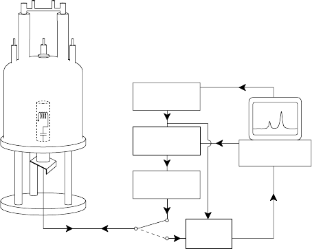
Fig. 9.11. Typical setup of an NMR spectrometer.
pulse the magnetization M
z
is turned into the x − y plane and locked paral-
lel to B
1
in a frame rotating about B
0
with ω
L
= γB
0
. During application
of the B
1
locking field the magnetization M
ρ
in the rotating frame relaxes
and M
ρ
(τ
e
) can be measured by monitoring the FID at the end of the pulse.
Thus the relaxation time T
1ρ
is obtained. It reflects the spin relaxation in the
locking field being typically some 10
−4
T. Variation of the locking field ampli-
tude gives access to studies of the field dependence of T
1ρ
. Corresponding to
the change in frequencies from the MHz to the kHz regime when SLR times
in the rotating instead of the laboratory frame are measured, considerably
longer motional correlation times can be studied. We note, that a technique
to measure SLR times also in the laboratory frame at frequencies down to
the kHz regime is field-cycling relaxometry [38], which, however, will not be
discussed here.
Fig. 9.10 (c) shows a spin echo pulse sequence which can be used to probe
the decay of the transverse magnetization component M
y
and thus to mea-
sure the transverse relaxation time T
2
.Aπ/2 pulse turns the magnetization
to the plane perpendicular to the external magnetic field where it starts pre-
cessing around this field. Its transverse component which is built by the sum
of the nuclear spins starts to decay because of a dephasing of the spins. The π
pulse at time τ
e
causes a refocussing of the spin system resulting in a so-called
spin echo signal at time 2τ
e
. Its height is proportional to the magnetization
M
y
(2τ
e
) which is measured for different evolution times τ
e
between the two
pulses. According to (9.18) measurements with different delay times τ
e
allow
one to probe the decay of the transverse components and thus to determine
T
2
.

380 Paul Heitjans, Andreas Schirmer, and Sylvio Indris
It is obvious from the discussion above that the basis of modern NMR
techniques is data acquisition after pulsed excitation and subsequent Fourier
transformation. The main components of a Fourier-NMR spectrometer are
shown in Fig. 9.11. The sample is located in the coil of a rf resonant circuit
which is part of the probe. This is located in the center of a superconductive
magnet. The nuclei in the sample are excited by rf pulses which are generated,
starting from a highly precise synthesizer, with a rf modulator and a high-
power amplifier. The response signal of the sample nuclei, being weaker by
many orders of magnitude, is directed via a rf switch to a phase sensitive
detector (PSD), whose reference signal is provided by the synthesizer. The
whole procedure which may include not only single pulses but complex pulse
sequences, as described above, is controlled by a computer.
9.4 Method of β-Radiation Detected NMR Relaxation
Technical improvements have made NMR relaxation techniques described in
Sect. 9.3 a tool of steadily increasing versatility. On the one hand, higher
magnetic fields have improved the signal-to-noise ratio by increasing the
Boltzmann factor. On the other hand, by using more sensitive amplifiers
and digitized signal recorders weaker signals can be measured and less sam-
ple material or smaller samples under extreme conditions (e. g. high-pressure
cells) can be used. An alternative approach is to replace the steps of the
NMR relaxation experiments by unconventional ones which avoid certain
limitations. In this section concepts of such a method, known as β-radiation
detected NMR (β-NMR [39]) relaxation, will be introduced and discussed.
The principle of β-NMR relaxation is the use of the β-decay radiation
asymmetry of polarized, short-lived β-emitters embedded in the solid in order
to monitor the nuclear polarization and its decrease due to longitudinal, i. e.
spin-lattice relaxation [11]. The two steps of the classical NMR relaxation
experiment (Sect. 9.3) are replaced by (i) on-line production of the short-
lived polarized probe nuclei with lifetimes τ
β
ranging from some 10 ms to some
100 s, and (ii) in-situ measurement of the β-asymmetry during a subsequent
time interval of a few lifetimes. The signal amplitude resulting from step (i)
is determined by the angular distribution of the emission probability W (θ)
of β-particles from an ensemble of polarized β-active nuclei into a solid angle
element at angle θ between polarization and emission direction
W (θ)=1+f ·
v
c
· A · cos θ. (9.19)
f is the dipolar polarization characterized by a linearly varying population of
the nuclear Zeeman levels, v is the electron velocity, c the velocity of light and
A is a constant for the specific β-decay. The β-decay radiation asymmetry
a
β
is given by the 0
◦
-180
◦
-difference of W (θ) and is a direct measure of
9NMRandβ-NMR Studies of Diffusion 381
d,p,Li
d,t, He
(a) (b)
(d)
(c)
(e)
p,d
target
& sample
Li (d,p) Li
78
Li(n, ) Lig
78
B (d,p) B
11 12
sampleC-foils
B (d,p) B
11 12
sample
target
target surfaceOP
B
n
12
B
12
Li
8
3
Li
7
d ( Li, Li) p
78
target
& sample
target
collimator
Fig. 9.12. Different techniques to produce polarized β-active nuclei in condensed
matter: (a) capture of polarized neutrons, (b) nuclear reaction with polarized accel-
erated particles, (c) particle reaction with selected recoil angle, (d) nuclear reaction
with beam-foil polarization, (e) particle reaction and polarization by optical pump-
ing (OP). In each case an exemplary nuclear reaction is given.
P = f
v
c
A, the experimentally accessible polarization. It reflects the dipolar
polarization f and any changes of it.
Variants of the β-NMR method can be classified according to the way the
short-lived β-emitters embedded in the sample are generated and polarized,
which may be done in one or two steps. The polarized β-emitters can be
produced on-line in nuclear reactions with polarized reactor neutrons or with
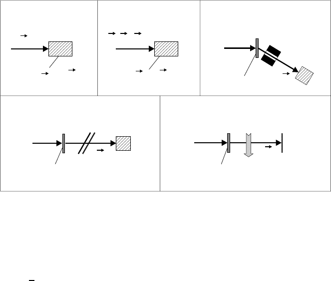
accelerator ions [39, 40]. Schemes of various production routes are shown in
Fig. 9.12. The generation and polarization by capture of polarized thermal
or cold neutrons (Fig. 9.12 (a)) will be dealt with in some detail below. With
accelerators different techniques may be applied (Fig. 9.12 (b)-(e)). First,
polarized incident particles may be used again (Fig. 9.12 (b)). In this case
one can profit from a sufficiently high polarization transfer to the short-lived
β-emitters in reactions of the type
7
Li(
−→
d,p)
8
−→
Li or
16
O(
−→
d,n)
17
−→
F [39]. The
accelerator ions have typically to have energies above 1 MeV. The β-emitters,
which can be β
+
as well as β
−
-emitters, have kinetic recoil energies in the
range of several keV and are stopped in the sample.
With unpolarized accelerator ions (Fig. 9.12 (c)-(e)) the short-lived β-
emitters emerging as recoil nuclei from the target foil have to be polarized
prior to implantation into the sample. This can be achieved either by a proper
selection of the recoil angle, into which the β-emitters escape from the reac-
tion (Fig. 9.12 (c)), or by polarizing the short-lived β-emitters after the reac-
382 Paul Heitjans, Andreas Schirmer, and Sylvio Indris
tion (Figs. 9.12 (d) and (e)). In the former case, the recoil angle selection of the
short-lived β-emitters corresponds to an angular momentum selection with a
polarization perpendicular to the reaction plane which is given by the trajec-
tories of the accelerator ion and the recoil nuclei [39,41]. Another technique to
obtain polarized β-emitters with unpolarized accelerator ions hitting the tar-
get is the use of hyperfine effects by passage of the β-emitters through carbon
foils which are tilted with respect to the beam direction [42,43] (Fig. 9.12 (d)).
A more established technique to polarize the recoil nuclei is optical pumping
which in combination with mass-separator extraction offers access to a large
variety of probe nuclei [44,45] (Fig. 9.12 (e)).
In all these nuclear techniques the polarization P , which in the classical
NMR experiments is determined by a Boltzmann factor and which gives rise
to a macroscopic magnetization M
0
(cf. (9.12)), is larger by many orders of
magnitude than a usual Boltzmann polarization and reaches some per cent.
Among the various β-NMR techniques sketched above, diffusion studies
have essentially been performed with β-NMR after capture of polarized neu-
trons (Fig. 9.12 (a)) and after a particle reaction with selected recoil angle
(Fig. 9.12 (c)). In the latter case the investigations were confined to bulk
metals and semiconductors [46–50] which is beyond the scope of this chap-
ter. Only very few diffusion studies were done using the optical pumping
technique (Fig. 9.12 (e)), e. g.
8
Li surface diffusion on Ru(001) [51] and on
Si(111) [52].
In the following the β-NMR technique after capture of polarized neutrons
will be further outlined [53]. The sample abundantly containing the nuclei
for the production reaction is placed in the field B
0
of an electromagnet and
irradiated with polarized cold neutrons. For example, in the case of the probe
nucleus
8
Li the reaction is
−→
n+
7
Li −→
8
−→
Li
∗
γ
s
−→
8
−→
Li
β
−
−→ 2α.
Established probe nuclei available with the neutron capture technique are:
8
Li (τ
β
=1.2s),
12
B(29ms),
20
F(16s),
23
Ne (57 s),
28
Al (3.2 min),
108
Ag
(3.5 min),
110
Ag (35 s),
116
In (20 s).
Special features of the β-NMR method with respect to relaxation mea-
surements are as follows. As outlined above, the polarization P is high and
independent of a Boltzmann factor. Thus low values of the magnetic field
(which provides the quantization axis) and high temperatures are accessible.
The magnetic field has to be stronger than parasitic internal and external
fields. The concentration of the probe nuclei is extremely small (typically 1
in 10
18
other nuclei) and as a consequence spin diffusion, i. e. the polariza-
tion transfer by resonant mutual spin flips of like nuclei, is suppressed due
to their large distance. Thus SLR by distant paramagnetic impurities, gen-
erally effective via spin diffusion in classical NMR relaxation measurements,
does not contribute to β-NMR relaxation. The highly diluted probe nuclei

9NMRandβ-NMR Studies of Diffusion 383
n-polarizer
macrobender
spin flipper
n-guide
chopper
electromagnet
analyzer
n-counter
sample
scintillators
Fig. 9.13. Experimental set-up of the β-NMR spectrometer at the reactor FRJ-2
of the Forschungszentrum J¨ulich.
relax individually and the β-asymmetry signal stems from an inhomogeneous
polarization average of the probe nuclei.
A technical advantage of β-radiation detected SLR is that no rf irradiation
is required. The field B
0
, correspondingly the Larmor frequency of the mea-
surement, is easily variable and skin effect problems do not arise. Bulk metal-
lic samples and/or metallic sample containers can be used. The latter is of-
ten desirable when corrosive materials are to be studied. The measurement of
SLR times T
1
is restricted to a time window of at most 0.01τ
β
<T
1
< 100τ
β
.
Fig. 9.13 shows the scheme of the β-NMR spectrometer at the research
reactor of the Forschungszentrum J¨ulich [53]. The set-up consists of a neu-
tron beam section with macrobender, neutron-polarizer and spin-flipper for
branching-off, polarizing and polarization switching of cold neutrons with a
broad wavelength distribution. The polarization switching is used in order
to cancel instrumental asymmetries of the NMR apparatus. Its main compo-
nents are chopper, collimator, sample in oven or cryostat, β-scintillator tele-
scope detectors and electromagnet. The chopper is used for the separation
of the production and the relaxation measurement periods of the short-lived
nuclei. The β-scintillator telescopes are mounted on the pole faces ‘North’
and ‘South’ and register the counting rates Z
N
and Z
S
at θ =0
◦
and 180
◦
,
respectively, which yield the experimental β-asymmetry
a
β
=
Z
N
− Z
S
Z
N
+ Z
S
. (9.20)
As pointed out above, a
β
is proportional to the nuclear polarization P . β-
NMR relaxation measurements are performed by monitoring transients a
β
(t)
after neutron activation pulses produced by the chopper. In simple cases
monoexponential transients
a
β
(t)=a
β0
· exp(−t/T
1
) (9.21)
with the SLR time T
1
are observed.
