Fischer W.B. Viral Membrane Proteins: Structure, Function, and Drug Design
Подождите немного. Документ загружается.

McCormick-Davis, C., Zhao, L.J., Mukherjee, S., Leung, K., Sheffer, D., Joag, S.V. et al. (1998). Chronology of
genetic changes in the vpu, env, and Nef genes of chimeric simian-human immunodeficiency virus (strain
HXB2) during acquisition of virulence for pig-tailed macaques. Virology 248, 275–283.
Mesleh, M.F. and Opella, S.J. (2003). Dipolar waves as NMR maps of helices in proteins. J. Magn. Reson. 163,
288–299.
Mesleh, M.F., Lee, S., Veglia, G., Thiriot, D.S., Marassi, M.M., and Opella, S.J. (2003). Dipolar waves map the struc-
ture and topology of helices in membrane proteins. J. Amer. Chem. Soc. 125, 8928–8935.
Mesleh, M.F., Veglia, G. DeSilva, T.M., Marassi, F.M., and Opella, S.J. (2002). Dipolar waves as NMR maps of
protein structure. J. Amer. Chem. Soc. 124, 4206–4207.
Miller, R.H. and Sarver, N. (1997). HIV accessory proteins as therapeutic targets. Nature Med. 3, 389–394.
Montal, M. (2003). Structure-function correlates of Vpu, a membrane protein of HIV-1. FEBS Lett. 552, 47–53.
Moore, P. B., Zhong, Q., Husslein, T., and Klein, M. L. (1998). Simulation of the HIV-1 Vpu transmembrane domain
as a pentameric bundle. FEBS Lett. 431, 143–148.
Navia, M.A., Fitzgerald, P.M., McKenner, B.M. et al. (1989). Three-dimensional structure of asparatyl protease from
human immunodeficiency virus HIV-1. Nature 337, 615–620.
Nevzorov, A.A. and Opella, S.J. (2003). A “magic sandwich” pulse sequence with reduced offset dependence for
high-resolution separated local field spectroscopy. J. Magn. Reson. 164, 182–186.
Opella, S.J. (1997). NMR and membrane proteins. Nat. Struct. Biol. NMR suppl., 845–848.
Opella, S.J., Stewart, P., and Valentine K. (1987). Protein structure by solid-state NMR spectroscopy. Q. Rev.
Biophys. 19, 7–49.
Opella, S.J., Nevzorov, A., Mesleh, M.F., and Marassi, F.M. (2002). Structure determination of membrane proteins
by NMR spectroscopy. Biochem. Cell Biol. 80, 597–604.
Pake, G. (1948). Nuclear resonance absorption in hydrated crystals: Fine structure of the proton line. J. Chem. Phys.
16, 327–336.
Park, S.H., Mrse, A.A., Nevzorov, A.A., Mesleh, M.G., Oblatt-Montal, M., Montal, M., and Opella, S.J. (2003).
Three-dimensional structure of the channel-forming trans-membrane domain of virus proteins “u” (Vpu) from
HIV-1. J. Mol. Biol. 333, 409–424.
Paul, M. and Jabbar, M.A. (1997). Phosphorylation of both phosphoacceptor sites in the HIV-1 Vpu cytoplasmic
domain is essential for Vpu-mediated ER degradation of CD4. Virology 232, 207–216.
Paul, M., Mazumder, S., Raja, N., and Jabbar, M.A. (1998). Mutational analysis of the human immunodeficiency
virus type 1 Vpu transmembrane domain that promotes the enhanced release of virus-like particles from the
plasma membrane of mammalian cells. J. Virol. 72, 1270–1279.
Prestegard, J.H., Al-Hashimi, H.M., and Tolman, J.R. (2001). NMR structures of biomolecules using field oriented
media and residual dipolar couplings. Q. Rev. Biophys. 33, 371–424.
Ritter, G.D., Jr., Yamshchikov, G., Cohen, S.J., and Mulligan, M.J. (1996). Human immunodeficiency virus type 2
glycoprotein enhancement of particle budding: Role of the cytoplasmic domain. J. Virol.
70, 2669–2673.
Schubert, U., Anton, L.C., Bacik, I., Cox, J.H., Bour, S., Bennink, J.R. et al. (1998). CD4 glycoprotein degradation
induced by human immunodeficiency virus type 1 Vpu protein requires the function of proteasomes and the
ubiquitin-conjugating pathway. J. Virol. 72, 2280–2288.
Schubert, U., Bour, S., Ferrer-Montiel, A. V., Montal, M., Maldarell, F., and Strebel, K. (1996a). The two biological
activities of human immunodeficiency virus type 1 Vpu protein involve two separable structural domains.
J. Virol. 70, 809–819.
Schubert, U., Bour, S., Willey, R.L., and Strebel, K. (1999). Regulation of virus release by the macrophage-tropic
human immunodeficiency virus type 1 AD8 isolate is redundant and can be controlled by either Vpu or Env.
J. Virol. 73, 887–896.
Schubert, U., Ferrer-Montiel, A.V., Oblatt-Montal, M., Henklein, P., Strebel, K., and Montal, M. (1996b).
Identification of an ion channel activity of the Vpu transmembrane domain and its involvement in the regula-
tion of virus release from HIV-1-infected cells. FEBS Lett. 398, 12–18.
Schubert, U., Henklein, P., Boldyreff, B., Wingender, E., Strebel, K., and Porstmann, T. (1994). The human
immunodeficiency virus type 1 encoded Vpu protein is phosphorylated by casein kinase-2 (CK-2) at positions
Ser 52 and Ser 56 within a predicted alpha-helix-turn-alpha-helix-motif. J. Mol. Biol. 236, 16–25.
Schubert, U. and Strebel, K. (1994). Differential activities of the human immunodeficiency virus type 1-encoded Vpu
protein are regulated by phosphorylation and occur in different cellular compartments. J. Virol. 68, 2260–2271.
Schwartz, M.D., Geraghty, R.J., and Panganiban, A.T. (1996). HIV-1 particle release mediated by Vpu is distinct
from that mediated by p6. Virology 224, 302–309.
162 S.J. Opella et al.
Schwartz, S., Felber, B.K., Fenyo, E.M., and Pavlakis, G.N. (1990). Env and Vpu proteins of human immuno-
deficiency virus type 1 are produced from multiple bicistronic mRNAs. J. Virol. 64, 5448–5456.
Singh, D.K., McCormick, C., Pacyniak, E., Lawrence, K., Dalton, S.B., Pinson, D.M. et al. (2001). A simian human
immunodeficiency virus with a nonfunctional Vpu (deltavpuSHIV(KU-1bMC33)) isolated from a macaque with
neuroAIDS has selected for mutations in env and nef that contributed to its pathogenic phenotype. Virology 282,
123–140.
Spevak, W., Keiper, B.D., Stratowa, C., and Castanon, M.J. (1993). Saccharomyces cerevisiae cdc15 mutants arrested
at a late stage in anaphase are rescued by Xenopus cDNAs encoding N-ras or a protein with beta-transducin
repeats. Mol. Cell Biol. 13, 4953–4966.
Stephens, E.B., McCormick, C., Pacyniak, E., Griffin, D., Pinson, D.M., Sun, F. et al. (2002). Deletion of the
vpu sequences prior to the env in a simian-human immunodeficiency virus results in enhanced Env precursor
synthesis but is less pathogenic for pig-tailed macaques. Virology 293, 252–261.
Stephens, E.B., Mukherjee, S., Sahni, M., Zhuge, W., Raghavan, R., Singh, D.K. et al. (1997). A cell-free stock of simian-
human immunodeficiency virus that causes AIDS in pig-tailed macaques has a limited number of amino acid sub-
stitutions in both SIVmac and HIV-1 regions of the genome and has offered cytotropism. Virology 231, 313–321.
Strebel, K., Klimkait, T., and Martin, M.A. (1988). A novel gene of HIV-1, vpu, and its 16-kilodalton product.
Science 241, 1221–1223.
Strebel, K., Klimkait, T., Maldarelli, F., and Martin, M.A. (1989). Molecular and biochemical analyses of human
immunodeficiency virus type 1 vpu protein. J. Virol. 63, 3784–3791.
Torres, J., Kekal, A., and Arkin, I.T. (2001). Mapping the energy surface of transmembrane helix-helix detections.
Biophys. J. 81, 2681–2692.
Turner, B.G. and Summers, M.F. (1999). Structural biology of HIV. J. Mol. Biol. 285, 1–32.
Vondrasek, J., van Bukirk, C.P., and Wlodawer, A. (1997). Database of three-dimensional structures of HIV pro-
teinases. Nat. Struct. Biol. 4,8.
Veglia, G. and Opella, S.J. (2000). Lanthanide ion binding to adventitious sites aligns membrane proteins in micelles
for solution NMR spectroscopy. J. Amer. Chem. Soc. 47, 11733–11734.
Vincent, M.J., Raja, N.U., and Jabbar, M. A. (1993). Human immunodeficiency virus type 1 Vpu protein induces
degradation of chimeric envelope glycoproteins bearing the cytoplasmic and anchor domains of CD4: Role of
the cytoplasmic domain in Vpu-induced degradation in the endoplasmic reticulum. J. Virol. 67, 5538–5549.
Wang, J., Denny, J., Tian, C., Kim, S., Mo, Y., Kovacs, F. et al. (2000). Imaging membrane protein helical wheels.
J. Magn. Reson. 144, 162–167.
Waugh, J.S. (1976). Uncoupling of local field spectra in nuclear magnetic resonance: Determination of atomic
positions in solids. Proc. Natl. Acad. Sci. USA 73, 1394–1397.
Wiertz, E.J., Jones, T.R., Sun, L., Bogyo, M., Geuze, H.J., and Ploegh, H.L. (1996). The human cytomegalovirus
US11 gene product dislocates MHC class I heavy chains from the endoplasmic reticulum to the cytosol. Cell
84, 769–779.
Willbold, D., Hoffman, S., and Rosch, P. (1997). Secondary structure and tertiary fold of the human immunodefi-
ciency virus protein U (Vpu) cytoplasmic domain in solution. Eur. J. Biochem. 245, 581–588.
Willey, R.L., Maldarelli, F., Martin, M. A., and Strebel, K. (1992). Human immunodeficiency virus type 1 Vpu
protein regulates the formation of intracellular gp160-CD4 complexes.
J. Virol. 66, 226–234.
Wlodawer, A. (2002). Rational approach to AIDS drug design through structural biology. Annu. Rev. Med. 53,
595–614.
Wlodawer, A., Miller, M., Jaskolski, M. et al. (1989). Conserved folding in retroviral proteases: Crystal structure of
a synthetic HIV-1 protease. Science 245, 616–621.
Wray, V., Federau, T., Henklein, P., Klabunde, S., Kunert, O., Schomburg, D. et al. (1995). Solution structure of the
hydrophilic region of HIV-1 encoded virus protein U (Vpu) by CD and 1H NMR spectroscopy. Int. J. Pept.
Protein Res. 45, 35–43.
Wray, V., Kinder, R., Federau, T., Henklein, P., Bechinger, B., and Schubert, U. (1999). Solution structure and
orientation of the transmembrane anchor domain of the HIV-1-encoded virus protein U by high-resolution
and solid-state NMR spectroscopy. Biochem. 38, 5272–5282.
Wu, C.H., Ramamoorthy, A., and Opella, S.J. (1994). High resolution heteronuclear dipolar solid-state NMR
spectroscopy. J. Magn. Reson., A109, 270–272.
Yaron, A., Hatzubai, A., Davis, M., Lavon, I., Amit, S., Manning, A.M. et al. (1998). Identification of the receptor
component of the IkappaBalpha-ubiquitin ligase. Nature 396, 590–594.
Structure and Function of Vpu from HIV-1 163

12
Structure, Phosphorylation, and
Biological Function of the HIV-1 Specific
Virus Protein U (Vpu)
Victor Wray and Ulrich Schubert
Knowledge describing the structure and function of the small regulatory human immuno-
deficiency virus type 1 (HIV-1) viral protein U (Vpu) has increased significantly over the last
decade. Vpu is an 81 amino acid class I oligomeric integral-membrane phosphoprotein that is
encoded exclusively by HIV-1. It can therefore be anticipated, that Vpu might contribute to
the increased pathogenic potential of HIV-1 when compared with HIV-2 that has so far had a
lower impact on the acquired immune deficiency syndrome (AIDS) pandemic. Various bio-
logical functions have been ascribed to Vpu: first, in the endoplasmic reticulum (ER) Vpu
induces degradation of CD4 in a process involving the ubiquitin–proteasome pathway and
phosphorylation of its cytoplasmic tail. In addition, there is also evidence that Vpu interferes
with major histocompatibility complex (MHC) class I antigen presentation and regulates Fas
mediated apoptosis. Second, Vpu augments virus release from a post ER compartment by a
cation-selective ion channel activity mediated by its transmembrane (TM) anchor. The phos-
phorylation of the molecule is mediated by the ubiquitous protein kinase caseinkinase 2
(CK-2) within a central conserved dodecapeptide at positions Ser
52
and Ser
56
located in a
flexible hinge region between two helical domains. Structural information, provided experi-
mentally mainly by solution- and solid-state nuclear magnetic resonance (NMR) spec-
troscopy and made possible through the availability of synthetic and recombinant material,
have shown that the biological activities of Vpu are localized in two distinct domains that are
mainly confined to the C-terminal cytoplasmic and N-terminal TM domains, respectively.
Similar to other small viral proteins that interact with membranes Vpu is a very flexible
molecule whose structure is exceptionally environment dependent. It assumes it’s most struc-
tured form in the hydrophobic environment in or at the membrane. An initial 20–23 residue
-helix in the N-terminus adopts a TM alignment while the cytoplasmic tail forms an
-helix-flexible--helix-turn motif, of which at least a part is bound parallel to the membrane
surface. Details of the arrangement of oligomeric forms of the molecule that are presumably
165
Victor Wray • Department of Structural Biology, German Research Centre for Biotechnology, Mascheroder
Weg 1, D-38124 Braunschweig, Germany. Ulrich Schubert • Institute for Clinical and Molecular Virology,
University of Erlangen-Nürnberg, Schlossgarten 4, D-91054 Erlangen, Germany.
Viral Membrane Proteins: Structure, Function, and Drug Design, edited by Wolfgang Fischer.
Kluwer Academic / Plenum Publishers, New York, 2005.
required for the ion channel activity, are emerging from recent theoretical calculations, while
this particular function is currently the area of pharmaceutical interest.
1. Introduction
Since the first description of the acquired immune deficiency syndrome (AIDS) in 1981
and the identification of human immunodeficiency Virus (HIV) as the causative ethnological
agent (Barre-Sinoussi et al., 1983) enormous progress has been made in understanding the
pathogenesis of human lentiviruses. Nonetheless, our knowledge of the contribution of par-
ticular virus factors to the induction of immunodeficiency is still far from complete. In gen-
eral, all replication competent retroviruses contain gag, pol, and env genes encoding structural
proteins and viral enzymes. Beside these retrovirus typical genes Lentiviruses, Spumaviruses,
and the human T-cell leukemia virus (HTLV) and its relatives encode small additional gene
products with regulatory functions in the viral life cycle. Unraveling the molecular structure
of these regulatory HIV proteins has proven essential for understanding and manipulating the
molecular mechanism of these viral factors (reviewed in Miller and Sarver, 1997).
Human immunodeficiency virus type 1 contains at least six regulatory genes, with the
Vpu unique to HIV-1. No structural homolog has been detected in primate Lentiviruses even
in closely related species such as HIV-2 or simian immunodeficiency virus (SIV), except for
the HIV-1 related isolate SIV
CPZ
(Huet et al., 1990). Depending on the particular HIV-1
isolate, Vpu is an 80- to 82-residue long type I anchored amphipathic membrane phospho-
protein with a functional and structural discernible domain architecture: first, Vpu augments
virus release from a post ER compartment by a cation-selective ion channel activity mediated
by its TM anchor (Ewart et al., 1996; Schubert et al., 1996a, b; for review see Lamb and
Pinto, 1997). Second, it affects the cell surface expression of several glycoproteins involved
in host immune response: well characterized is the Vpu induced degradation of the primary
virus receptor CD4 in the ER. This process requires the CK-2 dependent phosphorylation of
two conserved serine residues within the cytoplasmic tail of Vpu (Schubert et al., 1992, 1994;
Friborg et al., 1995; Paul and Jabbar, 1997) and the formation of multiprotein complexes con-
taining cellular factors such as h-TrCP and Skp1p (Margottin et al., 1998) that presumably
link the ubiquitin conjugating machinery to the cytoplasmic tail of CD4 leading to ubiquiti-
nation and finally proteolysis of CD4 by the 26S proteasome in the cytosol (Fujita et al.,
1997; Schubert et al., 1998). In addition, there is also evidence that Vpu decreases the ten-
dency for syncytia formation (Yao et al., 1993), reduces the transport of certain glycoproteins
(Vincent and Jabbar, 1995), interferes with an early step in the biosynthesis of MHC class I
molecules (Kerkau et al., 1997), and increases the susceptibility to CD95 (Fas) mediated
apoptosis (Casella et al., 1999) potentially by regulating the levels of Fas receptor at the cell
surface. While the importance of the Vpu induced interference of MHC-I and Fas pathways
for the HIV-1 replication cycle are rather obscure at the moment there are indications that the
Vpu induced CD4 degradation ensures high infectivity of HIV-1 by preventing incorporation
of envelope glycoproteins into budding virions, a function that is also supported by the acces-
sory protein Nef (Lama et al., 1999).
Like other accessory HIV gene products Vpu is not essential for virus replication in tis-
sue culture. However, it is conceivable that the two major biological functions of Vpu, down-
regulation of CD4 and augmentation of virus particle release, may contribute to the enhanced
pathogenic potential of HIV-1 when compared to its close relative, HIV-2 (Kanki et al., 1994;
166 Victor Wray and Ulrich Schubert
Marlink et al., 1994). Such a hypothesis is supported by in vivo studies indicating that Vpu
indeed increases HIV-1 pathogenicity in a SCID-hu mice model system (Aldrovandi and
Zack, 1996) or virus load and disease progression in a chimeric simian–human immunodefi-
ciency viruses (SHIV)/monkey model (Li et al., 1995).
Clearly, if Vpu is going to serve as a new target for anti-retroviral therapy and, thus,
play any role in the combinatory treatment of AIDS, a prerequisite is the understanding of the
relationship between its function and the molecular structure. Over the last few years, both
aspects of Vpu have been intensely investigated such that these details are currently furnish-
ing areas of pharmaceutical interest (Ewart et al., 2002).
2. Structure and Biochemistry of Vpu
Shortly after the discovery of Vpu (Cohen et al., 1988; Strebel et al., 1988), the protein
was characterized as a membrane phosphoprotein that is co-translationally integrated into the
ER membrane with the suspected class I topology and a reported molecular mass of 16 kDa
(Strebel et al., 1989). Besides theoretical prediction of an amphipathic protein sequence
(Strebel et al., 1989) nothing was known about the molecular structure of the protein at the
time of its first biochemical and functional characterization. To shed more light onto these
topics, we were interested in the determination of its atomic structure, the biochemical char-
acterization of the Vpu phosphorylation, and ultimately in the development of in vitro assays
for Vpu functions with the help of chemical synthesis of Vpu peptides.
A substantial amount of biochemical data exists that characterizes Vpu as a type I
oriented integral oligomeric membrane phosphoprotein composed of an amphipathic
sequence of 81 amino acids comprising a hydrophobic N-terminal TM anchor proximal to a
polar C-terminal cytoplasmic domain (Strebel et al., 1989; Klimkait et al., 1990; Maldarelli
et al., 1993). The latter contains a highly conserved dodecapeptide from Glu-47 to Gly-58 that
is conserved among all Vpu sequences of known HIV-1 isolates (Huet et al., 1990; Chen
et al., 1993) and contains two seryl residues in positions 52 and 56 which are phosphorylated
by CK-2 in a positive cooperative manner in HIV-1 infected cells (Schubert et al., 1992,
1994). Earlier studies by proteinase K digestion of membrane integrated Vpu indicated that
the membrane anchor is located at the N-terminus and contains less than 30 residues
(Maldarelli et al., 1993). Reconstitution of the synthetic membrane anchor of Vpu in planar
lipid bilayers identified a cation-selective ion channel activity (Schubert et al., 1996b) which
was also demonstrated for full-length Vpu in bilayers (Ewart et al., 1996) and in amphibian
oocytes (Schubert et al., 1996). The idea that Vpu functions as an ion channel was further-
more supported by the observation that Vpu enhances membrane permeability when
expressed in pro- and eukaryotic cells (Gonzales and Carrasco, 1998).
Our structural studies on Vpu involved the establishment of a general NMR approach
for the study of short membrane-associated peptides using a combination of high-resolution
NMR in solution and evaluation of the resulting structures by solid-state NMR in oriented
bilayers. These techniques in combination with solid phase peptide synthesis afford a very
powerful and straightforward approach to determining structural details of membrane-bound
Vpu peptides. This strategy has the advantages that peptide synthesis provides a ready source
of customized NMR probes, which can be easily and selectively labeled with
15
N.
Alternatively, the availability of such hydrophobic peptides is usually limited when recombi-
nant DNA techniques are applied, due to the low yield of mostly toxic and insoluble
Structure, Phosphorylation, and Biological Function of HIV-1 specific Vpu 167
TM peptides when expressed in bacteria, or to their general tendency to form high order
aggregates.
Over the last 10 years, considerable effort has been expended on determining the struc-
ture of Vpu as a prerequisite for understanding the molecular mechanism of its biological
activities. Initial difficulties in producing recombinant material, and the realization that the
molecule represents a type I integral membrane protein (Maldarelli et al., 1993) whose struc-
ture is environment-dependent, led to early studies being conducted on synthetic material
(Henklein et al., 1993). Our early attempts to produce recombinant Vpu failed as the protein,
when expressed in its authentic form, exhibited a high degree of cytotoxicity, probably
because of its activity on biological membranes. In addition, it formed high ordered aggre-
gates (Schubert et al., 1992). Further, we demonstrated that the recombinant Vpu could be
isolated from bacteria in a membrane-bound state, which was a bona fide substrate for in vitro
phosphorylation studies. However, the total yield of expressed protein in Escherichia coli was
far too low for production of purified material for subsequent NMR experiments (Schubert
et al., 1992). Later studies by others where successful in producing recombinant Vpu as a
fusion protein that formed inert inclusion bodies in E. coli (Ma et al., 2002). The failure of
such peptides, either produced synthetically or by recombinant techniques, to crystallize
precluded X-ray crystallographic studies and hence structural studies has relied on both
solution- and solid-state NMR spectroscopy as the main approach for producing models of
the structural domains in the molecule.
Biochemical investigations revealed that Vpu is an amphiphatic class I oligomeric inte-
gral membrane phosphoprotein that has 27 hydrophobic amino acids at its N-terminus which
function as a membrane anchor and a C-terminus of 54 hydrophilic and charged amino acids
that comprise the cytoplasmic domain. This latter domain was the first to receive attention.
Despite considerable differences in the amino acid sequences among Vpu proteins from differ-
ent HIV-1 isolates, the prediction of secondary structure suggested a strong conservation of a
supposed -helix-flexible--helix-turn motif for this domain (Schubert et al., 1994). Initial
circular dichroism data of a series of nine overlapping peptides corresponding to the cytoplas-
mic domain (Wray et al.,1995) and of larger fragments (Henklein et al., 1993) indicated the
presence of only transitory amounts of stable structure in aqueous solution alone while addition
of trifluoroethanol (TFE), a solvent that tends to favor secondary structure through stabilization
of intramolecular interactions and simulates a membrane-like milieu in solution (Buck, 1998),
afforded experimental evidence of the presence of limiting structures with two helices in
regions 28–52 and 58–72 of the cytoplasmic domain (Wray et al., 1995) separated by a loop
containing the two phosphorylation sites at Ser
52
and Ser
56
(Coadou et al., 2002).
The exact nature of the solution structure of the cytoplasmic domain (Federau et al.,
1996) was established using standard two-dimensional homonuclear
1
H NMR techniques
(Wüthrich, 1986) in combination with restrained molecular dynamics (MD) and energy min-
imization (EM) calculations (Brünger, 1992). The combined experimental data for the cyto-
plasmic domain of Vpu indicate Vpu
32–81
and a mutant in which the phosphoacceptors were
exchanged for Asn, in 50% aqueous TFE at pH 3.5, are predominantly monomeric and adopt
similar well-defined helix-interconnection-helix-turn conformations in which the four regions
are bounded by residues 37–51, 52–56, 57–72, and 73–78 (Federau et al., 1996). Identical
regions of secondary structure were determined from the
13
C and H chemical shifts of
2
H-/
13
C-/
15
N-labeled peptides analyzed in lipid micelles by multidimensional heteronuclear NMR
spectroscopy and indicated the start of the first cytoplasmic helix was at or near residue 31
(Marassi et al., 1999; Ma et al., 2002). Both helices are amphiphatic in character, but show
168 Victor Wray and Ulrich Schubert
different charge distributions. In general, the cytoplasmic region is N-terminally positively
charged, passes through a region of alternating charges in the first helix, and then becomes
negatively charged. The flexibility of the interconnecting hinge region permits orientational
freedom of the helices and comprises a highly conserved dodecapeptide (Schubert et al.,
1992). The presence of cis–trans isomerism of Pro
75
manifests itself as a doubling of cross-
peaks of Ala
74
and Trp
76
in the 2D
1
H spectra.
A
1
H NMR investigation of a peptide related to Vpu
37–81
in an organic-free high-salt
aqueous solution (Willbold et al., 1997) showed two helical secondary structures similar to
those in TFE and a less well-defined helix from 75–79 that was a turn in TFE. In order to be
compatible with the CD data in water these structures must be less stable than those in TFE.
Interestingly a small number of long-range Nuclear Overhauber Effects (NOEs) provided
evidence of a tertiary fold. Clearly this feature is very susceptible to the solution conditions
and is absent in TFE. The appearance of such weak tertiary structure in membrane-like
environments still requires verification in the context of the full-length molecule.
Attention was then focussed on the N-terminal hydrophobic domain of Vpu, which is
primarily associated with an ion channel activity either by itself (Schubert et al., 1996b) or in
the context of full-length Vpu (Ewart et al., 1996; Ma et al., 2002), and the orientation
of the various secondary structure elements of the full-length protein with respect to the
membrane. MD/EM calculations using NOE data generated as above for the soluble synthetic
peptide Vpu
1–39
in 50% TFE indicated a compact well-defined U-shaped tertiary structure
involving a short helix (residues 10–16) on the N-terminal side and a longer helix (22–36) on
the C-terminal side. The side chains of the aromatic residues, Trp
22
and Tyr
29
, in the latter
helix are directed toward the center of the molecule around which the hydrophobic core of the
folded molecule is positioned (Wray et al., 1999). In contrast to helices of the cytoplasmic
region, the helix region in the N-terminus is present at the lowest TFE concentrations and
may even be present in the absence of membrane mimetic although of limited solubility
(Wray et al., 1999). The tertiary structure however was inconsistent with the formation of ion-
conductive membrane pores in planar lipid bilayers (Schubert et al., 1996b) as this U-folded
N-terminus is unlikely to be able to span the bilayer. Consequently proton-decoupled
15
N
cross-polarization solid-state NMR spectroscopy has been employed to investigate full-length
Vpu and its isolated domains oriented in phospholipid bilayers using either synthetic
discretely
15
N-labeled amino acids (Wray et al., 1999; Henklein et al., 2000) or uniformly
15
N-labeled recombinant material (Marassi et al., 1999; Ma et al., 2002; Ho Park et al., 2003).
Details of these solid-state NMR approaches are reviewed more comprehensively in
Chapter 13 by Bechinger and Chapter 11 by Opella, respectively, in this volume. In brief the
15
N chemical shift data and line widths of N-terminal domain molecules are consistent with
a TM alignment of a helical polypeptide, implying that the nascent helices in the folded solu-
tion structure reassemble to form a linear -helix involving residues 6–29 that lies parallel to
the bilayer normal with a tilt angle of 30 (Wray et al., 1999) and placing Trp
22
near to the
Glu
28
-Tyr-Arg-motif (important for helix termination, anchorage and pore selectivity,
Sramala et al., 2003) at the membrane-cytoplasm interface. Detailed simulations of the
novel
1
H–
15
N dipolar coupling/
15
N chemical shift distribution afford an average tilt angle of
approximately 13 (Ho Park et al., 2003). A somewhat smaller value of 6.5 1.7 has been
concluded from independent site-specific Fourier transform Infrared dichroism data for
13
C-labeled Vpr
1–31
peptides (Kukol and Arkin, 1999).
For the cytoplasmic domain both solid-state NMR approaches (Marassi et al.,
1999; Henklein et al., 2000) agree that the first helical cytoplasmic domain, residues 31–51,
Structure, Phosphorylation, and Biological Function of HIV-1 specific Vpu 169
interacts strongly with the bilayer and assumes an orientation parallel to the membrane
surface. In contrast, data for the second C-terminal helix in an isolated fragment (Vpu
51–81
)
showed little interaction with the membrane (Henklein et al., 2000), while interpretation of
data from a comparison of uniformally
15
N-labeled cytoplasmic fragments for this helix is
ambiguous (Marassi et al., 1999). Interestingly synchrotron radiation-based X-ray reflectiv-
ity methods applied to Langmuir monolayers of mixtures of Vpu and an appropriate phos-
pholipid (Zheng et al., 2001) offers significant insight into structural changes that occur at the
membrane upon changing protein concentration and surface pressure. Thus the tilt angle of
the TM domain decreases with increasing pressure and at medium pressure both cytoplasmic
helices lie on the surface of the phospholipid headgroups. At low pressure the increased tilt
angle of the membrane domain disrupts the position of the cytoplasmic helices and forces
them into the bulk water phase with presumably concurrent loss in structure. In contrast at the
highest pressures measured where there was insufficient space for both helices to lie on the
surface the second helix is forced off the surface to form a two-helix bundle. Although these
experimental parameters may not reflect physiological conditions in a comprehensive way,
they do emphasize the various possibilities that are inherent in the system and that are pre-
sumably strongly influenced by environmental conditions, protein phosphorylation state,
membrane composition, and protein–protein interactions.
3. Biochemical Analysis of Vpu Phosphorylation
Following the first report that Vpu is post-translationally modified by phosphorylation
(Strebel et al., 1989) we searched for consensus sequences for eukaryotic protein kinases
within the Vpu protein and identified the seryl residues in positions 52 and 56 as two poten-
tial phosphorylation sites that correspond to the consensus
S
/
T
XX
D
/
E
, the minimal sequence
recognized by the ubiquitous casein kinase-2 (CK-2) (Schubert et al., 1992). The two CK-2
phosphorylation sites are conserved in all known Vpu sequences and represent the consensus
S
52
GN(E/D)S(E/D)G(E/D)
59
. The assumption that Vpu is a substrate for CK-2 in vivo was
first supported by our observation that phosphorylation of Vpu in HIV-1 infected T-cells can
be blocked by inhibitors specific for CK-2 (Schubert et al., 1992). Furthermore, using bacte-
rial expressed recombinant Vpu as a substrate for in vitro phosphorylation studies we were
able to show that membrane-bound full-length Vpu can be phosphorylated by purified CK-2
or by CK-2 containing extracts from mammalian cells. For identification of phosphoacceptor
sites in Vpu and for biochemical characterization of the kinase reaction we employed syn-
thetic peptides of the cytoplasmic tail of Vpu. Initially, we investigated phosphorylation of
synthetic peptides comprising the hydrophilic, polar C-terminal domain of Vpu from position
I
32
to L
81
. We were able to demonstrate that a peptide, Vpu
32–81
, containing the wild-type
sequence was phosphorylated in vitro by purified recombinant enzyme CK-2 or by whole cell
extract of mammalian cells, and that inhibitors specific for CK-2 can block the in vitro phos-
phorylation. In contrast, a corresponding mutant peptide, Vpum
2/6
, was spared by CK-2 con-
firming that phosphorylation occurs at both predicted phosphoacceptor sites. Serine residues
in positions 52 and 56 were replaced by asparagine in the sequence of the mutated peptide
Vpum
2/6
. Since both, serine and asparagine, have similar effects on secondary structure
according to the “structure derived correlation matrix (SCM)” described by Niefind and
Schomburg (1991), these replacements should not influence the structure of the protein
170 Victor Wray and Ulrich Schubert
backbone, a prediction which was later confirmed by
1
H NMR spectroscopy (Wray et al., 1995;
Federau et al., 1996). Direct evidence was provided that CK-2 targets the phosphoacceptor
sites individually in both positions. For this purpose, the K
m
values of CK-2 to three 54 amino
acid peptides comprising the entire hydrophilic part and containing single serine to asparagine
transitions in either position 52 or 56 were established (Vpu52, Vpu56, and Vpuwt (Schubert
et al., 1994)). The 3-fold higher K
m
value of CK-2 to Vpu56 revealed a preferential phospho-
rylation of S
56
over S
52
. This would be in accordance with a positive cooperative mechanism
of CK-2-phosphorylation of Vpu in which phosphorylation of the residue S
56
occurs first.
Subsequently, phosphoserine in position 56 stimulates phosphorylation of the second phos-
phoacceptor S
52
. This model of sequential CK-2 phosphorylation was recently supported by
the findings from Paul and Jabber (1997) that dual phosphorylation of serine residues in both
positions, 52 and 56, but not individual phosphorylation of either one of these residues are
sufficient to induce Vpu mediated proteolysis of CD4. These in vitro analyses provided the
basis for follow up studies demonstrating that in the context of HIV-1 the Vpu induced degra-
dation of CD4 is strictly dependent on the phosphorylation of both acceptor sites, serine 52
and 56 (Schubert and Strebel, 1994). In contrast, the virus release function of Vpu, is not con-
trolled by phosphorylation, and furthermore, this activity depends on the ability of the mem-
brane anchor of Vpu to form cation-selective ion channels. This observation led to the model
of the two distinct functional and structural domain architecture of Vpu (Schubert et al.,
1996a).
Although phosphorylation is of crucial importance for the regulation of Vpu function
its structural consequences have received scant attention. Solution NMR studies have been
restricted to monitoring changes that occur on phosphorylation in the fragment 41–62 in
water and 50% TFE at pH 3.5 and 7.2 (Coadou et al., 2002). Distinct changes are observed
in the chemical shift and NOE patterns which correspond to some loss of helix propensity in
the region 42–49 as well as a change toward a more -strand-like structure for residues 50–62
with a corresponding displacement of the C-terminal helix. The relevance of these to the
membrane-bound protein are difficult to assess, particularly as the sensitivity of current solid-
state NMR methods did not disclose any significant changes in the orientation of the first
cytoplasmic helix upon phosphorylation (Henklein et al., 2000) while the consequences for
the entire domain awaits investigation.
Thus the experimental evidence demands a dynamic two domain model for the
membrane-associated structure of monomeric full-length Vpu shown in Figure 12.1 that
corresponds in many aspects to those already reported in the literature by us (Wray et al.,
1999; Henklein et al., 2000) and others (Marassi et al., 1999). Most recently attention has
been focused on the ion channel activity of Vpu and its similarity to other viral ion channels
(for review see Fischer and Sansom, 2002). Although direct experimental evidence of the
structure of the ion channel are unavailable a considerable number of MD investigations of
various Vpu fragments embedded in octane/water and lipid bilayer systems (for recent results
see Cordes et al., 2002 and references therein and Lopez et al., 2002) combined with con-
ductance measurements provide evidence for water-filled five-helix bundles, the relative ori-
entations of their monomeric units and rationale of the weak cation selectivity. Progress in
this area is considered in Chapter 14 by Lemaitre et al. (this volume).
In summary, although the monomeric model provides details of the topology and
positions of secondary structure in the bound state it is clear that considerable more experi-
mental and theoretical work is required if a meaningful rationalization is to be achieved
of Vpu in its functional forms in vivo where it exists as a phosphoprotein in multiprotein
Structure, Phosphorylation, and Biological Function of HIV-1 specific Vpu 171
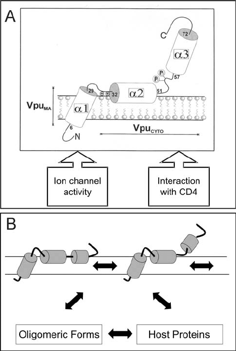
complexes involving CD4, -TrCp, and Skp 1 (Margottin et al., 1998) or in homo-oligomeric
non-phosphorylated forms required for its ion channel activity in the cell membrane.
Improvements in experimental technologies, particularly solid-state NMR, X-ray diffraction,
FTIR dichroism and cryoelectron microscopy, as well as improvements in computational
technology should provide the keys to our future understanding and facilitate approaches to
antiviral drug design.
172 Victor Wray and Ulrich Schubert
Figure 12.1. Model of the membrane-bound structures of monomeric full-length Vpu (A) and its dynamic
forms (B).