Fischer W.B. Viral Membrane Proteins: Structure, Function, and Drug Design
Подождите немного. Документ загружается.


10
Computer Simulations of Proton
Transport Through the M2 Channel of
the Influenza A Virus
Yujie Wu and Gregory A. Voth
1. Introduction
The M2 channel of the influenza A virus is formed by a viral integral membrane pro-
tein. This channel is highly proton-selective and low pH-gated. It plays important roles in the
viral life cycle, which have been illustrated by many experiments as will be further elaborated
upon in the next section, making it of great interest to drug design and medicine. These char-
acteristics of the M2 channel have attracted a significant amount of research effort in the past
couple of decades. Today, experimental studies have covered various aspects, including, but
not limited to, virology, cellular biology, electrophysiology, molecular genetics, biochemistry,
and structural biology. Despite these efforts, some of the most important properties of this
important channel remain unclear. One key example is the detailed mechanism for proton
selectivity and gating. Furthermore, information concerning the explicit dynamics of proton
transport (PT) inside the channel is largely unavailable from experiments.
Recent computer simulation studies from our group have shed some light on these
issues. From the structural and dynamical properties of the protein and pore water to the
dynamics of an excess proton inside the channel, these studies have presented us with a more
detailed picture of the M2 system at the atomic level and helped to improve our understand-
ing. This chapter summarizes our recent computer simulation results on the M2 channel with
an emphasis on the PT process through the channel and the molecular mechanisms under-
lying its selectivity and gating properties.
In the following section, an overview of current experimental results is presented.
We then review the computer simulations of explicit PT through the M2 channel and discuss
its implication for the selectivity and gating mechanisms in the following section. Finally, we
present and discuss our latest computational results concerning the possible closed and open
structures of the channel and the proton conductance mechanism.
131
Yujie Wu and Gregory A. Voth • Department of Chemistry and Henry Eyring Center for Theoretical
Chemistry, University of Utah, 315 S. 1400 E. Rm 2020, Salt Lake City, Utah.
Viral Membrane Proteins: Structure, Function, and Drug Design, edited by Wolfgang Fischer.
Kluwer Academic / Plenum Publishers, New York, 2005.
2. Overview of Experimental Results for the M2 Channel
2.1. The Roles of the M2 Channel in the Viral Life Cycle
The M2 channel is formed by the viral M2 protein that can be found in the membrane
of both the viral particle and the infected cells. When the pH at the N-terminal/extracellular
side of the channel goes lower than 5.8 or so, the channel opens and selectively transports
protons across the membrance from its N-terminal side to its C-terminal side (Pinto et al.,
1992; Chizhmakov et al., 1996).
This function of the M2 channel provides a pH-regulating mechanism that has been
found crucial in the viral life cycle. The dissociation of the viral matrix protein from the
ribonucleoprotein, which is part of the viral uncoating process, requires a low pH environ-
ment produced through the M2 channel (Bukrinskaya et al., 1982; Sugrue et al., 1990; Martin
and Helenius, 1991; Wang et al., 1993). Some influenza virus subtypes also need the M2
channel in the viral maturation process, where it elevates the intravesicular pH of the
trans-Golgi network, preventing the viral protein haemagglutinin from incorrect folding in an
otherwise low pH environment (Ciampor et al., 1992; Grambas and Hay, 1992; Grambas
et al., 1992; Takeuchi and Lamb, 1994). Experiments have shown that both parts of the viral
life cycle can be interrupted by blocking the M2 channel with the antiflu drug amantadine
(1-aminoadamantine hydrochloride, AMT) (Ciampor et al., 1992; Grambas and Hay, 1992;
Grambas et al., 1992; Sugrue et al., 1990).
2.2. The Architecture of the M2 Channel
The gene for the M2 peptide has been cloned and sequenced, revealing a primary
structure containing 97 amino acids (Lamb and Choppin, 1981; Lamb et al., 1981; Hull
et al., 1988). The peptide can fold into three structural domains: a 24-residue N-terminal/
extracellular domain, a 19-residue transmembrane (TM) domain, and a 54-residue C-terminal/
cytoplasmic domain (Lamb et al., 1985). The whole M2 protein is a parallel homotetramer of
the M2 peptide (Holsinger and Lamb, 1991; Sugrue and Hay, 1991; Pinto et al., 1977;
Sakaguchi et al., 1997; Bauer et al., 1999; Kochendoerfer et al., 1999; Salom et al., 2000;
Tian et al., 2002). The tetramer is held together mainly by noncovalent interactions. Inter-
subunit disulfide links, existing in the N-terminal domain (Holsinger and Lamb, 1991; Sugrue
and Hay, 1991; Castrucci et al., 1997; Kochendoerfer et al., 1999), further stabilizes the
tetrameric structure. The TM domain can fold into an -helix and is the main channel-
formation structure (Holsinger et al., 1994; Pinto et al., 1997). In the form of tetramer, the
helices are able to align an aqueous pore through which ions can be transported across
the membrane. A Cys-scanning mutagenesis study by Pinto et al. (1997) suggested that the
overall structure of the tetramer is a left-handed coiled-coil or helix bundle; and the tilt angle
of the -helices with respect to the membrane normal has recently been determinied for
the M2 protein in liposomes, revealing a value of 25 3 (Tian et al., 2002).
Interestingly, a synthetic 25-residue peptide (M2-TMP) with the amino acid sequence
corresponding to the residues 22–46 (encompassing the segment for the TM domain, residues
25–43) of the M2 peptide has been found to be able to form proton-selective channels in lipid
bilayers (Duff and Ashley, 1992). The M2-TMP channel is also AMT-sensitive and has simi-
lar ion selectivity and transport efficiency to the M2 channel (Duff and Ashley, 1992; Salom
et al., 2000). Furthermore, many evidences have indicated that they are also very similar in
132 Yujie Wu and Gregory A. Voth
structure. For example, it was found to form tetramers in lipid micelles (Kochendoerfer et al.,
1999; Salom et al., 2000). Circular dichroism and solid-state NMR data have shown that its
secondary structure is predominantly -helix (Duff et al., 1992; Song et al., 2000; Wang
et al., 2001). The helix tilt angle with respect to the membrane normal and the rotational angle
around the helix axis are roughly 30–40 and 50 respectively, as determined by both site-
directed infrared dichroism spectra (Kukol et al., 1999; Torres et al., 2000; Torres and Arkin,
2002) and solid-state NMR (Kovacs and Cross, 1997; Kovacs et al., 2000; Song et al., 2000;
Wang et al., 2001). The tetramer of M2-TMP was also confirmed to be left-handed so that the
hydrophilic residues can be oriented toward the pore lumen (Kovacs and Cross, 1997).
Therefore, the M2-TMP channel has been used as a simplified model for the M2 channel in
many studies.
2.3. Ion Conductance Mechanisms
The M2 channel is highly proton-selective—at least 10
6
-fold more conductive than
other cations (Chizhmakov et al., 2003; Mould et al., 2000); and it is low-pH gated—under-
going a 50-fold conductance increase from pH 8.2 to pH 4.5 (Wang et al., 1995; Mould et al.,
2000). Though the detailed structure responsible for these functions remains unclear, several
lines of evidence have suggested that a highly conservative residue His37 plays a crucial role.
This residue is the only one that has a pKa around 6 in the TM domain. Mutagenesias studies
have shown that replacing it with Ala, Glu, or Gly can result in a large increase in proton
conductance and loss of pH-induced gating behavior (Pinto et al., 1992; Wang et al., 1995).
Replacing it with Glu can also reduce its selectivity (Wang et al., 1995), while mutating it to
Cys completely abolishes its channel function (Shuck et al., 2000). The pH titration (Wang
et al., 1995) and Cu
2
inhibition experiments (Gandhi et al., 1999) also suggest that the His37
residue is implicated in the selective filter.
Two conductance mechanisms have been proposed. One is called the gating mechanism
(Sansom et al., 1997), which suggests that each of the His37 side chains may acquire an addi-
tional proton to be positively charged when the pH goes lower than the pKa. Then due to the
electrosatic repulsion, the side chains sway from each other, thus opening the otherwise occu-
luded pore to let the pore water penetrate through to form a continuous proton-conductive
water wire (proton wire). The other mechanism (known as the shuttle mechanism) suggests
that the His37 residues are directly involved in a proton relay such that they accept the excess
proton at one side while release a proton at the other side of the imidazole ring (Pinto et al.,
1997). In contrast to the gating mechanism, the bi-protonated intermediate is presumed to be
relatively short-lived and tends to release either the - or -hydrogen back to the pore water
to become neutral again (proton shuttling). To transport the next proton, the initial state might
be regenerated through tautomerization or flipping of the imidazole ring.
The existence of bi-protonated histidines in the open state of the M2-TMP channel has
been reported by a UV Resonance Raman (UVRR) spectroscopy study (Okada et al., 2001).
This may lend support to the gating mechanism. Futhermore, their data suggested that the
histidine residues would be fully bi-protonated since the addition of Sodium Dodecyl Sulfate
(SDS), which was expected to disrupt the bundle structure and expose the subunits to the low
pH buffer, did not yield a change in the corresponding Raman intensity. However, no evidence
was provided to show the subunits were really exposed to the buffer upon adding SDS.
Several recent experimental studies also indicated that Trp41 is another important
residue involved in the gating mechanism. This includes the UVRR experimental study
Computer Simulations of Proton Transport 133
mentioned above (Okada et al., 2001), which suggested that the Trp41 residue may have
cation– interaction with the bi-protonated histidine residues in the open state. A mutagene-
sis–electrophysiological study found that mutating Trp41 to Phe, Cys, or Ala results in
“leaky” channels that can transport protons outward (i.e., from the C-end to the N-end), sug-
gesting that Trp41 may be the actual channel gate (Tang et al., 2002). Cross and coworkers
determined that the distance between the N
-His37 and C
-Trp41 should be less than 3.9 Å
for the closed channel (Nishimura et al., 2002), implying the involvement of Trp41, and fur-
thermore they proposed a closed structure with the (t-160, t-105)* conformation for His37
and Trp41, in which the Trp41 residues form the actual channel gate.
3. Molecular Dynamics Simulations of
Proton Transport in the M2 Channel
The gating mechanism was proposed through a restrained molecular dynamics (MD)
simulation (Sansom et al., 1997), where the four charged histidines were observed to move
away to open the pore. However, a later MD simulation without restraints illustrated that a
fully biprotonated state destabilizes the overall channel structure while a doubly biprotonated
state leads to a deformed, yet closed, structure (Schweighofer and Pohorille, 2000). The
latter result suggests that a fully biprotonated channel is unlikely and the shuttle mechanism
seems more favorable.
The above simulations mainly focused on the structure and/or dynamics of the protein
and pore water molecules, and the excess proton was not included explicitly. No doubt,
explicit PT simulation in the M2 channel may yield more information regarding the mecha-
nism and the PT dynamics.
3.1. Explicit Proton Transport Simulation and Properties
of the Excess Proton in the M2 Channel
Explicit PT simulations in the M2 channel were successfully performed by our group
(Smondyrev and Voth, 2002) with the aid of a Multistate Empirical-Valence Bond (MS-EVB)
model for PT in aqueous systems (Schmitt and Voth, 1998, 1999a,b, 2000; Cuma et al., 2000,
2001; Day et al., 2002). The biomolecular system for these simulations included an excess
proton, the TM domain, and a fully solvated dimyristoylphosphatidyl choline (DMPC)
bilayer. The system was simulated under the condition of constant temperature and volume,
and no restraints were used. With a presumption that one stable biprotonated His37 might
somewhat open the channel for protons, the TM domain was constructed to contain one His
and three neutral His’s. These histidine residues were treated as chemically stable in the MD
simulations—they can neither accept nor donate a proton. Seven roughly 1 nsec MD simula-
tion trajectories were produced from different starting configurations, in all of which the
excess proton was placed inside the channel near the N-end.
134 Yujie Wu and Gregory A. Voth
*The notation— (t-160, t-105)—means that the conformations of His37 and Trp41 are the t-160 and t-105 rotamers,
respectively. The nomenclature for rotamers used here follows the Penultimate Rotamer Library (Lovell et al.,
2000). In the text, when the monoprotonation state of his histidine is taken into account, the symbol or is
added—for example, (t-160, t-105, ␦)—to indicate the histidine is - or -monoprotonated, respectively.
Detailed pictures on the dynamics and solvation structure of the excess proton in
the M2 channel were obtained from these simulations. Here the most important results
are reviewed. It was found that the excess proton in the channel still favors an Eigen-like sol-
vation structure most of the time. This property is similar to that of the bulk water. However,
the overall diffusion coefficient of the excess proton was reduced by the channel by up to a
factor of three; in some cases, it was even immobilized for long periods. It was confirmed that
the excess proton in the channel is transferred via the so-called Grotthuss structural diffusion
mechanism as in bulk water, where the excess proton’s solvation structure rather than the
hydronium is propagated in space (Tuckerman et al., 1995; Day et al., 2000).
It was also found that the dynamics of the protein has dramatic influence on the excess
proton’s motion in the channel: A frozen channel was observed in the simulations to
effectively immobilize the otherwise transferring excess proton (Figure 10.1). The same
phenomenon has also been observed in explicit PT simulations of the gramicidin A channel
(unpublished data). Though this result may not be too surprising, it is not fully understood.
A recent simulation study on the excess proton in a leucine–serine synthetic channel (LS2)
has reported the significance of the channel’s local microenvironment on the excess proton’s
hydration properties (Wu and Voth, 2003b). Further studies on this important topic are
currently underway in our group.
3.2. Implications for the Proton Conductance Mechanism
Though detailed side chains’ structure and dynamics of the amino acid residues are not
the target of the Smondyrev–Voth work, the simulation results do present a different view of
the conductance mechanism in terms of PT. The MD simulation trajectories have reached
roughly 1 nsec. Within this timescale, the excess proton was observed to pass through the
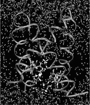
channel at different times in three of the seven trajectories. A simulation snapshot when the
excess proton was passing through the gating region by hopping through a transient water
wire is given in Figure 10.2. Concerted widening of the pore radius was observed in the three
trajectories when the proton was passing through the gating region. This may be ascribed to
Computer Simulations of Proton Transport 135
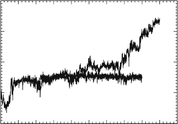
0
50
100
150
200
250
300
350
400
450 500
time
(p
sec
)
–20
–10
0
10
20
z
CEC
(Å)
Original simulation
Frozen channel
Figure 10.1. The z coordinate of the protonic center of excess charge (CEC) as a function of time for two
simulations, in one of which the channel was frozen. Notice how the CEC was effectively immobilized in the frozen
channel.
the conformational change of the side chains of the His37 residues. Although the applied TM
electric field (100–200 mV) may complicate these conclusions, this result lends direct sup-
port to the gating mechanism. However, the picture here also seems rather different from that
described previously. The MD simulations suggest that one (or maybe two) biprotonated his-
tidine may be sufficient for opening the channel for protons while still keeping it closed for
other ions. Moreover, it seems that the presence of one positive charge near the constrictive
region formed by the His37 residues does not to lead to a barrier too high for protons to pass
through.
4. Possible Closed and Open Conformations
Comparing the M2 channel model used in our previous work (Smondyrev and Voth,
2002) with the available NMR backbone structure determined by Cross et al. (2001) showed
good agreement in the helix tilt angles (Wu and Voth, 2003a). However, the rotational angles
deviate substantially. Though this result is already quite good for computer simulations
without previous knowledge of experimental structural data, a better starting structure for fur-
ther MD simulations is certainly desirable. Furthermore, it is important to include accurate
conformations of the key residues involved in the proposed conductance mechanism.
4.1. A Possible Conformation for the Closed M2 Channel
As mentioned in Section 2.3, Cross and coworkers have recently proposed a conforma-
tion of His37 and Trp41—(t-160, t-105)—for the closed M2 channel (Nishimura et al., 2002).
Nevertheless, based on the same structural information and our computational results,
136 Yujie Wu and Gregory A. Voth
Figure 10.2. An MD simulation snapshot showing the excess proton (white ball indicated by arrow) passing the
gating region by hopping through a transient water wire. The backbone of the channel is displayed by coils. The
water molecules are shown as the small angled sticks. The dots in between the two water layers are the DMPC lipids.
an alternative conformation—(t60, t90, ␦)—has recently been proposed (Wu and Voth,
2003a). This result was obtained via a thorough scan over the conformational space followed
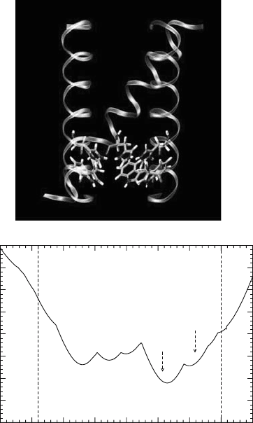
by energetic and functional assessment. A representative structure of this conformation
is shown in Figure 10.3a while Figure 10.3b shows the pore radius as a function of the
channel axis. From these figures, it can be seen that a constrictive region is formed by both
His37 and Trp41.
It was found that this conformation is consistent with nearly all relevant published
experimental results. For example, the mutagenesis, results by Pinto and colleagues (Tang
et al., 2002) can be interpreted with this conformation: The -hydrogen of His37 is pointed
toward the indole ring of Trp41, implying a hydrogen– interaction that can stabilize each
Computer Simulations of Proton Transport 137
–20
–15
–10
–5
0
5
10
15
2
0
z
(
Å
)
0
1
2
3
4
5
6
7
8
Pore radius (Å)
N-end C-end
His37
Trp41
(a)
(b)
Figure 10.3. (a) A typical structure of the (t60, t90, ␦) conformation of His37 and Trp41 for the closed channel
and (b) the corresponding pore radius profile. The backbone of the channel is displayed by coils. For the sake of
clarity, one helix and one pair of His37 and Trp41 are not shown.
other’s conformation; changing the tryptophan to Phe, Cys, or Ala would abolish this inter-
action, destabilizing the conformation of His37 and resulting in a leaky channel. In contrast,
if the Trp41 residues are mutated to Tyr, the conformation of His37 can be maintained
because the Tyr residues can provide hydroxyl groups to form hydrogen bonds with the
-hydrogens of the His37 residues. This explains why mutating Trp41 to Tyr does not lead
to a leaky channel.
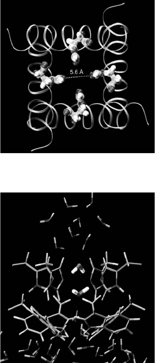
In this proposed closed conformation, the four -nitrogens of the His37 residues
are not protonated and are pointed to one another (Figure 10.4). The distance between two
138 Yujie Wu and Gregory A. Voth
Figure 10.4. The view of the same structure in Figure 10.3 from the C-end. Notice the close distance between the
two diagonal -nitrogens.
Figure 10.5. A snapshot from a MD simulation in which the backbone and heavy atoms of His37 and Trp41 were
position-restrained and the His37 and Trp41 residues took the (t60, t90, ␦) conformation. Notice the two pore waters
near the His37 residues have opposite orientation.
-nitrogens in the diagonal is within the range of 4.8–6 Å. This structure can be a good
chelating site for Cu
2
, explaining why the M2 channel can be inhibited by Cu
2
ions
(Gandhi et al., 1999).
The constrictive region formed by His37 and Trp41 is narrow enough to prevent cations
effectively larger than an excess proton from passing through the channel. To seek an answer
to how it can stop proton transport, an MD simulation has been carried out. The positions of
the backbone atoms and the heavy atoms of His37 and Trp41 were restrained in the simula-
tion to stabilize the conformation, while the other atoms were free to move. An interesting
pore water structure in the constrictive region was seen in the simulation (Figure 10.5). The
pore waters near His37 have opposite orientations. Similar pore water structure has been
found in an MD simulation of aquaporin and has been used to explain why that channel does
not transport protons (Tajkhorshid et al., 2002). This conformation also leads to an explana-
tion for the channel’s gating behavior: The opposite orientations of the two water molecules
interrupt the proton wire through which the excess proton can hop; and recovery of the pro-
ton wire must involve at least one His37 residue being protonated. The simulation also shows
that the conformation of Trp41 prevents His37 from being exposed to the bulk water at the
C-end, explaining why protons cannot be transported outward in a wild type M2 channel.
4.2. A Possible Conformation for the Open M2 Channel
Efficient proton shuttling by His37 requires its -nitrogen to be pointed toward the
N-end of the channel so that both - and -nitrogens can simultaneously form hydrogen bonds
with pore water. Furthermore, the -nitrogen should be deprotonated so that it can accept
a proton coming from the N-end of the channel. For these reasons, the orientation of His37
in our proposed closed structure does not necessarily imply the shuttle mechanism. It was
therefore necessary to find an open structure into which the closed structure can evolve upon
protonation. We found a small rotation of the histidine’s
2
angle from 60 to 0 and slight
adjustment of Trp41’s dihedral angles can lead to a conformation with the pore wide enough
for water to penetrate through (Figure 10.6) (Wu and Voth, 2003a). Our density functional
theory calculations showed a pair of histidine and tryptophan in this conformation has much
lower energy than that obtained by rotating the histidine’s
2
angle from 60 to 180, which
can yield a conformation having the same pore radius.
The close contact between His37 and Trp41 in this open structure makes it possible to
have a cation– interaction between the two residues. This is in accord with the UVRR study
(Okada et al., 2001). To examine how this structure may open the channel and at the same
time maintain the proton-selectivity, we performed an MD simulation in which the positions
of the backbone and the heavy atoms of the His37 and Trp41 residues were restrained and all
His37 residues were biprotonated. Figure 10.7 shows the pore water structure of a snapshot
after equilibration. The figure illustrates that the pore radius at the constrictive region is large
enough to allow a water molecule to fit in while small enough to prevent ions larger than an
excess proton from passing through efficiently. It can also be clearly seen that the pore water
molecules are highly ordered and their orientations forbid proton transport from the N-end to
the histidine residues. This can be ascribed to the four positively charged histidine residues.
From this result, it may be concluded that the speculative open conformation can open the
channel without loss of proton-selectivity and that it is not very likely for the open channel
to have fully biprotonated histidines unless considerable conformational change of the
backbone occur during protonation.
Computer Simulations of Proton Transport 139
4.3. Further MD Simulations with the Closed Structure
In order to examine the stability of the closed conformation, we have recently per-
formed two MD simulations with the Amber 99 force field. One of them has position-
restraints only on the backbone atoms, while the other has no restraint. The conformational
stability can be evaluated through the calculated root mean squared deviations (RMSD) as
functions of time for the side chain atoms of His37 and Trp41 (Figure 10.8). Interestingly, the
conformation is more stable in the restrained simulation, while it is not in the free simulation.
Visual examination of the final structure also confirmed that the conformation of His37 and
Trp41 was maintained very well in the restrained simulation, while one pair of His37
and Trp41 in the free one lost their original conformation. The high stability in the restrained
simulation suggests that dihedral potentials of Amber 99 force field for the two amino acids
140 Yujie Wu and Gregory A. Voth
–20
–15
–10
–5
0
5
10
15
20
z (Å)
0
1
2
3
4
5
6
7
8
Pore radius (Å)
Trp41
N-end C-end
His37
(a)
(b)
Figure 10.6. (a) The conformation of His37 and Trp41 for the speculative open channel and (b) the corresponding
pore radius profile. The backbone of the channel is displayed by coils. For the sake of clarity, one helix and one pair
of His37 and Trp41 are not shown.